New red blood cell and reticulocyte parameters
and reference values for healthy individuals
and in chronic kidney disease
Novos parâmetros e valores de referência eritrocitários e reticulocitários normais
e na doença renal crônica
Patrícia S. Scherer1; Daniela Moraes2; Terezinha P. Munhoz1, 3; Vanessa Sgnaolin4
1. Hospital São Lucas of Pontifícia Universidade Católica do Rio Grande do Sul (PUCRS); 2. Fundação Estadual de Produção e Pesquisa em Saúde (FEPPS); 3. Faculdade de Farmácia, PUCRS; 4. Programa de Pós-Graduação em Gerontologia Biomédica, PUCRS.
First submission on 23/12/14; last submission on 23/12/14; accepted for publication on 23/02/15; published on 20/04/15
ABSTRACT
Introduction: The importance of local references values has been well described in the literature; this is because the characteristics of
the population may inluence the laboratory tests. Objective: To establish the reference range for traditional and extended red blood cell parameters and reticulocyte indices in order to investigate its application in patients with chronic kidney disease (CKD). Materials
and methods: 249 blood donors (125 males and 124 females) were selected to establish the reference values. The hemodialysis sample
consisted of 62 patients with terminal CKD (48 male and 14 female). All analyzes were performed using the Sysmex XE-5000 automated
hematology analyzer. Results: Differences between reference values was observed in relation to gender: red blood cells (RBC), hemoglobin
(HGB), hematocrit (HCT), mean corpuscular hemoglobin concentration (MCHC), percentage of hyperchromic red blood cells (%HYPER), percentage of microcytosis (%MICRO), percentage of macrocytosis (%MACRO), absolute reticulocyte count (RET), reticulocyte hemoglobin content (RET-He), immature reticulocyte fraction (IFR), low luorescence reticulocytes (LFR), medium luorescence reticulocytes (MFR), and high luorescence reticulocytes (HFR). Individuals with CKD presented RBC, HGB, HCT, MCHC, red cell distribution width expressed as coeficient of variation (RDW-CV), percentage of hypochromic red blood cells (%HYPO), percentage of reticulocytes (RET%), RET (female group), IFR, LFR, MFR, and HFR results compatible with the anemic state, which can be observed in 91.8% of patients. All studied parameters were in the area under the curve (AUC) > 0.4. In male group, %HYPO (AUC: 0.806) and IFR (AUC: 0.762) presented higher AUC values, while female group presented %HYPO (AUC: 0.806), %HYPER (AUC: 0.815), and IFR (AUC: 0.660). Conclusion: The medical advancement, the development of new techniques and hematological parameters have revealed important information about functional integrity of bone marrow, diagnosis of anemia and recombinant human erythropoietin monitoring therapy used in hemodialysis patients.
Key words: reference values; red blood cell indices; reticulocytes; chronic renal failure.
INTRODUCTION
Automated analysis of red blood cells (RBC) is performed using the impedance methodology for obtaining red blood cell count and determination of its volume, and spectrophotometry to measure the concentration of hemoglobin (HGB)(1). From
these measurements, the RBC indices are calculated to obtain the mean corpuscular volume (MCV), mean corpuscular hemoglobin (MCH) and mean corpuscular hemoglobin
concentration (MCHC)(2). Currently, additional analyzes are
performed, allowing the evaluation of extended RBC parameters, as percentage of hypochromia: percentage of hypochromic red blood cells (%HYPO), percentage of hyperchromic red blood cells (%HYPER), percentage of microcytosis (%MICRO), and percentage of macrocytosis (%MACRO), as well as its precursor cell, the reticulocyte.
The technique used for reticulocyte counts underwent a remarkable evolution, especially with the introduction of
automated counting by low cytometry, which promoted an increase in accuracy and precision, when compared to manual counts(3, 4). Likewise, there was the introduction of reticulocytes
indices that analyze the maturity characteristics of reticulocytes by measuring the luorescence intensity of the cell, classifying reticulocytes for high, medium and low luorescence, depending on the degree of cell maturation(5, 6). Another parameter inserted,
evaluates the reticulocyte hemoglobin content (RET-He), and relects the amount of iron available for hemoglobin production in bone marrow, thus it is proposed as a marker of iron stores in the erythrocyte(7, 8).
The clinical application of new parameters is clear and have been demonstrated in the differential diagnosis of microcytic
anemia(9), evaluation of thalassemia(9, 10), and differentiation of iron
deiciency anemia in patients with chronic diseases(11), providing
previously unattainable information on erythropoiesis(12). The
assessment of response to therapy with erythropoietin and iron supplementation in chronic renal failure patients undergoing hemodialysis is an important target for treatment follow-up and requires new markers.
OBJECTIVE
The objective of this study was to establish the reference range for the traditional and extended erythrocyte parameters and reticulocytes indices in a healthy population, in order to investigate its use in patients with chronic kidney disease (CKD).
MATERIALS AND METHODS
Study design
The study was conducted at the Clinical Pathology Laboratory of Hematology Department, together with the Nephrology and Blood Bank services at Hospital São Lucas of Pontifícia Universidade Católica do Rio Grande do Sul (PUCRS), Brazil. To establish the reference values, we selected 249 blood donors, which were 125 male and 124 female. The hemodialysis sample consisted of 62 CKD terminal patients of both gender (48 male and 14 female), later, these patients were classiied for statistical analysis in anemic and non-anemic patients. According to the deinition of the World Health Organization (WHO), hemoglobin below 12 g/dl for women and 13 g/dl for men is indicative of anemia(13).
All included individuals aged 18 years or older and signed the Informed Consent Form.
Laboratory variables
Samples were collected in vacuum tubes with ethylenediamine tetraacetic acid (EDTA) anticoagulant and processed within 4 hours after collection.
All hematologic analyzes were performed using the Sysmex XE-5000 automated hematology analyzer (Sysmex, Kobe, Japan), which allows the evaluation of traditional (erythrocyte count, HGB, hematocrit [HCT], MCV, MCH, MCHC and red cell distribution width expressed as coeficient of variation [RDW-CV]) and extended (red blood cell hemoglobin content [RBC-He], %HYPO, %HYPER, %MICRO, and %MACRO) erythrocyte parameters, and reticulocytes indices (absolute reticulocyte count [RET] and percentage of reticulocyte [RET%], low luorescence reticulocytes [LFR], medium luorescence reticulocytes [MFR], high luorescence reticulocytes [HFR], immature reticulocyte fraction [IFR], and [RET-He]). In the reticulocyte channel, blood cells are stained with a luorescent dye, polymethine, which is speciic for identiication of ribonucleic acid (RNA)/deoxyribonucleic acid (DNA), and then analyzed by low cytometry.
The daily quality control was performed using the three levels of commercial control Sysmex e-Check. The interlaboratory quality was monitored by participating in Laboratory Proiciency Testing program (Proiciência em Ensaios Laboratoriais [PELM]) of the Brazilian Society of Clinical Pathology (Sociedade Brasileira de Patologia Clínica).
Data analysis
The reference values calculation was carried out according to the Clinical and Laboratory Standards Institute (CLSI) guidelines C28-using the MedCalc Statistical Software version 13.3 (Ostend,
Belgium)(14). The other statistical analyzes were performed using
the Statistical Package for the Social Sciences (SPSS) version 20.0 for Windows (Chicago, EUA). Kolmogorov-Smirnov test was used to verify the Gaussian distribution of traditional and extended red cell and reticulocyte parameters. The parameters with normal distribution were presented as mean and standard deviation; furthermore, in cases where it was indicated to reject normality, the results were reported as median and interquartile
range. Student t-test and Mann-Whitney U-test were performed
Ethical aspects
This study was approved by the Scientiic Committee of the Hospital São Lucas of PUCRS, and by the Research Ethics Committee of PUCRS, under protocol No. 09/04527.
RESULTS
Reference values
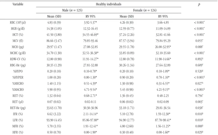
During the study period, the results of 249 healthy patients were included in the data set, represented by 125 male and 124 female. The reference values were calculated according to CLSI guidelines and are presented in Table 1.
In the comparison between genders, the parameters with p <
0.05 values must be interpreted in different ways, corresponding to RBC, HGB, HCT, MCHC, %HYPER, %MICRO, %MACRO, RET,
RET-He, IFR, LFR, MFR, and HFR. However, the other parameters (MCV, MCH, RDW-CV, RBC-He, %HYPO, and RET%) should be considered as a single reference value for both men and women.
Comparison between healthy individuals and CKD
patients
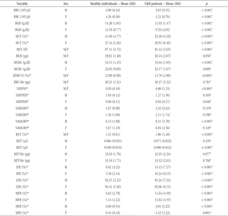
The results of traditional and extended erythrocyte parameters and reticulocytes indices of healthy individuals were compared with patients on hemodialysis (Table 2).
The previously described parameters were observed (Table 1) as having a different interpretation between men and women. The groups are made up as follows: men only, composed of 125 healthy individuals and 47 with CKD; only women, 124 healthy and 14 with CKD; and both, men and women, represented by 249 healthy individuals and 62 with CKD.
TABLE 1 − Evaluation of traditional and extended erythrocyte parameters and reticulocyte indices of healthy individuals (n = 249)
Variable Healthy individuals p
Male (n = 125) Female (n = 124)
Mean (SD) RV 95% Mean (SD) RV 95%
RBC (106/μl) 4.83 (0.39) 3.92-5.37* 4.26 (0.30) 3.66-4.85 < 0.001#
HGB (g/dl) 14.38 (1.05) 12.32-16.45 12.59 (0.77) 11.09-14.09 < 0.001#
HCT (%) 41.50 (3.80) 34.55-46.69* 37.24 (2.26) 32.81-41.66 < 0.001#
MCV (l) 86.66 (3.47) 79.85-93.46 87.57 (3.94) 79.84-95.29 0.055#
MCH (pg) 29.97 (1.47) 27.08-32.85 29.55 (1.78) 26.00-32.95* 0.088#
MCHC (g/dl) 34.70 (1.30) 32.51-36.38* 33.85 (0.89) 32.10-35.60 < 0.001#
RDW-CV (%) 12.80 (0.90) 11.91-14.27* 12.80 (0.78) 11.90-14.66* 0.892§
RBC-He (pg) 30.35 (1.29) 27.81-32.88 30.26 (1.34) 27.64-32.89 0.609#
%HYPO 0.20 (0.10) 0.10-0.78* 0.20 (0.10) 0.10-1.09* 0.520§
%HYPER 1.00 (0.20) 0.80-1.30* 0.90 (0.20) 0.70-1.10* < 0.001§
%MICRO 1.40 (1.15) 0.51-4.39* 1.30 (0.98) 0.31-6.55* 0.027§
%MACRO 5.90 (0.95) 4.71-9.54* 5.45 (0.98) 4.21-9.15* < 0.001§
RET (%) 1.32 (0.64) 0.68-2.71* 1.36 (0.45) 0.48-2.25 0.794#
RET (μl) 0.07 (0.02) 0.02-0.11 0.06 (0.02) 0.02-0.09 0.003#
RET-He (pg) 33.63 (1.70) 30.30-36.96 33.18 (1.71) 29.81-36.54 0.038#
IFR (%) 6.62 (3.22) 2.11-14.14* 5.10 (2.78) 1.59-12.30* 0.010§
LFR (%) 93.90 (4.45) 85.86-97.88* 94.90 (2.77) 87.70-98.41* 0.010§
MFR (%) 5.70 (3.55) 1.91-12.41* 4.80 (2.60) 1.56-11.25* 0.008§
HFR (%) 0.50 (0.70) 0.00-1.90* 0.30 (0.40) 0.00-1.60* 0.029§
SD: standard deviation; RV: reference value; RBC: red blood cells; HGB: hemoglobin; HCT: hematocrit; MCV: mean corpuscular volume; MCH: mean corpuscular hemoglobin; MCHC: mean corpuscular hemoglobin concentration; RDW-CV: red cell distribution width expressed as coefficient of variation; RBC-He: red blood cell hemoglobin content; %HYPO: percentage of hypochromic red blood cells; %HYPER: percentage of hyperchromic red blood cells; %MICRO: percentage of microcytosis; %MACRO: percentage of macrocytosis; RET: absolute reticulocyte count; RET-He: reticulocyte hemoglobin content; IFR: immature reticulocyte fraction; LFR: low fluorescence reticulocytes; MFR: medium fluorescence reticulocytes; HFR: high fluorescence reticulocytes.
TABLE 2 − Comparison between traditional and extended erythrocyte parameters and reticulocyte indices of healthy individuals and CKD patients Variable Sex Healthy individuals – Mean (SD) CKD patients – Mean (SD) p
RBC (106/μl) M 4.80 (0.34) 3.65 (0.55) < 0.001#
RBC (106/μl) F 4.26 (0.30) 3.21 (0.76) < 0.001#
HGB (g/dl) M 14.38 (1.05) 11.03 (1.47) < 0.001#
HGB (g/dl) F 12.59 (0.77) 9.59 (2.05) < 0.001#
HCT (%)* M 41.09 (4.77) 33.39 (4.59) < 0.001§
HCT (%)* F 37.24 (2.26) 28.91 (6.46) < 0.001§
MCV (l) M/F 87.11 (3.73) 91.42 (5.65) < 0.001#
MCH (pg) M/F 29.81 (1.48) 30.24 (2.07) 0.058#
MCHC (g/dl) M 34.51 (1.25) 33.04 (1.05) < 0.001#
MCHC (g/dl) F 33.85 (0.89) 33.17 (1.07) 0.009#
RDW-CV (%)* M/F 12.80 (0.90) 14.70 (2.00) <0.001§
RBC-He (pg) M/F 30.31 (1.31) 30.37 (2.22) 0.781#
%HYPO* M/F 0.20 (0.10) 0.80 (1.25) <0.001§
%HYPER* M 1.03 (0.12) 1.27 (1.56) 0.169§
%HYPER* F 0.90 (0.11) 0.83 (0.27) 0.040§
%MICRO* M 1.67 (0.99) 2.32 (2.62) 0.519§
%MICRO* F 1.56 (1.60) 2.11 (1.74) 0.198§
%MACRO* M 6.13 (1.08) 9.21 (5.70) < 0.001§
%MACRO* F 5.67 (1.10) 6.84 (2.56) 0.149§
RET (%)* M/F 1.31 (0.61) 1.86 (1.40) < 0.001§
RET (μl) M 0.066 (0.025) 0.071 (0.033) 0.310#
RET (μl) F 0.058 (0.019) 0.080 (0.043) < 0.001#
RET-He (pg) M 33.63 (1.70) 32.95 (3.24) 0.077#
RET-He (pg) F 33.18 (1.71) 33.32 (2.01) 0.768#
IFR (%)* M 6.62 (3.22) 14.13 (7.27) < 0.001§
IFR (%)* F 5.58 (2.44) 16.34 (9.15) < 0.001§
LFR (%)* M 93.37 (3.22) 85.34 (7.34) < 0.001§
LFR (%)* F 94.41 (2.46) 83.66 (9.15) < 0.001§
MFR (%)* M 6.02 (2.78) 11.64 (4.59) < 0.001§
MFR (%)* F 5.14 (2.22) 12.02 (4.55) < 0.001§
HFR (%)* M 0.60 (0.54) 3.01 (3.22) < 0.001§
HFR (%)* F 0.45 (0.43) 4.32 (5.22) 0.001§
CKD: chronic kidney disease: SD: standard deviation; M: male; F: female; RBC: red blood cells; HGB: hemoglobin; HCT: hematocrit; MCV: mean corpuscular volume; MCH: mean corpuscular hemoglobin; MCHC: mean corpuscular hemoglobin concentration; RDW-CV: red cell distribution width expressed as coefficient of variation; RBC-He: red blood cell hemoglobin content; %HYPO: percentage of hypochromic red blood cells; %HYPER: percentage of hyperchromic red blood cells; %MICRO: percentage of microcytosis; %MACRO: percentage of macrocytosis; RET: absolute reticulocyte count; RET-He: reticulocyte hemoglobin content; IFR: immature reticulocyte fraction; LFR: low fluorescence reticulocytes; MFR: medium fluorescence reticulocytes; HFR: high fluorescence reticulocytes.
* Kolmogorov-Smirnov test (p < 0.05); # Student’s t-test; § Mann-Whitney U-test. Individuals with CKD showed RBC, HGB, HCT, MCHC, RDW-CV, %HYPO, RET%, RET (female group), IFR, LFR, MFR, and HFR results compatible with the anemic state, which can be observed in 91.8% of these patients.
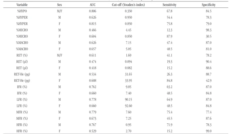
ROC curve
The results of ROC curve analysis are shown in the Figure.
FIGURE – ROC curve
ROC: receiver operating characteristic curve; %HYPO: percentage of hypochromic red blood cells; M: male; F: female; %HYPER: percentage of hyperchromic red blood cells; %MICRO: percentage of microcytosis; %MACRO: percentage of macrocytosis; RET: absolute reticulocyte count; RET-He: reticulocyte hemoglobin content; IFR: immature reticulocyte fraction; LFR: low fluorescence reticulocytes; MFR: medium fluorescence reticulocytes; HFR: high fluorescence reticulocytes.
individuals classiied as anemic, and female (n = 138), with 33 anemic women. In addition, a third group was composed of
both sexes (n = 310), with 90 anemic. All the studied parameters
showed area under the curve (AUC) > 0.4. The discriminative ability to predict anemia were: for the male group %HYPO (AUC: 0,806), IFR (AUC: 0,762) with their respective fractions LFR (AUC: 0,778), MFR (AUC: 0,779), and HFR (AUC: 0,767); for the female group %HYPO (AUC: 0,806), %HYPER (AUC: 0,815), IFR (AUC: 0,660) with their respective fractions LFR (AUC: 0,660), MFR (AUC: 0,673), and HFR (AUC: 0,529) (Table 3).
DISCUSSION
The importance of local references values has been well described in the literature, showing that variables, such as constitutional factors (gender, age and genetic variation) and extrinsic factors (posture, physical exercises, dietary habits, region of the studied population, use of caffeine and alcohol, use of drugs for therapeutic purposes or not, and pregnancy) inluence the population characteristics in which the tests will be applied in everyday life(15). WHO has suggested
the establishment of local reference values for laboratories and, along with the International Federation of Clinical Chemistry (IFCC) and CLSI, describe the reference value as the result obtained by observation or quantitative measurement of an analyte in an selected individual, based on well-deined criteria(16).
In our study, we observed the difference of the reference values between men and women in traditional red cell parameters (RBC, HGB, HCT, and MCHC), showing the importance of obtaining different ranges between genders. Similarly, for the extended red cell parameters and the reticulocytes indices (%HYPER, %MICRO, %MACRO, RET, RET-He, IFR, LFR, MFR, and HFR) was also veriied the need for different ranges between genders, reinforcing the correct classiication of reference range in hematological parameters.
According to WHO, anemia is deined as hemoglobin level < 13 g/dl in men and < 12 g/dl in women(13), one of the
most frequent and early complications in CKD(17). This study
showed that individuals with CKD have HGB lower than healthy individuals, as well as RBC and HCT values, showing signiicant statistical difference between them (p < 0.05). Studies report that
it is recommended to keep the HGB levels between 11-12 g/dl for patients with CKD, and is also essential to maintain normal iron stores before starting erythropoietin therapy(18, 19).
Another parameter that has shown high sensitivity for functional iron deiciency detection in patients with chronic
1- Specificity 1- Specificity 1.0 0.8 0.6 0.4 0.2 0.0 1.0 0.8 0.6 0.4 0.2 0.0 1.0 0.8 0.6 0.4 0.2 0.0 1.0 0.8 0.6 0.4 0.2 0.0 1.0 0.8 0.6 0.4 0.2 0.0 1.0 0.8 0.6 0.4 0.2 0.0 1.0 0.8 0.6 0.4 0.2 0.0 1.0 0.8 0.6 0.4 0.2 0.0 1.0 0.8 0.6 0.4 0.2 0.0 1.0 0.8 0.6 0.4 0.2 0.0 1.0 0.8 0.6 0.4 0.2 0.0 1.0 0.8 0.6 0.4 0.2 0.0 1.0 0.8 0.6 0.4 0.2 0.0 1.0 0.8 0.6 0.4 0.2 0.0 1.0 0.8 0.6 0.4 0.2 0.0 1.0 0.8 0.6 0.4 0.2 0.0 1.0 0.8 0.6 0.4 0.2 0.0 1.0 0.8 0.6 0.4 0.2 0.0 1.0 0.8 0.6 0.4 0.2 0.0 1.0 0.8 0.6 0.4 0.2 0.0
%HYPO (M/F) %HYPER (M) %HYPER (F)
%MICRO (M)
%MACRO (F)
%MACRO (M) %MICRO (F)
RET% (M/F)
RET (F) RET-He (M) RET-He (F) RET (M)
IFR (M) LFR (M)
LFR (F) MFR (M) MFR (F)
IFR (F)
HFR (M) HFR (F)
1- Specificity 1- Specificity 1- Specificity 1- Specificity 1- Specificity 1- Specificity 1- Specificity 1- Specificity 1- Specificity 1- Specificity 1- Specificity 1- Specificity 1- Specificity 1- Specificity 1- Specificity 1- Specificity 1- Specificity 1- Specificity Sensitivity Sensitivity Sensitivity Sensitivity Sensitivity Sensitivity Sensitivity Sensitivity Sensitivity Sensitivity Sensitivity Sensitivity Sensitivity Sensitivity Sensitivity Sensitivity Sensitivity Sensitivity Sensitivity Sensitivity
0.0 0.2 0.4 0.6 0.8 1.0
0.0 0.2 0.4 0.6 0.8 1.0 0.0 0.2 0.4 0.6 0.8 1.0
0.0 0.2 0.4 0.6 0.8 1.0
0.0 0.2 0.4 0.6 0.8 1.0 0.0 0.2 0.4 0.6 0.8 1.0
0.0 0.2 0.4 0.6 0.8 1.0 0.0 0.2 0.4 0.6 0.8 1.0 0.0 0.2 0.4 0.6 0.8 1.0 0.0 0.2 0.4 0.6 0.8 1.0
0.0 0.2 0.4 0.6 0.8 1.0 0.0 0.2 0.4 0.6 0.8 1.0
0.0 0.2 0.4 0.6 0.8 1.0 0.0 0.2 0.4 0.6 0.8 1.0
0.0 0.2 0.4 0.6 0.8 1.0 0.0 0.2 0.4 0.6 0.8 1.0 0.0 0.2 0.4 0.6 0.8 1.0 0.0 0.2 0.4 0.6 0.8 1.0
0.0 0.2 0.4 0.6 0.8 1.0
TABLE 3 − ROC curve, cutoff, sensitivity and specificity
Variable Sex AUC Cut-off (Youden’s index) Sensitivity Specificity
%HYPO M/F 0.806 0.350 67.8 84.5
%HYPER M 0.626 0.950 54.4 78.3
%HYPER F 0.815 0.850 75.8 79.0
%MICRO M 0.466 4.45 12.3 98.3
%MICRO F 0.604 0.850 87.9 30.5
%MACRO M 0.626 7.15 47.4 87.0
%MACRO F 0.657 5.05 48.5 81.0
RET (%) M/F 0.611 1.69 41.1 78.2
RET (μl) M 0.474 0.094 19.3 90.4
RET (μl) F 0.418 0.082 15.2 88.6
RET-He (pg) M 0.534 31.65 26.3 88.7
RET-He (pg) F 0.608 33.95 84.8 42.9
IFR (%) M 0.762 9.85 63.2 87.0
IFR (%) F 0.660 7.40 48.5 84.8
LFR (%) M 0.778 90.15 64.9 87.0
LFR (%) F 0.660 92.60 48.5 84.8
MFR (%) M 0.779 7.60 75.4 77.4
MFR (%) F 0.673 7.25 45.5 87.6
HFR (%) M 0.767 0.95 71.9 78.3
HFR (%) F 0.529 2.70 15.2 99.0
ROC: receiver operating characteristic; AUC: area under the curve; M: male; F: female; %HYPO: percentage of hypochromic red blood cells; %HYPER: percentage of hyperchromic red blood cells; %MICRO: percentage of microcytosis; %MACRO: percentage of macrocytosis; RET: absolute reticulocyte count; RET-He: reticulocyte hemoglobin content; IFR: immature reticulocyte fraction; LFR: low fluorescence reticulocytes; MFR: medium fluorescence reticulocytes; HFR: high fluorescence reticulocytes.
renal failure is the %HYPO. Bovy et al.(20) study demonstrated
a signiicant increase in the value of %HYPO parameter in iron deicient patients, as well as in the present study, in which individuals with CKD had an increased value of this parameter in relation to the healthy individuals group, indicating the presence of hypochromic red cell in hemodialysis patients, conirmed by the of the area under the curve analysis (AUC: 0.806), and was considered a good marker for iron deiciency.
The reticulocytes indices show a direct view of the bone marrow and the supply and use of iron, along with a response to erythropoietin therapy(21). In our study, RET% and RET (female
group) in individuals with CKD showed statistical difference
(p < 0.001) compared to healthy subjects, showing that in these
patients, the bone marrow is trying to compensate for the HGB and iron decrease, increasing the number of reticulocytes in peripheral blood. However, there was no statistical difference in the RET-He parameter, unlike observed by Brugnara et al.(7), who reported the
use of RET-He as a reliable marker for identifying the presence of iron deiciency with an AUC 0.913.
In this study there was an increase of immature reticulocyte fractions (IFR, LFR, MFR, and HFR) in patients with CKD, as observed by João et al.(6) and Choi et al.(22), who reported
that iron deiciency anemia is associated with an increase to approximately twice of IFR and reticulocyte fraction with average levels of luorescence, and an increase four times of the reticulocytes with high luorescence levels. The luorescence intensity is directly related to the RNA intracellular level, and, therefore, with the degree of reticulocytes maturation, suggesting that iron deiciency anemia is associated with an increase in the proportion of IFR, related to increased erythropoietic activity of bone marrow.
and HFR), ensuring reliability of the results obtained and potential clinical application(23).
CONCLUSION
With the advancement of medicine over the years and the emergence of new hematological techniques that bring more reliability to the results of parameters described in the literature (RBC, HGB, HCT, MCV, MCHC e RDW-CV), as well as for new extended erythrocyte parameters and reticulocyte indices, increasingly mentioned in studies, it has been obtained important
RESUMO
Introdução: A importância dos valores de referências locais tem sido bastante descrita na literatura, isso porque características
da população podem influenciar os testes laboratoriais. Objetivo: Estabelecer o intervalo de referência para parâmetros
eritrocitários tradicionais e estendidos e índices reticulocitários a fim de investigar sua aplicação em pacientes com doença
renal crônica (DRC). Materiais e métodos: Dos doadores de sangue, 249 pacientes foram selecionados para estabelecimento dos
valores de referência (125 homens e 124 mulheres); dos pacientes em hemodiálise, a amostra foi composta por 62 indivíduos com DRC terminal (48 homens e 14 mulheres). Todas as análises foram realizadas no avaliador hematológico Sysmex XE-5000.
Resultados: Foi observada uma distinção entre os valores de referência em relação ao gênero: células vermelhas do sangue
(RBC), hemoglobina (HGB), hematócrito (HCT), concentração de hemoglobina corpuscular média (CHCM), porcentagem de eritrócitos hiper-hemoglobinizados (%HIPER), porcentagem de microcitose (%MICRO), porcentagem de macrocitose (%MACRO), contagem absoluta de reticulócitos (RET), contagem relativa de reticulócitos (RET-He), fração de reticulócitos imaturos (IFR), reticulócitos de baixa fluorescência (LFR), reticulócitos de média fluorescência (MFR) e reticulócitos de alta fluorescência (HFR). Os indivíduos com DRC apresentaram resultados de RBC, HGB, HCT, CHCM, coeficiente de variação do tamanho dos eritrócitos (RDW-CV), %HIPO, RET%, RET (grupo das mulheres), IFR, LFR, MFR e HFR compatíveis com o estado anêmico, que pode ser observado em 91,8%. Todos os parâmetros estudados apresentaram área sob a curva (AUC) > 0,4. Para o grupo dos homens, a %HIPO (AUC: 0,806) e a IFR (AUC: 0,762) apresentaram melhores valores de AUC; já para o grupo das mulheres foram a %HIPO
(AUC: 0,806), a %HIPER (AUC: 0,815) e a IFR (AUC: 0,660). Conclusão: Avanço da medicina e surgimento de novas técnicas
e parâmetros hematológicos têm revelado informações importantes sobre a integridade funcional da medula, o diagnóstico das anemias e a monitorização terapêutica da utilização de eritropoetina humana recombinante nos pacientes em hemodiálise.
Unitermos: valores de referência; índices de eritrócitos; reticulócitos; insuficiência renal crônica.
REFERENCES
1. Ermens AA, Hoffmann JJ, Krockenberger M, Van Wijk EM. New erythrocyte and reticulocyte parameters on CELL-DYN Sapphire: analytical and preanalytical aspects. Int J Lab Hematol. 2012; 34(3): 274-82.
2. Wintrobe MM. The size and hemoglobin content of the erythrocyte: methods of determination and clinical application. J Lab Clin Med. 1990: 115: 374-87.
3. Savage RA, Skoog DP, Rabinovitch A. Analytic inaccuracy and imprecision in reticulocyte
counting: a preliminary report from the College of American Pathologists reticulocyte project. Blood Cells. 1985; 11: 97-112.
4. National Committee for Clinical Laboratory Standards. Reticulocyte counting by low cytometry; proposed guideline. Villanova, PA; 1993: NCCLS document H44-P.
5. Molina JR, Sanchez-Garcia J, Torres A, et al. Reticulocyte maturation parameters are reliable early predictors of hematopoietic engraftment information about the functional integrity of the medulla, the diagnosis of anemia and myelodysplastic syndromes(24) and
monitoring of hemodialysis patients treated with recombinant human erythropoietin(25, 26).
after allogeneic stem cell transplantation. Biol Blood Marrow Transplant. 2007; 13(2): 172-82.
6. João AR, Pinto S, Costa E. Subpopulações dos reticulócitos e fração de reticulócitos imaturos como indicadores de aumento da eritropoese em doentes com anemia por deiciência de ferro. Rev Bras Hematol Hemoter. 2008; 30(3): 188-92.
7. Brugnara C, Schiller B, Moran J. Reticulocyte hemoglobin equivalent (Ret He) and assessment of iron-deicient states. Clin Lab Haematol. 2006; 28(5): 303-8.
8. Kim JM, Ihm CH, Kim HJ. Evaluation of reticulocyte haemoglobin content as marker of iron deiciency and predictor of response to intravenous iron in haemodialysis patients. Int J Lab Hematol. 2008; 30: 46-52.
9. Urrechaga E. Red blood cell microcytosis and hypochromia in the differential diagnosis of iron deiciency and β-thalassaemia trait. Int J Lab Hematol. 2009; 31: 528-34.
10. Urrechaga E. Discriminant value of %microcytic/%hypochromic ratio in the differential diagnosis of microcytic anemia. Clin Chem Lab Med. 2008; 46: 1752-8.
11. Rehu M, Ahonen S, Punnonen K. The diagnostic accuracy of the percentage of hypochromic red blood cells (%HYPOm) and cellular hemoglobin in reticulocytes (CHr) in differentiating iron deiciency anemia and anemia of chronic diseases. Clin Chim Acta. 2011; 412: 1809-13.
12. Hoffmann JJ, Van Den Broek NM, Curvers J. Reference intervals of extended erythrocyte and reticulocyte parameters. Clin Chem Lab Med. 2012; 50(5): 941-8.
13. World Health Organization. Iron deiciency anemia: assessment, prevention and control. A guide for programme managers. Geneva: WHO; 2001.
14. CLSI. Defining, establishing, and verifying reference intervals in the clinical laboratory; approved guideline-third edition. CLSI document C28-A3. Wayne, PA: Clinical and Laboratory Standards Institute; 2008.
15. Ferreira CES, Andriolo A. Intervalos de referência no laboratório clínico. J Bras Patol Med Lab. 2008; 44(1): 11-6.
16. Bukhari KT, Zafar H. Reference values of reticulocyte counts in ive age groups of healthy infants at Rawalpindi, Pakistann. J Pak Med Assoc. 2013; 23: 9.
17. Bastos MG, Bregman R, Kirsztajn GM. Doença renal crônica: frequente e grave, mas também prevenível e tratável. Rev Assoc Med Bras. 2010; 56(2): 248-53.
18. National Kidney Foundation: KDOQI Clinical Practice Guideline and Clinical Practice Recommendations for anemia in chronic kidney disease: 2007 update of hemoglobin target. Am J Kidney Dis. 2007; 50(3): 471-530.
19. Ribeiro-Alves MA, Gordan PA. Diagnosis of anemia in patients with chronic kidney disease. J Bras Nefrol. 2014; 36: 9-12.
20. Bovy C, Gothot A, Delanaye P, Warling X, Krzesinski JM, Beguin Y. Mature erythrocyte parameters as new markers of functional irondeiciency in haemodialysis: sensitivity and speciicity. Nephrol Dial Transplant. 2007; 22: 1156-62.
21. Hörl WH. Iron therapy for renal anemia: how much needed, how much harmful? Pediatr Nephrol. 2007; 22: 480-9.
22. Choi JW, Pai SH. Reticulocyte subpopulations and reticulocyte maturity index (RMI) rise as body iron status falls. Am J Hematol. 2001; 67(2): 130-5.
23. Martinez EZ, Louzada N, Francisco P, Basílio B. A curva ROC para testes diagnósticos. Cad Saúde Colet. 2003; 11(1):7-31.
24. Lesesve JF, Daliphard S, Callat MP, Lenormand B. Increase of immature reticulocyte fraction in myelodysplastic syndromes. Clin Lab Haematol. 2004; 26(4): 301-2.
25. Bani G, Del Fabbro M. Behaviour of reticulocyte counts and immature reticulocyte fraction during a competitive season in élite athletes of four different sports. Int J Lab Hematol. 2007; 29(2): 127-31.
26. Bani G, Dolci A, Schönhuber H, Costantino B. Values of the parameter immature reticulocyte fraction in elite athletes. Clin Lab Haematol. 2004; 26(3): 241-2.
MAILING ADDRESS
Vanessa Sgnaolin