Recommendations for quality assurance in
multiparametric flow cytometry: first consensus of the
Brazilian Group of Flow Cytometry (GBCFLUX)
Recomendações para garantia da qualidade em citometria de fluxo multiparamétrica:
primeiro consenso do Grupo Brasileiro de Citometria de Fluxo (GBCFLUX)
Rodolfo P. Correia1; Ana Carolina A. Bortolucci1; Annelise C. W. Lopes2; Alex F. Sandes3; Ana Paula de Azambuja4; Marta A. Viana5; Maria M. Sales6;
Mihoko Yamamoto7; Nydia S. Bacal1, 8 (on behalf of GBCFLUX)
1. Hospital Israelita Albert Einstein. 2. Hospital das Clínicas da Faculdade de Medicina da Universidade de São Paulo (HCFMUSP); Instituto Hemomed – São Lucas Cell Therapy Group. 3. Grupo Fleury. 4. Hospital de Clínicas da Universidade Federal do Paraná (HCUFPR). 5. Laboratório DASA. 6. HCFMUSP.
7. Escola Paulista de Medicina/Universidade Federal de São Paulo (EPM/Unifesp). 8. Centro de Hematologia de São Paulo.
First submission on 10/08/15; last submission on 28/08/15; accepted for publication on 31/08/15; published on 20/12/15
ABSTRACT
The Brazilian Group of Flow Cytometry (Grupo Brasileiro de Citometria de Fluxo [GBCFLUX]), founded on April 24, 2010, is composed of experts in low cytometry (FC) area who have the common objective of contributing to technical and scientiic advances in Brazilian clinical and research laboratories. Among GBCFLUX working groups, the Quality Control (QC) subcommittee is responsible for discussing data in the literature and contributes to the quality assurance of the pre-analytical, analytical and post-analytical process in FC. The QC subcommittee’s actions began through meetings and lectures, in which data from the literature were reviewed and discussed with all participating members of the GBCFLUX. In a second step, it was decided to draw up a text of technical and scientiic consensus recommendations, informative and educative, for dissemination to all FC working groups in Brazil. To this effect, a questionnaire with objective responses was designed and sent to 35 recognized Brazilian institutions, in order to evaluate the QC proile of these institutions. Thus, the QC technical-scientiic recommendations, which will be described in this updating article, are intended to ensure the process quality, technical standardization, and reproducibility of results in FC.
Key words: low cytometry; quality control.
INTRODUCTION
Since its founding on April 24, 2010, the Brazilian Group of Flow Cytometry (Grupo Brasileiro de Citometria de Fluxo [GBCFLUX]) brings together experts in order to discuss and promote technical and scientiic advances in Brazilian clinical and research laboratories. The GBCFLUX is composed of working committees and, among them, the low cytometry (FC) quality control (QC) subcommittee, which is responsible for reviewing data of the literature, discussing and proposing recommendations that ensure the reliability of results and minimize potential technical failures, inherent to the method.
The subcommittee determined that a consensus of technical and scientiic recommendations on QC should be drawn up and disclosed to all FC working groups in Brazil. To this purpose, a questionnaire with 67 objective questions was sent to 35 recognized Brazilian institutions. From the 35 groups, 27 answered the questionnaire (73%), and the responses were compiled and presented at the 15th GBCFLUX meeting, in which
participants and members decided what would be mandatory, recommended and optional for all QC topics discussed.
Therefore, this article aims to inform the role of the Brazilian group of FC, as well as the literature recommendations, which could act as a guide for participants, establishing the
minimum necessary to guarantee the process quality, technical standardization, and reproducibility of results in FC.
QUALITY CONTROL OF THE PRE-ANALYTICAL
PHASE
In the laboratory environment, the pre-analytical phase is from the moment of the examination order until the sample arrival at the laboratory. According to the literature, 70% of errors in the clinical laboratory are directly related to the pre-analytical phase(1).
Anticoagulants for peripheral blood (PB) and
bone marrow (BM)
The characteristics of the anticoagulant used for PB and BM collection are described in Table 1. For immunophenotypic study,
we recommend using ethylenediaminetetraacetic acid (EDTA) or heparin.
Collection medium for cerebrospinal fluid (CSF),
cavity fluids, fine-needle aspiration, and tissue
fragments
CSF may be collected in a dry tube without anticoagulant, however, due to the high rate of cell degeneration, it is recommended to collect directly on special medium such as Transix® (Cytomark,
UK), or Roswell Park Memorial Institute (RPMI) culture medium, with 5% fetal bovine serum (FBS)(3)(Table 2).
TABLE 3 − Recommended volume of biological samples for analysis by FC
Material Volume (mL) Observation
PB Minimum 5
-BM Maximum 2 Avoid dilution with PB
CSF and cavity luids Minimum 5* Except for pediatric patients * because it is a material that usually has low cellularity, the higher the volume collected, the greater the assurance of complete immunophenotypic study.
FC: low cytometry; PB: peripheral blood; BM: bone marrow; CSF: cerebrospinal luid.
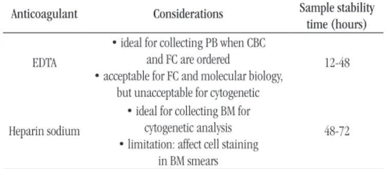
TABLE 1 −Characteristics of the anticoagulants used in PB
and BM collection for immunophenotyping by FC(2)
Anticoagulant Considerations Sample stability time (hours)
EDTA
• ideal for collecting PB when CBC
and FC are ordered
• acceptable for FC and molecular biology, but unacceptable for cytogenetic
12-48
Heparin sodium
• ideal for collecting BM for cytogenetic analysis • limitation: affect cell staining
in BM smears
48-72
PB: peripheral blood; BM: bone marrow; FC: low cytometry; ETDA: ethylenediaminetetraacetic acid.
TABLE 2 − Conditions for CSF collection and storage for analysis by FC(3)
Collection medium for CSF
CSF ratio: collection medium
Storage temperature
Stability time
Transix® 1:4 4ºC (2ºC to 8ºC) 48-72 hours
RPMI 5% FBS 1:1 4ºC (2ºC to 8ºC) 18 hours
CSF: cerebrospinal luid; FC: low cytometry; RPMI: Roswell Park Memorial Institute; FBS: fetal bovine serum.
For cavity liquids, collection in a dry tube is recommended; for ine-needle aspirates, lymph node tissue fragments is indicated the use of isotonic transport medium, RPMI for example. Lymph node in sterile saline should be quickly transported to the FC technical sector.
Sample identification and exam ordering form
It is mandatory to carry out sample identification before or during the material collection, and it must contain at least two identifiers that individualize the patient. The material should always be sent to the laboratory accompanied by the medical order and a duly complete form with the following information: a) service name, assistant doctor, telephone number; b) type of material, date and time of collection; c) the examination order with medical history, diagnosis hypothesis, and previous treatment; d) clinical data related to the investigation moment (diagnosis, relapse, control or
survey of minimal residual disease); and e) other laboratory data relevant to the diagnosis.
Volume of samples
Table 3 summarizes the recommended volume of samples commonly analyzed by FC.
Temperature and transport conditions
The samples must be sent to the laboratory immediately after collection and, in the case of PB and BM, they must be properly conditioned at a temperature of 18ºC to 25ºC(2). CSF, lymph nodes,
and tissue fragments must be kept refrigerated at 2ºC to 8ºC during transportation(3). It is important that the temperature is controlled,
Receipt of materials and rejection and restrictions
criteria
In the laboratory, during the receipt of the material, it is essential to check medical form data, and to evaluate for adverse conditions related to the collection, conditioning and transport(4).
Cell viability below 75% (evaluated by trypan blue, propidium iodide, 4’,6-Diamidino-2-phenylindole Dihydrochloride [DAPI], DRAQ-5® or 7 amino-actinomycin D [7-AAD]), intense hemolysis,
and presence of clots signiicantly compromise the quality of the immunophenotypic study, and should be considered as rejection criteria for the material. However, it is important not to reject noble samples (BM, CSF, cavity luids, and tissue fragments), but rather to process and analyze them rigorously to release complete or partial results.
It is required to describe any restriction information in the inal report, for example, describing the presence of clot – “suboptimal sample due to the presence of clot”.
QUALITY CONTROL OF
THE ANALYTICAL PHASE
Quality control of the equipment (FC)
The equipment functions must be checked daily, corrective and preventive actions (maintenance) must be deined and documented by the laboratory together with the company responsible for technical assistance.
The equipment should be placed on a physical structure without vibration and in a room with a temperature set between 19ºC and 25ºC. Ideally, the temperature registration and documentation occur during the periods of the day when the equipment is connected.
In the daily boot process, or when the equipment is in use, it is mandatory to check the luidic systems, pressure, vacuum and laser power(5). Although this assessment is automatically carried
out by the equipment, monitoring, recording and documentation are required.
The same is applied to the evaluation of light scattering and luorescence detection system, which laser alignment and photomultipliers (PMTs) performance are evaluated with speciic products for each equipment and supplier(5, 6).
It is important that the values obtained are easily accessible and iled in a historical sequence related to each equipment. It
is recommended to create spreadsheets and average/median and standard deviation (SD) graphs analysis, the same procedure for the registration and the traceability of historical occurrences and maintenance is applied.
To validate new equipment, checking and documentation of linearity, reproducibility, resolution and sensitivity of the equipment are optional when the company responsible for technical support of the equipment certiies these items(7).
The equivalence of analytical systems must be evaluated every six months, in the laboratories with different equipment performing the same tests(5). For quantitative assays, such as
CD4 and CD8 lymphocytes quantitative measure, and CD34+
progenitor cells quantitative measure, the use of statistical programs for comparison and/or historical coeficient of assay variation is recommended, and an acceptable limit established for each service.
For qualitative tests, such as immunophenotypic characterization of leukemias and lymphomas, the use of the categorical agreement for the inal diagnosis as a comparison criteria is recommended; different equipment must generate the same inal diagnosis composed by negative and positive expressions
of the markers used.
Periodicity of color compensation
For services that use combinations of standardized and validated luorescence, the daily veriication of color compensation of the low cytometer is recommended before starting the routine. Daily compensation of the luorescence is not recommended, but rather to verify if the predeined compensation is in accordance with the acceptability limits established and documented for each service.
It was decided that the periodicy for a new compensation matrix should be related to laser alignment performance, the variation of luorescence channels (∆PMT) and the performance of a tube labeled with ideal luorochromes combinations ideal for checking the compensation. If the evaluated parameters are within the acceptability limit, there is no need to apply a new compensation matrix. However, it is important to apply a new matrix if the values are outside the acceptable limits after preventive and corrective maintenance of the equipment.
For services using tandem luorochromes, it is necessary that the color compensation be speciic for each luorochrome-conjugated antibody and for each product lot(5, 8). For example,
as well as for CD117 PE-Cy7 marker. It is important to mention that when introducing a new tandem marker into the routine, it is necessary to apply a new compensation matrix adding the new product concerned. The previous electronic compensation matrix cannot be used for the calculation, since there may be variations in the luorescence intensity during the period between compensations.
Internal quality control
This involves ensuring that the obtained results are accurate, precise, consistent and reproducible. This performance is checked, usually by commercial reagents with reference values that assess the technical process as a whole.
It is important that this veriication is performed before the starting or releasing routine, and that the laboratory monitors the performance, assesses trends, and applies corrective action when necessary. It is recommended that monitoring is carried out by scatter plots showing the relationship to the average, such as the Levey-Jennings chart, which allows using Westgard Rules
(Figure 1).
Care should be taken when using the instruction values provided by the manufacturer, because it is important to ensure that the acceptability limits are two SD. As an alternative to the instruction values, the laboratories may use the determinations history to establish the average and variations of two SD. The use of at least twenty consecutive determinations of the same product lot is recommended for this application.
Despite the problems involving the use of commercial reagents in FC, as low availability, high cost and short shelf-life, it is mandatory that two levels of internal control (clinical decision level) are performed daily, or when performing quantitative tests of CD4/CD8 lymphocytes and quantitative measure of CD34+
progenitor cells(9). In the absence of commercial reagents, the
use of the samples analyzed the previous day with known values and variations established and documented by the laboratory is suggested as an alternative. The acceptability limits can be deined through analysis of at least twenty samples on consecutive days.
For leukemia and lymphoma immunophenotyping panels, monitoring and monthly recording of the performance of the monoclonal antibodies used is recommended, assessing antigen expression in normal and abnormal populations of the sample concerned. This strategy is important to evaluate the performance of reagents during the use, mainly due to decrease or loss of luorescence, which, if not detected, may induce the interpretation
of false negative results.
Reagents
Regarding the reagents and the working solutions, for example, hemolysis reagents, phosphate-buffered saline (PBS), and solutions for labeling monoclonal antibodies, it is mandatory to validate them before releasing for use. If such reagents are prepared daily, then daily validation is also required.
Regarding the commercial kits, each service must have a written procedure consistent with the manufacturers’ recommendations, as well as values within the acceptable limit established by kits that have reference values. Any change in the procedure must be documented and validated by each service(10).
Monoclonal antibodies conjugated with
fluorochromes
The validation of monoclonal antibodies (MAb) must occur before their release for use in routine. Validaton needs to be perfomed according to the lot and the receiving date of
FIGURE 1 − Levey-Jennings charts representing the daily assessment of an analyte with mean value and ranges deined by the product manufacturer (mean 50% ± 8%)
A) chart with acceptable performance and values ranging around average; B) chart shows the trend in which the values progressively deviate from the mean. Corrective actions are required because more than eight consecutive values differ from the average; C) chart with values out of the acceptable upper limit. Corrective actions are required when three consecutive determinations present unacceptable performance, when there is a consecutive violation of two SD, when two SD are violated on the same day by two levels of control, and when a violation of three SD occurs.
SD: standard deviation.
(A)
(B)
products, according to the following considerations: a) if several MAb bottles from the same lot are received on the same day, the validation of only one bottle is enough to indirectly validate the other bottles; b) if the antibodies are from different lots, each lot must be validated regardless of the receiving date. The same applies to the products of the same lot received on different dates.
This strategy is required since the packaging conditions during transportation may inluence MAb quality, even if they are
from the same lot.
Among MAb validation criteria, titration is the most important factor and it is intended to deine the optimal concentration of antigen-antibody saturation binding. Although MAb manufacturer recommends an ideal volume for labeling, titration is important to minimize the background luorescence and the non-speciic binding, while optimizing the signal-to-noise ratio relationship(7).
For titration, it is recommended:
• following the same standardized technical procedure for sample preparation, as the concentration of labeled cells, reagents, hemolysis buffers and ixing;
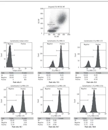
• the positive population should be as bright as possible (high luorescence intensity), since the negative population behave as the unlabeled sample. When applicable, it is recommended to use the so-called Peak Ratio (PR) calculation, which is to divide the positive population luorescence by the negative population luorescence, and the largest PR value is represented by the better distinction between populations (resolution), and therefore by the optimal concentration for use(11) (Figure 2);
• once titrated, the monoclonal antibody released for use may be used as a reference to validate other antibodies through the comparison between products. If the performance is similar and meet the criteria described above, established by each service, the “new” MAb may be released for use, respecting the same volume used in the comparative test; otherwise, the new Mab should be
titrated and an ideal volume for labeling is established.
Samples processing
It is suggested that the samples are processed immediately upon receipt, or as soon as possible after collection, respecting the maximum limit of 24 to 48 hours post-collection, depending on the anticoagulant used.
For samples with high proliferative and/or apoptosis level, for example, samples suspected of Burkitt’s lymphoma, diffuse large B-cell lymphoma, plasmocyte dyscrasia, BM collected after recent
chemotherapy and CSF, the immediate processing after collection and/or material receiving is mandatory(2).
Cell concentration for labeling
The recommended cell concentration for labeling samples with monoclonal antibodies conjugated with luorochromes is 5 × 10⁵ to 1 × 10⁶ cells/μl. For standard applications by working
groups, such as the Eurolow Consortium protocols, the volume 50 μl is recommended regardless of the cellularity(12). The Clinical
and Laboratory Standards Institute (CLSI) describes important details about the speciic cell concentration for different clinical applications(13, 14).
For rare events research and acquisition of million events in which the cell concentration exceeds 1 × 10⁶, concentration of the
FIGURE 2 − Example of MAb titration: 1 × 106 PB total cells labeled with IgG1 (mouse) – PE and different volumes/concentrations of CD3-PE, from the volume recommended by the manufacturer
The analysis of lymphocytes region (SSC × FSC) shows that the optimal volume ranges from 5.0 μl (1/4) to 1.25 (1/16), because in these concentrations there were enough luorescence intensity to identify the CD3 positive lymphocyte population from the other CD3 negative lymphocytes, which remained restricted to the region of unstained cells. The Peak Ratio values, 46.7 and 150.8 respectively, support the use of these volumes.
MAb: monoclonal antibodies; IgG1: immunoglobulin G1 class; PE: phycoerythrin; SSC INT: side scatter integral; FSC INT: forward scatter integral; PB: peripheral blood.
150 100 50 0 150 100 50 0 Count Count Negative Positive Positive [Ungated] FSC INT/SSC INT
1000 800 600 400 200 0 SSC INT
0 200 400 600 800 1000 FSC INT Lymphocytes
[Lymphocytes] isotype control
150 100 50 0 100 50 0 150 100 50 0 150 100 50 0 Count Count Count Count
106 106 106 106
106 106 106 106 106 106 106 106 106 106 106 106
106 106 106 106 106 106 106 106
Negative
Negative Negative Negative
Negative Positive
Positive Positive
Positive
Gate %Gate X-mode
Gate %Gate X-mode Gate %Gate X-mode Gate %Gate X-mode
Gate %Gate X-mode Gate %Gate X-mode
All 100.00 0.10
Negative 100.00 0.10
Positive 0 N/A
Peak ratio: 0
[Lymphocytes] 20 µl MAb (1)
20 µl MAb (1)
All 100.00 56.05
Negative 27.31 3.61 Positive 72.69 56.05
Peak ratio: 15.5
[Lymphocytes] 10 µl MAb (1/2)
Isotype control 10 µl MAb (1/2)
All 100.00 45.99 Negative 26.84 1.82 Positive 73.16 45.99 Peak ratio: 25.3
[Lymphocytes] 5 µl MAb (1/4)
5 µl MAb (1/4)
All 100.00 29.86
Negative 27.46 0.64 Positive 72.54 29.86
Peak ratio: 46.7
[Lymphocytes] 2.5 µl MAb (1/8)
2.5 µl MAb (1/8)
All 100.00 29.86 Negative 27.46 0.64 Positive 72.54 29.86 Peak ratio: 46.7
[Lymphocytes] 1.25 µl MAb (1/16)
1.25 µl MAb (1/16)
sample before labeling is recommended, as well as to titrate again the speciic markers for such applications(15). It is important to use
one control tube for each patient, in order to evaluate the low cytometer settings and the negativity and/or the autoluorescence level of the sample. Therefore, it is indicated to use a tube with sample and without monoclonal antibody (unlabelled tube) and/or a tube labeled with luorescence isotype controls. Additionally, the cell populations of the sample/tube itself labeled as luorescence internal control may be used(5, 13).
Data acquisition and analysis
For services that do not use post-labeling ixers, samples should be conditioned at temperature of 18ºC to 25ºC, protected from light, and acquired in the low cytometer, no later than two hours after labeling. If the service uses ixer, the samples should be conditioned at a temperature of 2ºC to 8ºC, and the acquisition carried out within 24 hours post-labeling. It is important to be alert to possible changes in the antigenic expression of the labels in ixed samples. It is recommended that the standardization of acquisition and data analyzing processes (settings, gates strategy, number of acquired events, and post-acquisition analysis on speciic software) is implemented, monitored and documented in the services. This action is important to minimize subjectivity and maximize the reproducibility of results(16).
It is worth noting that results interfacing technology, besides providing security to the process, avoids transcription errors of results. Thus, the services should invest in this tool for considerable gain in the quality process.
QUALITY CONTROL OF THE POST-ANALYTICAL
PHASE
Consistency analysis and release of the results
The consistency analysis of the results obtained by FC should be carried out prior to release of the inal result, and it consists of a critical review of immunophenotypic indings correlated with clinical data and other laboratory data, and double checking for verifying the analyzed and transcribed data(17).
In immunophenotypic evaluation of hematologic malignancies, it is recommended that the diagnostic conclusion is conducted together with the morphology and medical history, and, if necessary, to establish an active communication channel to the assistant doctor. When possible, it is advisable to associate the immunophenotypic results with the histopathological, cytogenetics
and molecular biology indings. For other applications, for example, lymphocyte immunophenotyping, quantitation of CD34+
progenitor cells and paroxysmal nocturnal hemoglobinuria, the application of consistency analysis established in the literature is recommended(14, 18).
Conditioning and retention period of biological
samples
After the report release, it is recommended to condition the biological samples at temperature of 2ºC to 8ºC for a period up
to three days(19). If performing new procedures and new labelings
with the material are required, it is mandatory to carry out a critical analysis of the results obtained due to possible changes in cell viability and antigenic expressions. When these changes are identiied, it is appropriate to describe them to described them in the report.
Data archiving and retention period
Archiving and backing up the data generated by the FC service (raw acquisition ile, analyzed iles, application forms, and working maps) must be iled in printed and/or electronic form for at least ive years(19, 20). It is recommended that the backup data is
performed daily.
External quality control
The external QC aims to assess the technical control of the process, through proiciency external programs and external commercials controls; the external QC samples are processed in the same way as the patient samples(19, 21).
It is essential that all tests performed in the service participate in external proiciency programs; ensuring test accuracy if the commercial products are unavailable, sending samples, in parallel, to another reference laboratory is recommended(1).
In situations where the test is not performed at another service (peculiar methodologies internally validated), the performance of the test/double blind evaluation is recommended. This consists of performing tests by two different analysts, and the result must necessarily be the same. Acceptability limits should be statistically established, monitored and documented by each service.
educational activities. These actions should be monitored in future tests, especially to investigate and ensure that the results of the patients analyzed in dates close to the external control have not been compromised due to detected error.
FINAL CONSIDERATIONS
This document was reviewed and approved by the Quality Control Subcommittee in Flow Cytometry (Subcomitê de Controle de Qualidade em Citometria de Fluxo) together with the GBCFLUX Scientiic Subcommittee. New versions and/or updates will be reported.
REFERENCES
1. Sales MM, Vasconcelos DDM. Citometria de luxo. Aplicações no laboratório clínico e de pesquisa. Seção I, Capítulo 6 – Controle de qualidade. São Paulo: Editora Atheneu; 2013.
2. Davis BH, Dasgupta A, Kussick S, Han JY, Estrellado A. Validation of cell-based luorescence assays: practice guidelines from the ICSH and ICCS - part II - preanalytical issues. Cytometry B Clin Cytom. 2013; 84(5): 286-90.
3. Kraan J, Gratama JW, Haioun C, et al. Flow cytometric immunophenotyping of cerebrospinal luid. Curr Protoc Cytom. 2008; Chapter 6: Unit 6 25.
4. National Institute for Allergy and Infectious Diseases/Division of AIDS guidelines for low cytometric immunophenotyping, version 1.0, Jan 1993, sec 1.06.
5. Oldaker TA. Quality control in clinical low cytometry. Clin Lab Med. 2007; 27(3): 671-85, viii.
It is important to report that learning this complex area must be continuous, and training and evaluation of technical skills are essential. The continuous improvement and monitoring processes of knowledge development must be monitored by the responsible doctor and technician.
ACKNOWLEDGEMENTS
The authors thank the GBCFLX members, who answered the QC questionnaires. We also thank all the participants and members of the GBCFLUX for their contribution in preparing this irst QC document.
RESUMO
O Grupo Brasileiro de Citometria de Fluxo (GBCFLUX), fundado em 24 de abril de 2010, é composto por especialistas da área de citometria de luxo (CF) que possuem o objetivo comum de contribuir para avanços técnico-cientíicos em laboratórios clínicos e de pesquisa brasileiros. Entre os grupos de trabalho do GBCFLUX, o subcomitê de Controle de Qualidade (CQ) é responsável por discutir dados da literatura e contribuir para a garantia da qualidade do processo pré-analítico, analítico e pós-analítico em CF. As ações do subcomitê de CQ iniciaram-se por meio de reuniões e palestras, nas quais dados da literatura foram revisados e discutidos com todos os membros participantes do GBCFLUX. Em uma segunda etapa, deiniu-se elaborar um texto de recomendações consensuais técnico-cientíicas, informativas e educativas para divulgação a todos os grupos que trabalham com CF no Brasil. Para tanto, foi elaborado um questionário com respostas objetivas, sendo enviado para 35 instituições brasileiras cadastradas, com a inalidade de avaliar o peril de CQ dessas instituições. Dessa forma, as recomendações técnico-cientíicas de CQ que serão descritas neste artigo de atualização têm o objetivo de contribuir para a garantia da qualidade do processo, a padronização técnica e a reprodutibilidade dos resultados em CF.
Unitermos: citometria de luxo; controle de qualidade.
6. Kalina T, Flores-Montero J, van der Velden VH, et al. EuroFlow standardization of low cytometer instrument settings and immunophenotyping protocols. Leukemia. 2012; 26(9): 1986-2010. 7. Tanqri S, Vall H, Kaplan D, et al. Validation of cell-based luorescence assays: practice guidelines from the ICSH and ICCS - part III - analytical issues. Cytometry B Clin Cytom. 2013; 84(5): 291-308.
8. Roederer M. Compensation in low cytometry. Curr Protoc Cytom. 2002; Chapter 1: Unit 1 14.
9. Department of Health and Human Services. Centers for Medicare and Medicaid Services. Clinical laboratory improvement amendments of 1988; inal rule. Fed Register. 1992(Feb28):7146 [42CFR493.1256]. 10. Caldwell CW. Analyte-speciic reagents in the low cytometry laboratory. Arch Pathol Lab Med. 1998; 122(10): 861-4.
of normal, reactive and malignant leukocytes. Leukemia. 2012; 26(9): 1908-75.
13. Clinical and Laboratory Standards Institute (CLSI). Clinical low cytometric analysis of neoplastic hematolymphoid cells; Approved Guideline, second edition. CLSI document H43-A2 (ISBN 1-56238-635-3). Wayne, Pennsylvania: Clinical and Laboratory Standards Institute; 2007. 19087-1898.
14. Clinical and Laboratory Standards Institute (CLSI). Enumeration of immunologically deined cell populations by low cytometry; Approved Guideline, second edition. CLSI document H42-A2 (ISBN 1-56238-640-9). Wayne, Pennsylvania: Clinical and Laboratory Standards Institute; 2007. 19087-1898.
15. Hedley BD, Keeney M. Technical issues: low cytometry and rare event analysis. Int J Lab Hematol. 2013; 35(3): 344-50.
16. Stelzer GT, Marti G, Hurley A, McCoy P, Jr., Lovett EJ, Schwartz A. U.S.-Canadian Consensus recommendations on the immunophenotypic analysis of hematologic neoplasia by low cytometry: standardization and validation of laboratory procedures. Cytometry. 1997; 30(5): 214-30.
17. Borowitz MJ, Bray R, Gascoyne R, et al. U.S.-Canadian Consensus recommendations on the immunophenotypic analysis of hematologic neoplasia by low cytometry: data analysis and interpretation. Cytometry. 1997; 30(5): 236-44.
18. Sutherland DR, Keeney M, Gratama JW. Enumeration of CD34+ hematopoietic stem and progenitor cells. Curr Protoc Cytom. 2003; Chapter 6: Unit 6 4.
19. Barnett D, Louzao R, Gambell P, De J, Oldaker T, Hanson CA. Validation of cell-based luorescence assays: practice guidelines from the ICSH and ICCS - part IV - postanalytic considerations. Cytometry B Clin Cytom. 2013; 84(5): 309-14.
20. Resolução – RDC/ANVISA nº 302, de 13 de outubro de 2005. Regulamento técnico para funcionamento de Laboratórios clínicos. 21. Department of Health and Human Services. Centers for Medicare and Medicaid Services. Clinical laboratory improvement amendments of 1988; final rule. Fed Register. 2003( Jan24):7166 [42CFR493.1256(d)(8)].
MAILING ADDRESS
Nydia Strachman Bacal