IX
Radiol Bras 2006;39(6):IX–XII
WHICH IS YOUR DIAGNOSIS?
Marcelo Souto Nacif1,4
, Amarino Carvalho de Oliveira Júnior2
, Denise Madeira Moreira3
, Mônica Regina Nagano4
, José Hugo Mendes Luz4
, Mauro Esteves de Oliveira4
, Arnaldo Rabischoffsky5
, José Feldman6
, Carlos Eduardo Rochitte7
Study developed at Hospital Procardíaco, Rio de Janeiro, RJ, Brazil. 1. Professor at Faculdade de Medicina de Teresópolis and IPGMCC Post-Graduation Course, MD, Radiologist at HUAP-UFF. 2. Coordinator for Service of Radiology and Diagnostic Imaging, Hospital Procardíaco. 3. Sub-Coordinator for Service of Radiology and Diagnostic Imaging, Hospital Procardíaco. 4. MDs, Radiologists at Hospital Procardíaco. 5. Coordinator for Service of Echocardiography, Hospital Procardíaco. 6. MD, Cardiologist. 7. MD, Advisor for Hospital Procardíaco. Mailing address: Prof. Dr. Marcelo Souto Nacif. Rua Álvares de Azevedo, 130, ap. 704/A, Icaraí. Niterói, RJ, Brazil 24220-042. E-mail: [email protected]
Female, 66-year-old patient, with 1.65 m in height, weighting 59 kg, 74 bpm, arte-rial blood pressure of 140/80 mmHg, has been referred to the Service of Radiology and Diagnostic Imaging of Hospital Pró-Cardíaco for evaluation by cardiac magnetic resonance imaging (MRI). Previous echocardiogram demonstrated a normal left ven-tricular (LV) global systolic function (ejection fraction - – Simpson = 63%).
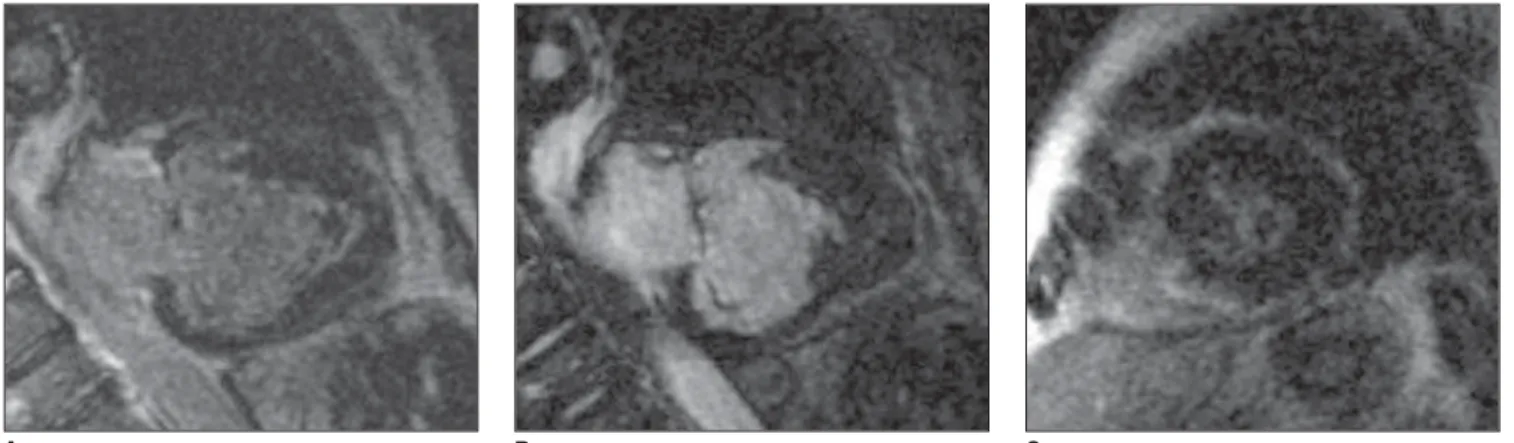
Figure 2. Electrocardiogram-gated acquisitions. Delayed enhanced image: longitudinal (A,B) and short axis (C).
A B C
D E
A B C
F
Figure 1. Electrocardiogram-gated acquisitions with cine Fiesta (SSFP), T1FGRET and T1FGRIR, at end-diastole, in the following planes: outflow tract (A), 2-chamber view (B), 4-chamber view (C), short-axis (D); (E) LV function map, and (F) aortic flow curve.
F
lo
w
(m
l/
s
e
c
)
X Radiol Bras 2006;39(6):IX–XII Images description
Figure 1 – Electrocardiogram-gated acquisitions with cine-Fiesta (SSFP), T1FGRET and T1FGRIR, at end-diastole, in following planes: outflow tract (A), 2-chamber (B), 4-chamber (C) and short-axis (D); (E) LV function map; and (F) aortic flow curve. Observe that the atrium and right ventricle (RV) present with nor-mal size, with preserved segmental and global functions. There is an apical fill-ing of the left ventricular cavity with pre-served LV medial and basal segmental functions. Minimum mitral regurgitation and increase in volume of the left atrium, besides mild aortic insufficiency.
Figure 2 – Electrocardiogram-gated acquisitions, longitudinal delayed en-hancement with calcification (thrombus) (A), longitudinal (B) and short-axis (C). Subendocardial perfusion defect on the apex and delayed subendocardial en-hancement of the LV apical filling surface. Presence of a central hypointense image in this region, which may correspond to an adhered thrombus/calcification.
Diagnostic: Endomyocardial fibrosis with gross calcifications and normal LV function. Mild aortic insufficiency.
COMMENTS
Endomyocardial fibrosis was first de-scribed by Davies, in 1948. It is a com-plex entity of yet undetermined etiology, endemic in several tropical countries, with varied forms of presentation, repre-senting, in some regions, the primary cause of restrictive myocardiopathy. Possible etiologic factors are: serotonin-rich diet, malnutrition, filariosis and viral diseases(1–6).
Endomyocardial fibrosis is character-ized (Chart 1) by formation of endocar-dial fibrous tissue, most frequently lo-cated in the apex of the affected ventricle, which, angiographically, characterizes the typical image of LV and RV apical amputation. Eventually, the fibrosis ex-tends to the myocardium, the presence of a massive endocardial calcification, whatever its etiology, being a rare find-ing in the literature and with possibility of becoming a diagnostic and prognos-tic marker of the disease(1,6–11).
Endomyocardial fibrosis clinical manifestations depend on the form and intensity of cardiac involvement. For difficulting the ventricular relaxation, the fibrosis may cause restrictive cardiac fail-ure. The presence of valvar insufficiency makes such manifestations even more noticeable. Such clinical manifestations differ according to the involvement of the cardiac chambers (predominance in right, left or both chambers)(1,3,9,12–15).
Imaging studies, chest x-ray, echocar-diogram, nuclear medicine, magnetic reso-nance imaging and hemodynamic study, identify ventricular shape changes and allow the recognition and determination of the degree of the chamber
involve-ment(4,6,11,16,17).
The echocardiogram demonstrates signs of restriction associated with a large atrial dilatation and ventricular chambers obstruction, and raises suspi-cion of endomyocardial fibrosis(4,14,18).
Echocardiogram should be the best propedeutic method for endomyocardial fibrosis diagnostic; however, consider-ing the rarity of this disease, it is not al-ways considered and there are evidences that echocardiography underestimates the fibrosis intensity and does not iden-tify mild forms of the disease(5,6,10–12,19).
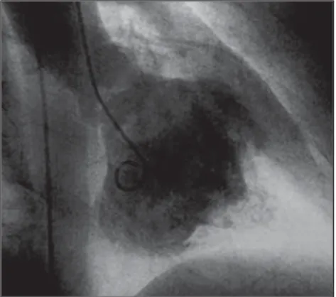
The hemodynamic study associated with angiography still is considered the golden standard for the diagnosis of endomyocardial fibrosis due only to its comprehensive use along years (Figure 3). However, it is important to observe that the cardiac MRI is already demon-strating its great diagnostic value(1,4,5,8,13).
The pressoric data with ventricular pressure curves presenting a “square root” pattern identify the cardiac restric-tion, while angiography demonstrates the apex and inflow tract amputation, outflow tract dilatation, atrial increase
and signs of atrioventricular valves in-sufficiency. The hemodynamic study re-sults also are important for evaluation of the degree of cardiac involvement, this factor being of prognostic importance(6,
8,13,17,18).
The relevance of the diagnosis of endomyocardial fibrosis originates from the fact the symptomatic presentation of this disease has a poor prognosis, a fact which has been documented since the first publications on this matter. We have observed that patients with endomyo-cardial fibrosis confined to LV are most frequently asymptomatic, and that the disease presents a fair evolution, even in those patients with a significant ven-tricular fibrosis. In these cases, the pres-ence of mitral insufficiency is a determin-ing factor for symptoms onset(11,15,17,18).
The surgery changes the natural his-tory of the disease, reducing symptoms and mortality. Because of their fair evo-lution, asymptomatic patients - espe-cially those with isolated LV involvement -, just should be maintained on clinical follow-up(10–13,16,18).
Signs of restricted ventricular filling
Alteration of the LV posterior wall motion dynamics – On the LV posterior wall one observes a fast protodiastolic motion followed by rectification in the remaining period, nearly always without the typical telediastolic expansion sec-ondary to the volume resulting from the atrial contraction. This is a sign of dias-tolic restriction, also frequently found in Chart 1 Endomyocardial fibrosis diagnosis.
• Identification of fibrosis in the ventricular cham-ber.
• Signs of restricted ventricular filling.
• Signs of cardiac valves involvement. • Secondary signs.
• It is essential to determine if the involvement is uni- or bilateral, balanced, or predominant in one of the ventricles.
XI
Radiol Bras 2006;39(6):IX–XII
constrictive pericarditis. Curiously, con-trary to what could be assumed, this LV posterior wall motion is not an exclusive feature of isolated or bilateral left ventricu-lar endomyocardial fibrosis, and may be observed in isolated presentations of right-sided endomyocardial fibrosis(5,11,15,17).
Differential diagnosis
In right-sided endomyocardial fibro-sis, a differential diagnosis with constric-tive pericarditis and Ebstein’s disease should be established. Some signs are common to the three diseases, like alter-ation in the interventricular septum move-ment, presystolic opening of the pulmo-nary vein, anterograde diastolic flow in the right ventricle outflow tract and right atrium dilatation. It is worth to emphasiz-ing that, in constrictive pericarditis, the tricuspid valve is structurally normal, and there is no reason for the left ventricle
Figure 5. Chest computed tomography, mediastinal window, where endocardial gross calcifications can be observed.
B A
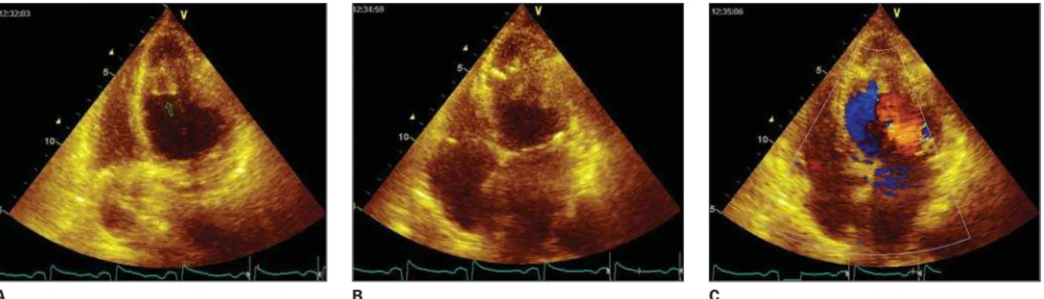
Figure 4. Echocardiogram with color Doppler demonstrating left atrium dilatation. LV apical filling, with preserved contraction in this region, and endocardial margins calcification. Normal LV systolic function, with decrease in the apical relaxation. Also, mitral ring calcification was found, with mild mitral and aortic insufficiencies.
outflow tract to dilate. In the Ebstein’s disease, there is no sign of restriction and the anterior leaflet of the tricuspid valve is redundant. Additionally, in Ebsteins’s disease, the tricuspid reflux is situated more apically, contrarily to endomyocar-dial fibroses, whose reflux is slow and of undefined localization(1,3,8,9,12,19).
In left-sided endomyocardial fibrosis, the differential diagnosis should be done with other mitral insufficiency etiologies, especially rheumatic fever. In this circum-stance, the differentiation is relatively easy because of the presence of mitral valve leaflets thickening and adherence. In dilated cardiomyopathies with func-tional regurgitation and eventual forma-tion of apical thrombus, the hypocontrac-tility is diffuse and the valve structural aspect is normal. In apical thrombosis of chronic chagasic myocarditis or ischemic cardiopathies, the wall is akinetic or
pre-sents dyskinetic movement, which does not occur in endomyocardial fibrosis(1,2,
4,5,11,16,18).
In the present case, the echocardio-gram (Figure 4) had already demonstrated signs of LV apical filling with preserved contraction and normal systolic function. Later, we could observe a chest CT of this patient (Figure 5), which demon-strated endocardial gross calcifications. Presently, cardiac MRI is a mainstay in the evaluation of the myocardium thick-ness and function, as well as the LV per-fusion, allowing the assessment of the myocardial enhancement, which is a rel-evant factor in the diagnosis of several cardiac diseases.
It is important to emphasize that, in our service, the technological development has allowed us to develop a multidisci-plinary work, integrating the several ex-amination methods in cardiology, and
in-volving radiologists in active clinical cussions and in the study of heart dis-eases, bringing an important contribution to the diagnostic and therapeutic deci-sions making process in this field.
REFERENCES
1. Barretto ACP, Luz PL, Oliveira SA, et al. Deter-minants of survival in endomyocardial fibrosis. Circulation 1989;80:I177–182.
2. Borggrefe M, Block M, Breithardt G. Identifica-tion and management of the high risk patient with dilated cardiomyopathy. Br Heart J 1994;72(6 Suppl):S42–45.
3. Chan WL, Chiang BN, Kong CW, Lee JB, Wang SP, Hsu TL. Endomyocardial fibrosis with mas-sive endocardial calcific deposits. Clin Cardiol 1987;10:541–545.
C
XII Radiol Bras 2006;39(6):IX–XII
4. Metras D. Endocardial fibrosis and its surgical treatment: Ivory Coast experience. In: Valiathan MS, Somers K, Kartha CC, editors. Endomyocar-dial fibrosis. Delhi: Oxford University Press, 1993; 211.
5. Richardson P, McKenna W, Bristow M, et al. Re-port of the 1995 World Health Organization/Inter-national Society and Federation of Cardiology. Task Force on the Definition and Classification of cardiomyopathies. Circulation 1996;93:841–842. 6. Wynne J, Braunwald E. The cardiomyopathies and myocarditis. In: Braunwald E, editor. Heart dis-ease. A textbook of cardiovascular medicine. Phila-delphia/London: WB Saunders/Elsevier, 2005. 7. Chopra P, Narula J, Talwar KK, Kumar V, Bhatia
ML. Histomorphologic characteristics of endo-myocardial fibrosis: an endoendo-myocardial biopsy study. Hum Pathol 1990;21:613–616. 8. Kinare SG, Deshpande JR. Endomyocardial fibrosis
outside Kerala: an autopsy study. In: Valiathan MS, Somers K, Kartha CC, editors. Endomyocardial fibrosis. Delhi: Oxford University Press, 1993; 141–149.
9. Moraes AV, Medeiros CCJ, Abensur H, et al. Eco-cardiografia transesofágica intraoperatória na cirur-gia cardíaca: experiência inicial. Rev Bras Eco 1992;15:5–11.
10. Oliveira SA, Pereira Barretto AC, Mady C, et al. Surgical treatment of endomyocardial fibrosis: a new approach. J Am Coll Cardiol 1990;16:1246– 1251.
11. Stillman AE, White RD. Adult heart disease ex-cluding myocardial ischemia and viability. In:
Edelman RR, Hesselink JR, Zlatkin MB, Crues JV, editors. Clinical magnetic resonance imaging. Philadelphia/London: WB Saunders/Elsevier, 2006;1016–1039.
12. D’Arbela PG, Mutazindwa T, Patel AK, Somers K. Survival after first presentation with endomyo-cardial fibrosis. Br Heart J 1972;34:403–407. 13. Dave T, Narula JP, Chopra P. Myocardial and
endocardial involvement in tuberculous constric-tive pericarditis: difficulty in biopsy distinction from endomyocardial fibrosis as a cause of restric-tive heart disease. Int J Cardiol 1990;28:245–251. 14. Mady C, Barretto ACP, Oliveira SA, Stolf N,
Bellotti G, Pileggi F. Evolution of the endocar-dial fibrotic process in endomyocarendocar-dial fibrosis. Am J Cardiol 1991;68:402–406.
15. Ribeiro PA, Muthusamy R, Duran CM. Right-sided endomyocardial fibrosis with recurrent pul-monary emboli leading to irreversible pulpul-monary hypertension. Br Heart J 1992;68:326–329. 16. Gupta PN, Valiathan MS, Balakrishnan KG,
Kartha CC, Ghosh MK. Clinical course of endo-myocardial fibrosis. Br Heart J 1989;62:450–454. 17. Pereira Barretto AC, Mady C, Arteaga E, et al. Quadro clínico da endomiocardiofibrose. Correla-ção com a intensidade da fibrose. Arq Bras Cardiol 1988;51:401–405.
18. Moraes CR. Early and late results of surgery for endomyocardial fibrosis. In: Olsen EGJ, Sekiguchi M, editors. Restrictive cardiomyopathy and ar-rhythmias. Tokyo: University of Tokyo Press, 1990;49.