Arq Neuropsiquiatr 2005;63(3-B):862-863
Neurosurgery Department of Hospital Cristo Redentor, Porto Alegre, RS, Brazil: 1MD, Resident; 2MD Neurosurgeon. Received 24 January 2005, received in final form 8 April 2005. Accepted 20 May 2005.
D r. Rodrigo Mendonça - Serviço de Neuro c i ru rgia do Hospital Cristo Redentor - Rua Domingos Rubbo 40 - 91040-000 Porto Alegre RS - Brasil. E-mail: [email protected]
The computerized tomography (CT) scan of epidural hematomas usually show hyperdense bi-convex shaped mass lesions. Isodense epidural he-matomas are rare features with few cases re p o rt-ed in the neuro s u rgical literature1 - 3. Diff e rent the-ories have been proposed to explain this atypical radiological finding, from low hematocrit values t o dilution of the epidural blood due to mixture with cerebrospinal fluid (CSF).
We present a case in which a bilateral epidural hematoma was isodense with the brain and could be easily misdiagnosed, leading to unapro p r i a t e treatment.
CASE
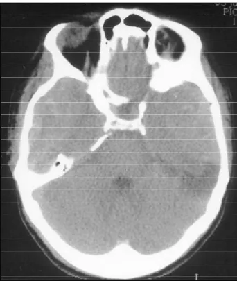
A 23-year-old man arrived the emergency room 3 hours after suffering head injury due to an assault, with con-ciousness impairment. There was a history of alcohol inges-tion and the initial GCS (glasgow coma score) was 14. T h e re were not clinical signs of skull base fracture. The patient presented a rapid clinical deterioration to coma-tous state and re q u i red intubation. A CT scan was per-f o rmed at this moment (Fig 1), and showed large bilater-al isodense epidurbilater-al hematomas. He was rapidly transfer-red to the operating room with GCS score 4 and dilated pupils. At this moment blood tests showed a hematocrit 29.4%, hemoglobin 9.6 g/dl and normal coagulation
studi-es. A bicoronal incision was made, with bilateral tempo-ral craniectomy. Operative findings included partially clot-ted bilateral epidural hematomas and an extensive skull base fracture, with active bleeding from the inner ear. T h e re was no dural laceration. The patient had a favor-able outcome, with remarkfavor-able clinical improvement in the immediate post-operative period, being discharg e d in good neurological condition (GOS 5) after 19 days.
DISCUSSION
Isodense epidural hematomas are rare radiolo-gical findings. In the series of 151 patients re p o rte d by Ta p i e ro et al., 40% presented hyperacute epidu-ral hematomas but none of these were isodense with the brain4. The densities of subdural and epi-dural hematomas on CT scans are related to the attenuaton values of the clot, as a function of the e ry t h rocyte and hemoglobin protein concentra-tion and in a lesser extent related to the iron con-tent of the hemoglobin molecule5. Serum hemog l o-bin concentrations ranging from 9 to 11 g/dl have a p p roximately the same density of the brain on CT scans. The clotting mechanisms are not essent i a l for the CT attenuation, but clot retraction with se-paration of serum and absorption of fluid acts in-c reasing the foin-cal hemoglobin in-conin-centration and the density of acute clots1.
BILATERAL ISODENSE EPIDURAL HEMATOMA
Case report
Rodrigo Mendonça
1, Telmo T.F. Lima
1, Leandro I. Dini
2, Cláudio L.L. Krebs
2ABSTRACT - We present a case of a severe head injuried 23 year-old male patient. The initial CT scan disclo-sed bilateral epidural hematoma, isodense with the brain, thus being a pitfall in diagnosis. Brief case re p o rt , image and literature rewiew are presented.
KEY WORDS: epidural hematoma, head injury, computed tomography.
Hematoma epidural isodenso bilateral: relato de caso
RESUMO - Apresentamos o caso de um homem de 23 anos com traumatismo craniano grave. A TC de crânio d e m o n s t rou um volumoso hematoma epidural bilateral, isodenso com o cére b ro, sendo uma arm a d i l h a ao diagnóstico. São apresentados um breve relato, estudo da imagem tomográfica e revisão da literatura.
Arq Neuropsiquiatr 2005;63(3-B) 863
The “hyperacute” extradural hematomas are usually hyperdense, with some small areas iso or hypodense within the lesion4. The possible causes of these combined densities are the presence of fresh, unclotted blood (which has a low attenua-tion coefficient), a low hematocrit or a mix of
blo-od with CSF due to dural lacerations. Another p roposed mechanism is the continuous washout of the blood within the hematoma throught the diploic veins after a skull fracture4.
In the present case, the CT findings could be ex-plained by the anemia or by the presence of the skull base fracture. The dura was intact, thus the isodense hematoma could not be related to the CSF mixture .
It is important to state that early diagnosis and t reatment of extra-axial traumatic hematomas may result in a important decline in morbidity and mort a-lity and the misdiagnose is a potentially fatal situa-tion. Neuro s u rgeons should be aware of the above discussed condition, so prompt recognition and tre-atment can be achieved with better outcomes.
A k n o w l e d g e m e n t s– Special thanks to the Dr. Mar-co Antonio Stefani and Dr. Álvaro Ernani Georg for the revision of this report.
REFERENCES
1. A r rese I, Lobato RD, Gómez PA, Nunez A P. Hyperacute epidural hae-matoma isodense with the brain on computed tomography. Acta Neuro-chir (Wien) 2004;146:193-194.
2. May PL, Miles JB. Acute isodense extradural haematoma. Br J Neuro-surg 1989;3:221-224.
3. Rieth KG, Schwartz FT, Davis DO. Acute isodense epidural hematoma on computed tomography. J Comput Assist Tomogr 1979;3:691-693. 4. Ta p i e ro B, Richer E, Laurent F, Guibert-Tranier F, Caille M.
Post-trauma-tic extra-dural haematomas. J Neuroradiol 1984;11:213-226. 5. G re e n b e rg J, Cohen WA, Cooper PR. The “hyperacute” extraaxial
in-tracranial hematoma: computed tomographic findings and clinical sig-nificance. Neurosurgery 1985;17:48-56.