Universidade de Aveiro 2012
Secção Autónoma de Ciências da Saúde
POLIANA
POLLIZELLO LOPES
CIMENTOS ÓSSEOS ACRÍLICOS MODIFICADOS
COM ENCHIMENTO BIOATIVO E BIODEGRADÁVEL
ACRYLIC
BONE
CEMENTS
MODIFIED
WITH
Universidade de Aveiro 2012
Secção Autónoma de Ciências da Saúde
POLIANA
POLLIZELLO LOPES
CIMENTOS ÓSSEOS ACRÍLICOS MODIFICADOS
COM ENCHIMENTO BIOATIVO E BIODEGRADÁVEL
ACRYLIC
BONE
CEMENTS
MODIFIED
WITH
BIOACTIVE AND BIODEGRADABLE FILLERS
Dissertação apresentada à Universidade de Aveiro para cumprimento dos requisitos necessários à obtenção do grau de Doutor em Engenharia Biomédica, realizada sob a orientação científica da Doutora Maria Helena Figueira Vaz Fernandes, Professora Associada do Departamento de Engenharia Cerâmica e do Vidro da Universidade de Aveiro
Apoio financeiro da FCT e do FSE no âmbito do III Quadro Comunitário de Apoio.
o júri
presidente Prof. Doutor António Carlos Mendes de Sousa
professor catedrático da Universidade de Aveiro
Prof. Doutor Fernando Jorge Mendes Monteiro
professor catedrático da Faculdade de Engenharia da Universidade do Porto
Prof. Doutor Nelson Fernando Pacheco da Rocha
professor catedrático da Universidade de Aveiro
Prof. Doutor João Filipe Colardelle da Luz Mano
professor associado da Universidade do Minho
Prof. Doutora Maria Helena Figueira Vaz Fernandes
professora associada da Universidade de Aveiro (Orientadora)
Prof. Doutor António Manuel Silvério Cabrita
professor auxiliar da Faculdade de Medicina da Universidade de Coimbra
Prof. Doutor José Martinho Marques de Oliveira
Agradecimentos/ Acknowledgements
I wish to thank, first and foremost, my supervisor, my family, and my friends, for different, but decisive reasons.
Thank you Prof. Maria Helena Fernandes for sharing the knowledge and advice, and for giving me the opportunity to continue my studies in such an interesting area of knowledge, having always as landscape background the wonderful city of Aveiro.
I would also like to express my thanks to Professor Maria Helena Raposo Fernandes and her group - School of Dental Medicine, University of Porto - for their collaboration in studies of in vitro biocompatibility.
I must comunicate my gratitude to the technical support given by Ana Sofia Marques Ribeiro, Célia Cristina Moreira Pereira Miranda, Maria Joao de Pinho Bastos and Marta Carmona Ferro.
Thank you my colleagues and friends in my research group Ana Margarida Silva, Erika Davim and Nuno Almeida. I want to express my deepest gratitude to Ana Luisa Daniel da Silva, Bárbara Leite Ferreira and Nathalie Barroca whose advices and encouragements were essential in many instances of this journey.
I thank and dedicate to my wonderful parents, Ademir and Marlene, sister Patrícia, and my brother Patrik for the unconditional love and for supporting me all my life. Their effort and dedication were essential to make me get this far. My sincere thanks to my husband, Gonçalo. This thesis is the result of your encouragement, help and love.
To the Portuguese Foundation for Science and Technology (FCT) for my scholarships (Ref.SFRH/BD/27961/2006), the Centre for Research in Ceramic and Composite Materials CICECO and the University of Aveiro for funding and supporting this work.
palavras-chave Cimento ósseo acrílico, vidro bioativo, compósitos, polímero biodegradável, libertação de fármaco, ibuprofeno, resposta celular.
resumo O cimento ósseo acrílico é o único material utilizado para a fixação de próteses em cirurgias ortopédicas, surgindo como uma alternativa às técnicas não cimentadas. Cerca de um milhão de pacientes são anualmente tratados para a substituição total da articulação do quadril e do joelho. Com a maior expectativa de vida da população e o aumento do número de cirurgias realizadas por ano espera-se que o uso do cimento ósseo aumente substancialmente.
A fraca ligação do cimento ao osso é um problema comum que pode causar perda asséptica da prótese. Assim, torna-se necessário investir no desenvolvimento de cimentos ósseos alternativos que permitam promover maior estabilidade e melhor desempenho do implante.
O principal objetivo desta tese foi desenvolver um cimento ósseo bioativo, capaz de ligar-se ao osso, com propriedades melhoradas relativamente aos sistemas convencionais. A preparação dos materiais foi realizada por dois processos diferentes, a polimerização por via térmica e a polimerização por via química.
Inicialmente, utilizando o processo térmico, foram desenvolvidos compósitos de PMMA-co-EHA reforçados com vidro de sílica (CSi) e vidro de boro (CB) e comparados em termos do seu comportamento in vitro em meio acelular e celular. A formação de precipitados de fosfato de cálcio foi observada sobre a superfície de todos os compósitos indicando que estes materiais são potencialmente bioativos. Em relação à avaliação biológica o CSi demonstrou um efeito indutor da proliferação das células. As células apresentaram uma morfologia normal e alta taxa de crescimento quando comparadas com o padrão de cultura. Por outro lado ocorreu inibição da proliferação celular para o CB provavelmente devido à sua elevada taxa de degradação, levando a uma elevada concentraçao de iões de B e de Mg no meio de cultura.
O efeito do vidro nos cimentos curados por via química, incorporando um activador de baixa toxicidade, também foi avaliado. Os resultados sugerem que as novas formulações podem diminuir o efeito exotérmico na cura do cimento e melhorar as propriedades mecânicas (flexão e compressão). Outro estudo conduzido neste trabalho explorou a possibilidade de incorporar ibuprofeno (fármaco anti-inflamatório) no cimento, dando origem a um material capaz de ser simultaneamente, bioativo e promotor da libertação controlada de fármacos. Neste contexto foi evidenciado que o desempenho do cimento desenvolvido pode contribuir para minimizar o processo inflamatório associado a uma cirurgia ortopédica.
keywords Acrylic bone cement, bioactive glass, composites, biodegradable polymer, drug delivery, ibuprofen, cell response.
abstract Acrylic bone cement is the only material currently used for anchoring the prosthesis in orthopaedic surgery, being an alternative to non-cemented techniques. About one million patients worldwide are treated annually for total replacement of hips and knee joints. With the longer life expectancy of the population, and the increasing number of surgeries performed every year, the use of acrylic bone cements is expected to rise substantially.
The non bone bonding capability of the cement is a common problem which can cause aseptic loosening of the prosthesis. Thus, alternative cements must be developed to provide higher stability and better performance of the implant. The main objective of this thesis was to develop a bioactive bone cement, with bone bonding capability, and better properties than the conventional cement. Two different methods of preparation were used in this study, polymerization by chemical route (self-cured) and polymerization by thermal route (heat cured). Initially, through the thermal route, PMMA-co-EHA composites filled with a silicate glass (CSi) and a borate glass (CB) were developed and compared in terms of their in vitro behaviour, both in acellular and in cellular media. The growth of spherical calcium phosphate aggregates was observed in acellular medium on all composite surfaces indicating that these materials became potentially bioactive. Considering the biological assessment, the CSi demonstrated an inductive effect on the proliferation of cells. The cells showed a normal morphology and high growth rate when compared to standard culture plates. On the other hand, inhibition of cell proliferation occurred in the CB probably due to its high degradation rate, leading to high B and Mg ionic concentration in the cell culture medium.
The effect of glass in self cured cements, incorporating an activator of reduced toxicity, was also assessed in this work. The results suggested that the new formulations may lessen the exothermal effect on curing and improve the mechanical properties (bending and compressive). Another study conducted in this thesis explores the possibility of incorporating ibuprofen (anti-inflammatory drug) into the cement, aiming the development of a composite that simultaneously show bioactive behaviour and controlled drug release . It was evidenced that, regarding the drug release, the performance of the developed cements can contribute to blunt the inflammatory process associated to an orthopedic surgery.
Finally the solid phase of the bioactive self-curing acrylic cements was modified by different biodegradable polymers. The addition of the biodegradable fillers
CONTENTS
LIST OF ABREVIATIONS...IV LIST OF FIGURES...VII LIST OF TABLES...X CHAPTER 1 ... 1 HISTORY... 1 BIOMEDICAL APPLICATIONS... 2Total Hip Replacement ... 3
TYPICAL COMPOSITIONS... 5
Antibiotics... 6
PREPARATION OF BONE CEMENTS... 8
CHEMICAL REACTIONS AND SETTING PROCESS... 9
RESIDUAL MONOMER... 12
MECHANICAL PROPERTIES... 13
ANCHORAGE MECHANISMS... 14
MAIN DRAWBACKS OF COMMERCIAL BONE CEMENTS... 15
Aseptic Loosening ... 15
High Polymerization Temperature... 16
Release of MMA monomer ... 17
ALTERNATIVES TO THE STANDARD COMPONENTS... 17
Radiopaque Agents ... 17
Activators... 19
BIOACTIVE BONE CEMENTS... 20
Bioactive cements filled with calcium phosphates... 21
Bioactive cements filled with glasses... 24
CEMENTS FILLED WITH BIODEGRADABLE FILLERS... 27
CONCLUSIONS AND SCOPE OF THE THESIS... 30
REFERENCES... 32 CHAPTER 2 ... 41 SECTION I...41 ABSTRACT... 41 INTRODUCTION... 42 EXPERIMENTAL... 42 Glass preparation ... 42
Preparation of the composites ... 42
In vitro assay in SBF ... 43
RESULTS AND DISCUSSION... 43
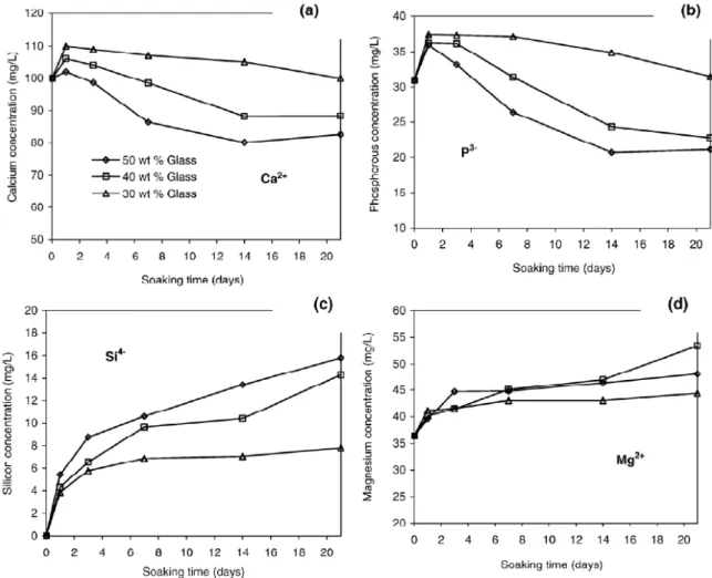
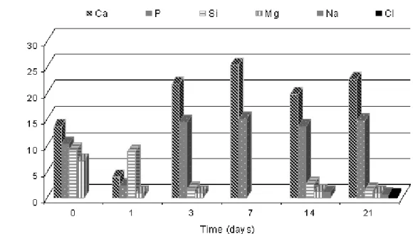
Changes in SBF composition ... 43
Formation and characterization of the surface layer... 45
CONCLUSIONS... 49
REFERENCES... 49
INTRODUCTION... 52
MATERIALS AND METHODS... 53
Preparation of the glasses... 53
Preparation of the composites ... 53
In vitro bioactivity ... 54
Biocompatibility studies ... 54
MG63 osteoblast-like cells ... 54
Human bone marrow cells ... 55
Cell viability/proliferation... 55
Alkaline phosphatase activity... 55
SEM and CLSM microscopy... 56
Statistical analysis ... 56
RESULTS AND DISCUSSION... 56
In vitro Bioactivity... 56 Biological Assessment... 61 CONCLUSIONS... 65 REFERENCES... 65 CHAPTER 3 ... 69 ABSTRACT... 69 INTRODUCTION... 70
MATERIALS AND METHODS... 71
Cement preparation... 71
Determination of curing parameters... 72
Residual monomer content ... 73
In vitro bioactivity ... 73
Mechanical Properties ... 73
Water uptake and weight loss ... 74
Osteoblastic cytocompatibility... 74
Cell viability/proliferation... 75
Alkaline phosphatase activity... 75
Scanning electron microscopy... 75
Statistical analysis ... 76
RESULTS AND DISCUSSIONS... 76
Curing Parameters... 76
Residual Monomer content... 78
In vitro bioactivity ... 78
Mechanical Properties ... 81
Water uptake and weight loss ... 83
Osteoblastic cytocompatibility... 85 Cell viability/proliferation... 85 Osteoblastic differentiation ... 86 CONCLUSIONS... 90 REFERENCES... 91 CHAPTER 4 ... 95 ABSTRACT... 95 INTRODUCTION... 96
Materials... 97
Preparation of the cements ... 98
Setting parameters ... 98
Residual monomer content ... 99
Assessment of in vitro bioactivity... 99
In vitro drug release... 100
Statistical analysis ... 100
RESULTS AND DISCUSSIONS... 100
Curing Parameters... 100
Residual Monomer content... 101
In vitro bioactivity ... 102 Ibuprofen Release ... 107 CONCLUSIONS... 110 REFERENCES... 111 CHAPTER 5 ... 115 ABSTRACT... 115 INTRODUCTION... 116
MATERIALS AND METHODS... 117
Materials... 117
Cements preparation ... 118
Thermal behaviour and crystallization ... 119
Setting parameters ... 119
Residual monomer ... 120
Mechanical behaviour ... 120
Water uptake, weight loss and surface evaluation... 120
Statistical analysis ... 121
Osteoblastic cytocompatibility... 121
Human bone marrow cell cultures ... 121
Cell viability/proliferation... 122
Alkaline phosphatase activity... 122
CLSM and SEM observation ... 122
Statistical analysis... 122
RESULTS AND DISCUSSION... 123
Thermal behaviour and crystallization ... 123
Setting Parameters and Residual Monomer ... 126
Mechanical Properties ... 128
Degradation and surface analysis ... 130
Osteoblastic cytocompatibility... 133
CONCLUSION... 137
REFERENCES... 137
CHAPTER 6 ... 141
CONCLUSIONS... 141
LIST OF ABBREVIATIONS
AA Acrylic acid
ALP Alkaline phosphatase ANP Acryloyl-N-phenylpiperazine
ASTM American society for testing and materials AW-GC Apatite and wollastonite glass-ceramic BaSO4 Barium sulphate
BBC Bioactive bone cement
BET Brunauer–Emmett–Teller method
BIEM 2-(2-bromoisobutyryloxy) ethyl methacrylate Bis-GMA Bisphenol-α-glycidyl methacrylate
BPEM 2-(2-bromopropionyloxy) ethyl methacrylate BPO Benzoyl peroxide
BZN 4,4-bis-dimethylamino benzydrol CLSM Confocal laser scanning microscopy CS NP Chitosan nanoparticles
DML 4-N,N-Dimethylaminobenzyl laurate DMOH N,N-dimethylaminobenzyl-alcohol DMT N,N-dimethyl-4-toluidine
DSC Differential scanning calorimetry EDS X-ray spectroscopy
EHA 2-ethyl hexylacrylate HA Hydroxyapatite
HCA Hydroxycarbonate apatite
HDBCs Hydrophilic, partially degradable and bioactive cements HEMA Hydroxyethyl methacrylate
H NMR Proton nuclear magnetic resonance HOB Human osteoblast-like cells HQ Hydroquinone
IB Ibuprofen
ICP Inductively coupled plasma IHQM 2,5-diiodo-8-quinolyl methacrylate IPMA 4-iodophenol methacrylate
LD50 Lethal dose, 50%
α -MEM α-Minimal Essential Medium
4-META 4-methacryloxyethyl trimellitate anhyhydride MMA Methyl methacrylate
MNP Methacryloyl-N-phenylpiperazine
MTT 3-(4,5-dimethylthiazol-2-yl)-2,5-diphenyltetrazolium bromide NSAIDs Non-steroidal anti-inflammatory drugs
PBS Phosphate-buffered saline PCL poly(ε-caprolactone) PEMAnBMA poly(ethylmethacrylate-n-butylmethacrylate) PHAs poly(hydroxyalkanoates) PHB poly(3-hydroxybutyrate) PHBV poly(3-hydroxybutyrate-co-3- hydroxyvalerate) PLA poly-L-lactic acid
PMMA poly(methyl methacrylate) PPF poly(propylene fumarate) PTFE poly(tetrafluoroethylene)
QCS NP Quaternary ammonium chitosan derivative nanoparticles SBF Simulated body fluid
SCA Corn starch/cellulose acetate blends SEM Scanning electron microscopy SSA Specific surface area
TCP Tricalcium phosphate
α-TCP α- tricalcium phosphate β-TCP β-tricalcium phosphate
Tg Glass transition temperature THA Total hip arthroplasty THRs Total hip replacements TKRs Total knee replacements Tm Melting temperatures Tmax Maximum temperature
TMS Tetramethylsilane TPB Triphenyl bismuth TPSs Thermoplastic starches
WL Weight loss WU Water uptake
Xc Degree of crystallinity XRD X-ray diffraction ZrO2 Zirconium dioxide
LIST OF FIGURES
CHAPTER 1
Figure 1: Biomedical applications of a bone cement. ... 3
Figure 2: Components of the total hip arthroplasty (THA) system... 4
Figure 3: Main components of a typical acrylic cement ... 6
Figure 4: Cement preparation by hand mixing and vacuum mixing. ... 8
Figure 5: Temperature versus time curve of a curing acrylic bone cement. ... 10
Figure 6: Chemical structure of the activators mentioned. ... 19
Figure 7: Mechanism of apatite formation on glass... 25
CHAPTER 2 SECTION I Figure 1: Variation of ionic concentrations of (a) calcium, (b) phosphorous (c) silicon, and (d) magnesium in SBF, during the incubation period... 44
Figure 2: XRD patterns of the surface of composite (a) prepared with 50% and (b) 30% (wt) of glass, during the immersion period. ... 45
Figure 3: SEM micrographs of the surface of composites C5 and C4 before and after soaking in SBF for 3 and 21 days. ... 46
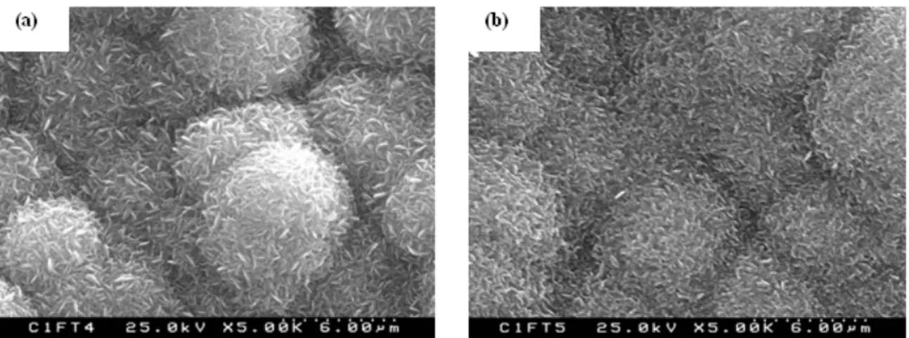
Figure 4: Detail SEM micrographs of the HA layer formed on the surface of composite C5 after soaking in SBF for (a) 14 and (b) 21 days. ... 47
Figure 5: EDS pattern of the surface of composite C5 during immersion period... 48
SECTION II Figure 1: SEM micrographs, for the CSi composite (a) before immersion, (b) after 3 days and (c) after 7 days in SBF. For CB, (d) before immersion, (e) after 3 days and (f) after 7 days. Bars 60 µm (insert: bar = 6 µm). ... 57
Figure 2: EDS results, for CSi (a) before immersion, (b) after 3 days and (c) after 7 days. For CB (d) before immersion, (e) after 3 days and (f) after 7 days... 58
Figure 3: DRX patterns of composite (a) CSi and (b) CB. ... 59
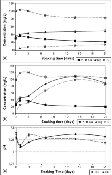
Figure 4: Variation of ionic concentration in SBF due to immersion of (a) CSi and (b) CB, and (c) pH evolution with time... 60
Figure 5: Behaviour of MG63 osteoblast-like cells cultured up to 7 days over CSi and CB composites. (A) Cell viability/proliferation, estimated by MTT assay, (*) Significantly different from control culture. CLSM images at 7 days, on (B) CSi and (C) CB... 61
Figure 6: CLSM observation, for CSi composites seeded with human bone marrow cells and cultured for (A) 1 hour, (B,C) 24 hours, (D) 7 days, (E) 14 days and (F) 21 days. For CB composite (G) 1 hour, (H, I) 24 hours, (J) 7 days, (K) 14 days and (L) 21 days. A, B, G and H: bar, 60 µm; C – F and I – L: bar, 500 µm. ... 62
Figure 7: Cell viability/proliferation by MTT assay (A), and alkaline phosphatase activity (B) of
human bone marrow cells grown over CSi and CB for 21 days. (*) Significantly different from
control culture. ... 63
Figure 8: SEM appearance of human bone marrow cells cultured for 21 days over CSi composite, inset: EDS spectrum of the mineralized structures, and CB composite ... 63
CHAPTER 3 Figure 1: Curing parameters for cements filled with silicate glass (a) and cements filled with borate glass (b). ... 76
Figure 2: Residual monomer content for the studied cements. ... 78
Figure 3: SEM micrographs of the cements after soaking in SBF and EDS spectra. ... 79
Figure 4: XRD patterns of surface cements MSi3 (a), MSi5 (b), MB3 (c) and MB5 (d). ... 80
Figure 5: Variation of ionic concentration in SBF due to immersion of cements filled with a silicate glass (a, b) and cements filled with a borate glass (c, d)... 81
Figure 6: Water uptake (a), weight loss (b) and pH values (c) for the investigated formulations. . 83
Figure 7: Cell viability/proliferation (A) and alkaline phosphatase activity (B) of human osteoblastic bone marrow cells cultured over PMMA and the glass-filled cements, for 21 days.*Significantly different from PMMA ... 86
Figure 8: SEM observation of PMMA and the glass-filled cements colonized with human osteoblastic bone marrow cells, at days 14 and 21. Over PMMA, at day 14, cells were barely seen, but partially covered the material surface at day 21 (A, B). On the Si-glass compositions, cells were clearly seen at day 14 (C, E, G) and, at day 21, formation of a thick cell layer was observed over MSi3 and MSi4 (D, F), but few cells were visible on MSi5 (H). The B-glass cements presented significantly lower cell growth. Cells were visible at day 14 on MB3 and BM4 (I, K), mostly forming small cell clusters, but not over MB5 (M); at day 21, the presence of cells was not evident on the three composites (J, L, N). Bar = 100 µm, except for B, D, F (200 µm)... 87
Figure 9: High magnification SEM images of PMMA and the glass-filled cements colonized with human osteoblastic bone marrow cells. The Si composites were able to form a mineralized matrix at day 14 (A – C) and MSi3 and MSi4 also showed a mineralized cell layer at day 21 (D, E); representative X-Ray spectrum of the globular mineral structures showed well evident Ca and P peaks (J). The MB3 and MB4 cements showed the formation of globular structures associated with the cell clusters at day 14 (G, H) and the x-Ray spectrum exhibited small Ca and P peaks (K). At day 21, PMMA also showed cell-associated globular structures (I), but they did not contain Ca and P peaks (L). A, C, E: Bar = 50 µm; B, F-I: Bar = 20 µm; D: Bar = 10 µm... 88
Figure 10: SEM images of MSi3 and MSi4 cements colonized with human osteoblastic bone marrow cells, showing that cells hardly colonized the pores present on the material surface. Images show the pores with a very smoth surface (A, D, E) and the cells growing around the pore (C, D), forming bridges (A, E) and covering the pores (B, C, arrows). A, D, E: Bar = 200 µm; B, C: Bar = 500 µm. ... 89
CHAPTER 4 Figure 1: Setting parameters for the investigated formulations ... 101
Figure 2: Residual monomer content for the studied cements, one month after polymerization.. 102 Figure 3: SEM micrographs of the bone cements surface (a) before immersion, (b) after 7 days and
Figure 4: XRD patterns of composites after 21 days of soaking in PBS ... 104
Figure 5: EDS profiles of the surface (a) IB5, (b) IB10 and (c) IB20. For each measurement a standard deviation of approximately 0.2 was determined. ... 105
Figure 6: SEM micrographs of the cements surface (a) after 3 days, (b, c) after 14 days in SBF; (d) EDS spectra... 106
Figure 7: Variation in ionic concentrations of Ca and P in SBF... 107
Figure 8: Ibuprofen release curves of the studied cements. ... 107
Figure 9: Release curve analysis and parameters according to power law (a) up to 7 days, (b) between 7 and 30 days. ... 108
CHAPTER 5 Figure 1: Chemical structure of used biodegradable polymers... 117
Figure 2: DSC thermograms of pure biodegradable polymers PHB and PHBV and studied cements... 123
Figure 3: Variation in the degree of crystallinity of the PHB and PHBV in the cements... 124
Figure 4: XRD patterns of (a) PHB and PHBV, (b) PHB20B and PHBV20Si. ... 125
Figure 5: Curing curves of the developed cements filled with (a) PHB and (b) PHBV. ... 126
Figure 6: Load-displacement curves for the cements (a) PHB10, (b) PHB20, (c) PHBV10 and (d) PHBV20. ... 128
Figure 7: Bending properties of all the prepared formulations. ... 129
Figure 8: Evolution of water uptake (WU) and loss of weight (WL) for the cement filled with PHB (a, c) and cement filled with PHBV (b, d). ... 131
Figure 9: SEM micrographs of cements before and after 28 days of immersion in PBS (a) PHB10Si, (b) PHB20Si, (c) PHB10B and (d) PHB20B, (e) PHBV10Si, (f) PHBV20Si, (g) PHBV10B and (h) PHBV20B... 132
Figure 10: Cell viability/proliferation (a) and ALP activity (b) of human bone marrow osteoblastic cells seeded over the cements for 21 days. *Significantly different from PMMA ... 134
Figure 11: Representative CLSM images of human bone marrow osteoblastic cells seeded over the cements for 4 and 14 days; bar = 400 µm. ... 135
Figure 12: Representative SEM images of the cements cultured with human bone marrow osteoblastic cells, at day 21. ... 136
LIST OF TABLES
CHAPTER 1Table 1: Revision rates for the fixation methods used in THA. ... 5 CHAPTER 2
SECTION I
Table 1: Chemical composition of the PMMA-based composites investigated (wt %)... 43 Table 2: Ion concentrations and pH of SBF and those of human blood plasma. ... 43 Table 3: Ca/P molar ratios, obtained by EDS, during the immersion of composite C5... 48 SECTION II
Table 1: Ionic Concentrations (mM) of SBF and Human Blood Plasma. ... 54 CHAPTER 3
Table 1: Glass composition (mol%), specific surface area and density. ... 71 Table 2: Composition of the solid and liquid phases of the developed cement (wt%). ... 72 Table 3: Mechanical properties of the samples. ... 82 CHAPTER 4
Table 1: Solid phase composition of the formulations produced (wt%) ... 98 Table 2: Drug release of cements, based on UV spectroscopy measurements... 110 CHAPTER 5
Table 1: Chemical composition of all cements developed (wt%). ... 118 Table 2: Values of setting parameters and Mr for the investigated cements... 127
CHAPTER 1
HISTORY AND CURRENT STATE OF ACRYLIC BONE CEMENTS
HISTORY
Bone cements are substances used to fix prosthesis to the bones often in joint replacements surgeries and to repair damaged or diseased areas of bones. Most of the bone cements commercially available and currently used in orthopedic procedures are acrylic cements. The basic component of the acrylic bone cement is methyl methacrylate (MMA) which is an ester and can polymerize to form poly(methyl methacrylate) (PMMA). Large scale chemical synthesis of MMA was achieved in the 1920s in the laboratories of Rohm and Haas, and one of the first biomedical applications of PMMA was the fabrication of dentures in 1935 [1, 2]. In the same period (1936) the company Kulzer found that prepolymerized PMMA powder (poly(methyl methacrylate)) could be partly dissolved in MMA, forming a dough that hardens when benzoyl peroxide (BPO) is added and the mixture is heated to 100 °C in a stone mould. Kulzer is at present the producer of the Palacos®, a commercial acrylic bone cement used in orthopaedic surgeries.
The first clinical use of this dough was in an attempt to close cranial defects in monkeys in 1938. Seven years later it was discovered that the polymerization of MMA could occur by itself at room temperature if a tertiary amine (N,N-dimethyl-4-toluidine, DMT) was added, leading to the establishment of a protocol for the chemical production of acrylic bone cements in 1943 [3, 4].
PMMA was first introduced in the orthopedic surgery by Dr. Jean Judet and his brother, Dr. Robert Jude. The Judet brothers developed a hip prosthesis made from PMMA, which was implanted in 1946 [5]. In 1951 Kaier and Jansen in Copenhagen were the first to use PMMA bone cements for the fixation of acrylic cups to the subchondral bone of the femoral head [6]. Nevertheless, it was Sir John Charnley who popularised their use in 1958 and presented the preliminary results of a new method for the fixation of joint prostheses to bone. The idea was to distribute the contact stresses between the implant and the bone over a large area by means of acrylic bone cement. This idea represented an important breakthrough in the field of orthopaedics and led to the development of a worldwide successful technique. The main advantages of the cemented prostheses lay in the excellent primary fixation, in the even load distribution between the implant and the bone, and in the fact that the technique allows a fast recovery of the patient [7].
The addition of antimicrobial agents to acrylic bone cements began as early as 1970. Engelbrecht and Buchholz started investigations on PMMA cement to determine its suitability as a
zirconium dioxide (ZrO2), have been added to the bone cement in order to provide radio-opacity
[11]. In the 1980s acrylic bone cements were also introduced to treat vertebral compression fractures caused by osteoporosis, skeletal metastases and angiomas [12].
Since then many types of bone cements have been developed. Nowadays, there are over 30 commercially available acrylic bone cement brands approved by the relevant regulatory authorities (such as the Food and Drug Administration, FDA, in the US and the Medical Devices Agency in the UK), for use in cemented arthroplasties [13]. In Portugal, the INFARMED is the Portuguese Regulatory Agency for pharmaceuticals, which is the institution in charge of guaranteeing that the legal requirements for the marketing of medicines are met.
BIOMEDICAL APPLICATIONS
The PMMA gained its popularity during World War II as a polymer for biomedical applications, when polymer fragments accidentally implanted in the eyes and other body tissues of pilots during aircraft crashes did not cause damage to the body [14]. When used for orthopedic applications, certain additions are made to PMMA and thus it receives the name of bone cement.
Bone cements have been used as a fixation medium in a number of joint replacements including knee, hip, shoulder, elbow, ankle, and wrist replacements. In recent years, its application has been expanded to vertebroplasty, a procedure in which cement is injected percutaneously into the vertebral body in order to stabilize fractures that occur primarily as a result of osteoporosis. Another variation of this procedure is khyphoplasty, during which a balloon is inserted percutaneously into the vertebral body, inflated to restore the height of the compressed vertebrae and subsequently filled with injected bone cement to stabilize the fracture. These newer applications of bone cement have been successful in relieving pain and restoring vertebral strength and function [15, 16]. Some applications of bone cements are depicted in Figure 1.
In 2002, Khan et al. [17] conducted a systematic review of the literature on treating patients with displaced intracapsular femoral neck fractures. The authors concluded that the publications tended to support the use of cemented hemiarthroplasty, suggesting a lower revision rate, less thigh pain and better mobility in patients whose prosthesis was cemented.
Figure 1: Biomedical applications of a bone cement.
Nowadays the major orthopaedic surgical procedures are total hip and knee joint replacements with 1 million performed worldwide annually. A large proportion of these are anchored to the contiguous cancellous bone in an acrylic bone cement bed [13]. The clinical success rate for cemented implant with 15 years exceeds 90%, especially those of the hip and knee in patients aged over 50 years [18]. In developed countries the acrylic bone cements are used in more than 90% of total hip surgeries [19, 20]. In Sweden, over the period 1979–2000, about 97% of primary total hip replacements (THRs) were cemented [21], and in the United States, 77% of primary total knee replacements (TKRs) were cemented. The majority of total replacements of other joints is also cemented. With the ‘‘graying’’ of the populations in many countries it is expected that the use of acrylic bone cements rise substantially [13].
Total Hip Replacement
A large range of rotary motion is permitted at the hip due to the fitting between femur and pelvis; the top of the femur terminates in a ball-shaped head that fits into a cup-like cavity (the acetabulum) within the pelvis. This joint is susceptible to fracture, which normally occurs at the narrow region just below the head, through the femoral neck. The hip may also become diseased by osteoarthritis; in this case small lumps of bone form on the rubbing surfaces of the joint, which causes pain as the head rotates in the acetabulum and a joint replacement is necessary. Damaged and diseased hip joints have been replaced with artificial joint successfully [22].
prosthesis fixation. However, neither cemented nor uncemented fixation excludes the likelihood of prosthesis loosening [23]. A schematic diagram of the total hip replacement is presented in Figure 2.
Figure 2: Components of the total hip arthroplasty (THA) system [22]
The hip implant fixation can be cemented, uncemented or hybrid [24, 25].
Cemented fixation: The acrylic bone cement is used to hold the femoral and acetabular
components in place. The cemented hip replacement relies on a stable interface between the prosthesis-cement-bone resulting in a faster rehabilitation. Although cemented implants have a long and distinguished track record of success, they are not ideal for everyone. This fixation method is more commonly recommended for older patients, for patients with conditions such as rheumatoid arthritis, and for younger patients with compromised health or poor bone quality and density.
Uncemented fixation: The fixation is made through direct contact to bone without the use of
cement. The implants are textured or have a surface coating (osteoconductive coating) providing bone growth into their surface. In general, these designs are larger and longer than those used with cement. Because they depend on new bone growth for stability, uncemented implants require a longer healing time than cemented replacements. This method is most often recommended for younger, more active patients and patients with good bone quality where bone ingrowth into the components can be predictably achieved.
Hybrid fixation: A hybrid total hip replacement has one component, usually the acetabular
socket, inserted without cement, and the other component, usually the femoral stem, inserted with cement. This technique was introduced in the early 1980s.
The superiority of either fixation method has not been proved conclusively because of the influences of confounding variables, such as patient age, sex, body weight, and diagnosis [26].
Most of the literature showed that better short and mid-term clinical and functional outcomes could be obtained from cemented femoral fixation than from uncemented femoral fixation [23]. Recent meta-analyses also support superior results of cement fixation when compared to uncemented fixation in large subsets of patient populations [27]. Table 1 presents the rate of revision prostheses according to the fixation method, which also proves that the cemented fixation still shows statistically the best results in terms of the whole THA population. In accordance still with the table uncemented prostheses have the worst performing, resulting in a higher incidence of revision for the studied period [28, 29].
Table 1: Revision rates for the fixation methods used in THA [28].
Revision rates by prosthesis type at one, three and five years for primary hip replacement procedures, undertaken between 1st April 2003 and 31st December 2009.
Prosthesis type Number of patients 1 year 3 years 5 years Cemented 99,359 0.6% 1.4% 2.0% Uncemented 62,937 1.3% 2.5% 3.4% Hybrid 31,662 0.9% 1.8% 2.7%
TYPICAL COMPOSITIONS
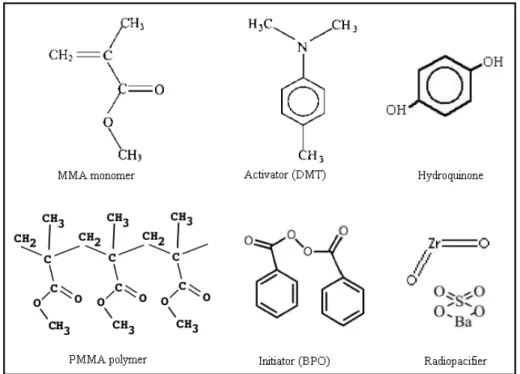
A typical acrylic bone cement is self-polymerising and consists of two components, a liquid monomer (methyl methacrylate, MMA) and a powder component (polymethylmethacrylate, PMMA). The two components are mixed in the appropriate proportions to form the bone cement. Other additives are included in these components for specific purpose [7, 30].
The monomer contains:
- Hydroquinone (HQ), an inhibitor, which prevents the monomer from prepolymerising spontaneously,
- N-N dimethyl-4-toluidine (DMT), an activator/accelerator, which speeds up the polymerisation reaction.
The powder component contains:
- Radiopaque agent, either barium sulphate (BaSO4) or zirconium dioxide (ZrO2), which
allows the bone cement to be observed on x-rays.
A number of commercial formulations can also include an antibiotic, such as gentamicin sulphate, that provides prophylaxis against infections, which can occur during surgery [8, 31]. Figure 3 illustrates the chemical structure of the main components of an acrylic cement.
Figure 3: Main components of a typical acrylic cement
There are some advantages to using two bone cement components instead of simply polymerize pure MMA monomer: The polymerization of MMA is too slow compared with the duration of surgery. The monomer has a very low viscosity and can easily diffuse into the blood stream. It is much easier to shape the doughy cement to fill the space between the prosthesis and bone. The use of less monomer and the presence of pre-polymerized PMMA decrease the amount of released heat and assist in heat dissipation, thus lowering the overall temperature. Pure MMA, upon polymerization into PMMA, has a volumetric shrinkage of 21% due to differences in the density of the MMA monomer and the PMMA polymer. This contraction is unacceptable and would lead to a large gap at the cement-bone interface, compromising the fixation of the prosthesis [32].
Antibiotics
Surgical operating rooms have sterile conditions, but even under these conditions some bacteria can pass through all of the protective barriers and contaminate the open body tissues during the
surgery. In order to prevent post-operative infections, some small quantities of antibiotics can be added into the bone cement [11].
An acrylic bone cement is a meshwork of PMMA chains. Antibiotics enclosed in these meshes are released by elution from the bone cement. The elution properties of cements correlate directly with the ability to absorb water, which is determined by the hydrophobicity of their components [8].
The first trials of adding antibiotic a bone cement were performed in late 60s and the first antibiotic loaded bone cement appeared in 1970. Gentamicin sulphate was chosen due to its wide spectrum antimicrobial activity, water solubility, thermal stability, low allergenicity and ability to confer long-term protection [9]. Currently most of the 18 different antibiotic-loaded bone cements available on the market, contain gentamicin sulphate [33].
The prophylactic effect of gentamicin-containing bone cement on postoperative infections in total hip arthroplasties was compared with that of systemically given antibiotics [34]. It was observed that the incidence of postoperative infections in the patients with gentamicin-containing bone cement was significantly less than the group which was treated with systemic antibiotic therapy. It was also reported that the presence of small amounts of antibiotics did not change the handling characteristics and did not reduce the strength of the cement below acceptable standards.
The liberation of antibiotics from the cement matrix and the effect of antibiotics on the properties of the cement are two important issues [11]. Antibiotics are typically released in two stages: there is a peak release followed by a long tail of low lost that continues for days or month. Approximately 90% of the drug may be retained inside the cement, being eluted only from the surface and from a network of cracks and voids in the bone cement by a dissolution–diffusion mechanism [35].
Several in vitro and in vivo studies have indicated bacterial growth on antibiotic-loaded bone cements with increased occurrence of gentamicin-resistant strains [36-38]. The increasing bacterial resistance to gentamicin has prompted renewed interest in the addition of further antibiotics to bone cements, such as tobramycin and cefuroxime [39, 40]. Multidrug targeting is assumed not only to be more powerful but also to prevent the emergence of resistant strains through the synergistic action of two antibiotics. In Europe, one multidrug-loaded bone cement containing gentamicin and clindamycin, Copal® is commercially available [33]. Combination of gentamicin and clindamycin in a bone cement formulation has a theoretical antimicrobial effect on more than 90% of the bacteria common to infected arthroplasties. The release of gentamicin seems to be enhanced by the release of clindamycin in this cement [41]. This may be an effect of the extra antibiotic, which acts as a soluble additive that leaves a network of voids behind, enhancing further release [42].
Multidrug targeting may be effective in preventing resistance but using it is a difficult option in bone cements, as the release of the different antibiotics depends on factors not easily controllable. For example vancomycin has a high molecular weight and shows poor release because it is trapped in the cement matrix [43]. Also, combinations of antibiotics must be carefully selected due to known cross-resistances. For this reason, it is not advisable to join the gentamicin and tobramycin. There is always the possibility of an antagonistic effect in the different ways by which the antibiotics act upon the bacterial life cycle [41].
PREPARATION OF BONE CEMENTS
Bone cements are prepared under operating room conditions, which consist of a temperature of 21-24 ºC and a relative humidity no less than 50%. When bone cement was first introduced the only available method was hand mixing; in this case the powder-containing pouch is cut by a sterile scissor, and the contents are put in a sterile bowl. Then the liquid ampoule is opened, and the content poured on the powder. They are mixed at atmospheric pressure until a homogeneous dough is obtained (1–3 min) [11, 44, 45]. This type of mixing method can introduce a significant amount of air into the mixture and a relatively high degree of trapped porosity (5-16%) in the set cement [46]. To overcome these drawbacks, new mixing techniques have been introduced such as vacuum-mixing and centrifugation, aiming to reduce the porosity of the cement [45, 47]. Preparation of a cement by the hand mixing process and by a vacuum mixer are shown in Figure 4.
Pores in the cement primarily result from air bubbles which have become entrapped during hand-mixing of the powder and liquid components, but it is also accepted that monomer evaporation at the high polymerization temperatures may contribute to produce embedded bubbles [48].
In vacuum-mixing, the bone cement is mixed while under a vacuum; which is supposed to eliminate the voids entrapped during the mixing process [49]. In centrifugation mixing the dough is immediately poured into a syringe that is then promptly placed in a centrifuge and spun with a speed of 2300 to 4000 rpm for 30-180 s [50], forcing out the air bubbles due to centrifugal forces [51].
After mixing, the cement is either manually placed into the cavity by means of “finger packing” or injected with a cement gun. The cement may also be pressurized at this time causing increased cement-bone interdigitation and providing a stronger interface [52].
CHEMICAL REACTIONS AND SETTING PROCESS
Mixing the two components (solid and liquid) produces the starting up of a typical addition polymerization reaction of the liquid monomer [7]. The MMA monomer can be polymerized through radicals formed by several different methods including the collision of two monomer molecules of sufficient energy or the decomposition of an initiator molecule by means of heat, light or chemical reaction [11, 53]. Bone cement polymerization is based on the free radical polymerization of MMA initiated by a redox system generated by reaction between the initiator (BPO) and the activator (DMT) which comprises three steps: initiation, propagation and termination.
The initiation step involves a reaction between the initiator and the activator causing the decomposition of the BPO, which splits into two fragments upon dissociation of the weak peroxy bond resulting in benzoyl radicals at room temperature. The second step is chain propagation, which basically consists in successive addition of monomer units to the active radicals already produced in the initiation stage. The free radical attacks one of the double bonds of the MMA monomer resulting in a larger free radical, then this new free radical attacks another MMA monomer and the chain propagates until a PMMA of relatively high molecular weight is formed. A consequence of this propagation phase is an increase in viscosity, due to the increasing concentration of polymer molecules and increasing molecular weight of the growing chains. Lastly, the chain termination can be achieved by combination of two chains (combination) or by hydrogen transfer reaction (disproportionation). The first method is the simplest way, wherein the two
When polymerization of the monomer is complete the pre-polymerized PMMA beads that form the powder (as described in "Typical compositions") are embedded into a solid PMMA matrix. The polymerization process is an exothermic reaction; in which heat is generated firstly when the benzoyl peroxide molecule is split, and secondly during the propagation stage of the reaction. The polymerization is very rapid and reaches completion in approximately 10–15 min, at which point the cement has set [55].
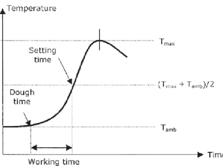
The variation of temperature with time, during the preparation of the material, can be monitored leading to a typical curve which indicates the setting process of the cement, Figure 5.
The time at which the mixed cement mass does not adhere to a surgically gloved finger is known as the dough time. This time is limited by the Standard Specification ISO5833 for acrylic resin cements [56] to a maximum of 5 minutes. The time elapsed from the moment at which the powder and liquid components are mixed until the cement is set, is known as the setting time. Setting time can be calculated as the time at which the temperature of the mass is the sum of the room temperature and maximum temperature divided by two. The ISO5833 establishes that the range time of the setting must be 5-15 minutes. The maximum temperature or peak temperature is produced by the exothermic propagation reactions which take place during polymerization. The cement sets before the peak temperature is reached. This value is limited to 90 ºC. Finally, the difference between the setting time and the dough time is called the working time, and it corresponds to the period of time during which the cement is workable and has to be implanted (or molded) [3, 7, 57].
The peak temperature recorded in vitro does not correspond to those actually reached in vivo. Clinical tests showed significantly lower intraoperative peaks at the bone-cement interface due to the thin layer of cement, the heat dissipation of the system via the implants and local blood circulation. The peak temperature in vivo is usually below the protein coagulation temperature, assumed to be around 56 ºC [4, 7]. Meyer et al. [58] found that the maximum temperature of the curing bone cement could be lowered by reducing the ambient temperature and the cement mantle thickness.
The setting time is sensitive to ambient temperature, thus, when the temperature of the operating room increases, the polymerization rate also increases and the dough hardens quicker [11]. For example, the bone cement Surgical Simplex® P has a setting time of 9 minutes at room temperature of 24 ºC, 12 minutes at 21 ºC and 15 minutes at 18 ºC [59]. Also the temperature of the powder and liquid components, of the implant and of the mixing equipment can markedly affect the setting time and the curing temperature. If the cement components are stored at temperatures lower or higher than that of the room, sufficient time must be allowed for them to reach the appropriate ambient operating room temperature before they are mixed, otherwise setting time will be correspondingly lengthened or shortened [60].
Others factors that can affect the curing properties of PMMA bone cements are:
The powder/liquid ratio of the cement, which is taken as the ratio between the weight of the
powder in g and the volume of the liquid in ml, has a very strong effect on the curing parameters. Increasing the powder/liquid ratio (by increasing the amount of PMMA powder or by decreasing the content of monomer), produces the decrease of the peak temperature [58, 61, 62]. These results can be understood in terms of the relative amounts of monomer present whose polymerization causes heat release and of the role played by PMMA beads (powder) that absorb heat [7]. The optimal ratio given is about 2:1 (w/v), which is used in most of the commercial bone cement formulations.
PMMA bead size, i.e. the average diameter and size distribution of PMMA beads also plays an
important role in the curing properties. Besides its structural role as a component of the cement matrix, PMMA beads serve as a heat sink, dissipating energy released by the exothermic polymerization of MMA. The incorporation of PMMA beads with larger mean diameters and widespread distributions of particle size has been reported to decrease the maximum temperature and delay the curing process [63, 64]. Pascual B. et al. [64] prepared formulations with different sizes of PMMA particles and their results indicated that the use of PMMA particles of 50-60 µm average diameter and size distribution of 10-140 µm reduced the peak temperature by about 30 ºC and increased the setting time by 5-6 min, in comparison with commercial systems CMW®
(diameter 21 µm and interval 5-50 µm) and Rostal® (diameter 31 µm and interval 10-60 µm), without any noticeable mechanical deterioration.
Initiator and activator, the rate of radical formation is dependent on the concentrations of
activator and initiator, being also necessary to take into consideration their effects on the setting parameters. Regarding the kinetics, increasing the amount of DMT and BPO increases the rate of polymerization and, consequently, the magnitude of the maximum polymerization temperature [64, 65]. Faster radical formation activates more monomers that act as nucleation sites for polymer chain growth and produces additional downstream effects such as: 1) acceleration of the overall polymerization process, decreasing setting time; 2) simultaneously formation of more individual polymer chains, decreasing the average molecular weight and affecting the mechanical properties of the cement. Vazquez et al. [66] reported that the peak temperature decreased with decreasing BPO concentration. The authors found that the difference in peak temperature for the formulation prepared with the highest concentration of BPO and the one prepared with the lowest concentration was approximately 10 ºC and the setting time increased with decreasing initiator concentration, with differences around 5 min.
RESIDUAL MONOMER
Although most of the monomer in bone cement polymerizes, there is a small portion that volatilizes and escapes from the surface during polymerization. Another portion of that monomer becomes entrapped in the polymeric matrix as residual monomer [67].
During the curing of the cement a substantial increase in the viscosity of the mixture takes place due to a partial dissolution of the PMMA in its monomer. The polymer chains from the PMMA become available for free radical polymerization and entanglements of these chains with newly formed ones occurs, leading to an intimate connection in the structure [9].
The mobility of the monomer is greatly hindered by the increase in viscosity, and thus polymerization process evolves with difficulties stopping after a certain time without consuming all the present MMA monomer. In the curing process even the maximum temperature attained is lower than the PMMA glass transition temperature (Tg = 100-120 ºC) which hinders the total conversion of monomer into polymer [57].
As a consequence of the increase in viscosity of the cement, there is always an amount of 2–6% of non-reacted monomer that remains entrapped in the cement matrix after setting due to the decrease of free radicals diffusion rate [4].
Unreacted MMA not only acts as a plasticizer, influencing the mechanical properties of the cement but also leaks from the cement mantle into the surrounding tissues, causing toxic effects and impairing bone remodeling [68].
MECHANICAL PROPERTIES
The bone cement mechanical properties are very important in terms of clinical success and they have been studied in great detail and reported by many authors in several reviews [18, 69-73].
The function of bone cement is to fill the free space between the prosthesis and the bone. In this application it acts as an intermediary phase, fixing the implant to the bone, transmitting the applied force and body weight uniformly to the tissue and functioning as a load-bearing material [74]. If the transferred stress is higher than the capacity of load distribution, the cement can be fractured and the prosthesis can fail [75]. It is therefore very important that the cement is able to maintain its mechanical properties over a long period of time in vivo.
Static mechanical properties such as compression, tensile, flexural and shear are the relevant parameters to be evaluated in terms of the biomedical applications [57]. The variation of these properties is related to differences in composition, mixing methods, aging, temperature and viscosity during cement application [32]. It is known that acrylic polymers are stronger in compression than in tensile, and exhibit a viscoelastic behaviour, which means that their properties strongly depend on temperature and strain rate [22].
The addition of radiopaque agents has a significant effect on the mechanical properties of acrylic bone cements, which depend on their size and morphology [7]. The presence of antibiotics diminishes its mechanical properties, although the reduction depends markedly on the amount of antibiotic added [76].
An important factor that affects the bone cement mechanical properties is the porosity of the samples. Pores can act as weak points concentrating tensions and initiating a fracture [57]. These pores may be attributed to air entrapped during mixing, monomer evaporation over polymerization and shrinkage around particles, giving rise to formulations that can have 2-10% pores volume fractions [77]. To reduce its formation new mixing techniques have been introduced such as vacuum-mixing and centrifugation.
The influence of body fluids and body temperature (37 °C) can be relevant to different properties of the cement. Sorption of water generally lowers the mechanical properties [7]. However, fracture mechanics studies show that the crack velocity is slower in water than in air, and that fracture toughness is about 15 to 20% higher in water than in air [78].
Low viscosity cements might not withstand the bleeding pressure in the femur with the consequence of blood entrapment within the cement representing potential areas of weakness with increased fracture risk. Normal or high viscosity cements in this regard seem to be more appropriate resulting in better long-term performance [3, 4]. High-viscosity bone cements have shown to offer a lower incidence of revision and aseptic loosening in total hip arthroplasties [11].
It has been reported that microcracks are usually developed in the interbead matrix just before failure and not through the pre-polymerized beads. These cracks propagate, and gross mechanical failure occurs [79]. The failure can also begin due to cracking induced by residual stress around pores or stress raisers. Some residual stresses are caused by the temperature differences arising after polymerization of bone cement [80].
ANCHORAGE MECHANISMS
The interfaces cement-bone and cement-prosthesis are considered the weak-link-zones in total hip arthroplasty [18]. Bone cements do not form chemical bonds either with the metallic implant or with the natural bone. They fix the prosthesis in the desired area by forming a mechanical interlock between the metallic implant and the bone, and transfer the load from one to the other. Bone cement diffuses into the microscopic irregularities of the bone cavity and provides a mechanical attachment to bone (interdigitation) [11].
The strength of the cement–bone interface and the success of an implant are related to the amount of interdigitation between the cement and the cancellous bone [81]. Cement pressurization improves cement intrusion into bone and this phenomenon may improve fixation, although it has been reported that it may also increase bone resorption and reduce bone formation [82]. The cement should be pressurized as early as possible within the rasped cavity (immediately after the dough stage if possible) [83]. The apparent strength of the cement–bone interface is significantly higher when the interface is loaded in shear rather than tensile loading [84].
The cement-implant interface is not very strong, similarly to what occurs with cement-bone. The attachment of bone cement to metallic implant is generally achieved by selecting an implant surface texture that creates a mechanical interlock with the cement or by an implant with a geometry that maintains stability. Higher surface roughness of the prosthesis leads to better fixation, since it allows for increased surface area contact with the cement as well as deeper interdigitation [85]. The interfacial bond strength also depends on the material of the prosthesis [86].
pre-coating cement [87, 88]. It was reported that PMMA coating increased the torsional fatigue strength of the metal–cement interface [89].
MAIN DRAWBACKS OF COMMERCIAL BONE CEMENTS
Aseptic Loosening
Aseptic loosening occurs when the implants become loose within the bone; a loose implant tends to be painful and frequently requires a revision surgery. Aseptic loosening is the main cause of failure of cemented total hip arthroplasties, being often associated with significant bone resorption, necessitating the use of special prostheses and bone grafting [90, 91]. During surgical revision of a loose cemented implant, a characteristic fibrous membrane is identified at the interface between the bone and the bone cement [92]. This fibrous membrane is laden with histiocytes and giant cells surrounding and engulfing cement, polyethylene and metallic debris [91].
The release of particles by the cement or by the prosthetic components can precede the mechanical instability and be the cause of loosening. It was shown that monocytes and macrophages responding to particles of bone cement are capable of differentiating into osteoclastic cells that resorb bone. Usually it is observed that no bone trabecula reaches the cement surface due to the presence of fibrous tissue [93]. This membrane can be caused by the toxicity of monomer release and the heat production of the polymerization, resulting in instability and movement at the interfaces [94]. These micromovements at the bone–cement and stem–cement interfaces can accelerate aseptic loosening.
Failure of PMMA increases bone resorption at the bone–cement interface of the prostheses. When this happens, new particles which are small enough to be phagocytized are produced. Phagocytosis of the particles results in the increased production of tumor necrosis factor by the macrophages, which may in turn lead to bone resorption and prosthetic loosening [95].
Loosening of the cemented prostheses involves not only the failure of the implant and/or the bone cement, but also the inflammatory response of the bone tissue against bone cement components. For example, it was shown that the inflammatory response to PMMA particles containing BaSO4 was greater than the response to plain PMMA particles of similar size [96].
The main factors involved in aseptic loosening and periprosthetic osteolysis are summarized below [97]:
Wear debris induced osteolysis: integration of the prosthesis into the surrounding bone can be
polyethylene, PMMA and metallic debris, leading to activation of osteoclastic activity. As a consequence, osteolysis and bone loss around the implant occur.
Micromovement of surfaces: implants that do not achieve adequate initial fixation will exhibit
micromotion in response to load. The greater the area of friction the more osteoclasts are activated causing osteolysis around the implant which leads to fatigue failure at interfaces. When the distance between bone and implant exceeds 150 µm, connective tissue membranes are formed between implant and bone as well as between implant and cement. These membranes hinder the osteo-integration of the prosthesis.
Inappropriate mechanical load and stress shielding: insertion of an implant leads to new
biomechanical relationships between various regions of the surrounding bone and the implant. As a consequence of stress shielding, bone apposition and higher bone density occur in regions around the implant receiving high loads, whereas regions receiving lower stress loading react with bone loss. Appropriate load transmission is thus an essential factor in maintaining bone volume. Optimal load transfer is influenced by the design and stiffness of the implant.
Post-operative immobilization: the post-operative decrease in weight bearing results in local
immobilization osteoporosis. Overall the post-operative bone loss mainly occurs in the first 6 months and can reach up to 50 % of the former bone stock.
Operative trauma: thermal and mechanical necrosis caused by surgical procedure, type of bone
cement and cementing techniques can alter bone quality.
High Polymerization Temperature
One of the main side effects of acrylic bone cements application is the rise of the temperature at the bone–cement interface during the polymerization of MMA. In bone cement formulations the powder part is already made of pre-polymerized PMMA particles, and this prevents the explosive polymerization reactions [11]. The highly exothermic polymerization process of the MMA, with a polymerization heat of 57 kJ per mole MMA, causes an increase of the local temperature [4]. The peak of temperature can vary from 80 to 124 ºC [11, 98].
According to the ISO5833 [56], standard for acrylic bone cements, the maximum temperature allowed in the setting reaction must be lower than 90 ºC (recorded using a device at room temperature). The levels for thermal tissue damages in bone are estimated to be between 48 and 60 ºC, and within this temperature range cell necrosis also depends on the exposure time [99]. In clinical hip or knee replacement, maximal interface temperatures as low as 48 °C and as high as 105 °C have been reported [100].
large bone window followed by acrylic cement reconstruction [101]. As the bone cement self-heats, the possibility of heat necrosis in the bone tissue exists. It was mentioned that the damage to the cells due to heat may be beneficial in reducing the rate of tumor recurrence [102].
Release of MMA monomer
As most of the organic monomeric chemicals, MMA itself is also toxic to the bone tissue. The release of MMA monomers from the cement into the circulating blood causes severe drop in blood pressure leading to an increase in the heart rate and impairing bone remodeling [11, 94]. This is caused by the direct chemical effect of MMA on blood vessels. The presence of MMA has been also associated with irritation of skin, eyes, and mucous membranes, allergic dermatitis, liver toxicity, fertility disturbances, arterial oxygen tension and possible cardiac arrest [103].
The proportion of residual monomer remaining in the polymerized bone cement is in the range of 2–6% just after hardening [4]. This percentage may decrease by up to 1-2% with time and then remain the same for years. Haas et al [61] measured the residual MMA monomer content to be 3.3% after 1h, 2.7% after 24h and 2.4% after 215 days under storage in an ambient air environment. Schoenfeld [104] found that most of the methyl methacrylate is released in the first hour and its toxicity disappears after 4 hours.
ALTERNATIVES TO THE STANDARD COMPONENTS
Radiopaque Agents
PMMA is not a radiopaque material, i.e., it is almost impossible to determine the borders of the cement applied during the surgery by ordinary x-ray imaging. Since 1972, radiopaque materials have been added to the bone cement in order to provide radio-opacity [11]. Addition of about 8-13% of barium sulfate (BaSO4) and 9-15% of zirconium oxide (ZrO2) to the powder part confers
higher opacity. Otherwise, the areas occupied by bone cement can be determined by using magnetic resonance imaging [105]. Additional opacifiers are often used for interventional procedures such as vertebroplasty, in which visibility is a key issue.
The presence of radiopaque materials may have some disadvantages. The lack of interaction between filler and matrix is the main reason for the detrimental effect of these particles on some mechanical properties and to the liberation of particles into the surrounding tissue [106, 107]. It was also observed that, osteolysis, i.e., bone resorption around bone cement application area, was more severe when radiopaque agents were used [108]. This situation was more evident for BaSO4
The development of radiopaque agents miscible with the polymer matrix as alternative routes for achieving radiopacity is an area of interest in bone cement field. The possibility to confer radiopacity by introducing an x-ray opaque iodine containing methacrylate in the liquid phase of the bone cement has been studied and 2,5-diiodo-8-quinolyl methacrylate (IHQM) was proposed as a new radiopaque agent. It was reported that the incorporation of IHQM yielded a decrease in the peak temperatures and a slight increase in the setting time. A content of 2 wt% of IHQM (over the total mass) was enough to render the cement radiopaque with acceptable values of curing parameters and enhanced mechanical properties [111]. IHQM provided significant improvements in tensile strength, toughness and ductility when comparing to both ZrO2 and BaSO4 containing
cements [106, 112]. The improvement of mechanical properties was due to both, the elimination of porosity associated to the BaSO4 particles and the reinforcing effect attributed to the
iodine-containing monomer [112].
The 4-iodophenol methacrylate (IPMA) was another compound synthesized to confer radiopacity, via in situ polymerization. Having a higher molecular weight than MMA, it leads to a decrease in the monomer concentration, resulting in shorter polymerization time, although Tmax was
approximately constant. A content of 15% IPMA conferred radiopacity equivalent to 10% BaSO4.
Regarding mechanical properties, the performance of the formulation with IPMA was better than that with BaSO4 [113, 114].
Organo-bismuth compounds such as triphenyl bismuth - TPB (a heavy metal containing organic compound which is relatively non-polar and thus hydrophobic or insensitive to moisture) were also studied as radiopaque agents by Deb et al. [107]. It was found that addition of TPB to the bone cement matrix up to 25% of the weight of the polymer did not affect the polymerization temperature and setting time. Performing the addition via dissolution in monomer phase, an increase in strain and reduction in brittleness was observed. The best mechanical properties were obtained for 10% TPB in solution.
More recently, two bromine containing monomers, 2-(2-bromoisobutyryloxy)ethyl methacrylate (BIEM) and 2-(2-bromopropionyloxy) ethyl methacrylate (BPEM), were synthesized and characterized as being good candidates to be used as radiopacifiers [115]. The addition of BPEM decreased the maximum temperature and increased the setting time, when compared with the radiolucent cement. It also decreased the glass transition temperature, enhanced the thermal stability, reduced the polymerization shrinkage and increased the compressive strength of the resultant material [116].