Determine Proliferation/Differentiation Fates in Human
Mesenchymal Stem Cells
Dilli Ram Bhandari1,2., Kwang-Won Seo1,2., Kyoung-Hwan Roh1,2., Ji-Won Jung1,2
, Soo-Kyung Kang3,
Kyung-Sun Kang1,2*
1Adult Stem Cell Research, College of Veterinary Medicine, Seoul National University, Seoul, Republic of Korea,2Laboratory of Stem Cell and Tumor Biology, Department of Veterinary Public Health, College of Veterinary Medicine, Seoul National University, Seoul, Korea,3Laboratory of Veterinary Biotechnology, College of Veterinary Medicine and BK21 Program for Veterinary Science, Seoul National University, Seoul, Korea
Abstract
Background:REX1/ZFP42 is a well-known embryonic stem cell (ESC) marker. However, the role of REX1, itself, is relatively unknown because the function of REX1 has only been reported in the differentiation of ESCs via STAT signaling pathways. Human mesenchymal stem cells (hMSCs) isolated from young tissues and cancer cells express REX1.
Methodology/Principal Finding:Human umbilical cord blood-derived MSCs (hUCB-MSCs) and adipose tissue-derived MSCs (hAD-MSCs) strongly express REX1 and have a lower activation status of p38 MAPK, but bone marrow-derived MSCs (hBM-MSCs) have weak REX1 expression and higher activation of p38 MAPK. These results indicated that REX1 expression in hMSCs was positively correlated with proliferation rates but inversely correlated with the phosphorylation of p38 MAPK. In hUCB-MSCs, the roles of REX1 and p38 MAPK were investigated, and a knockdown study was performed using a lentiviral vector-based small hairpin RNA (shRNA). After REX1 knockdown, decreased cell proliferation was observed. In REX1 knocked-down hUCB-MSCs, the osteogenic differentiation ability deteriorated, but the adipogenic potential increased or was similar to that observed in the controls. The phosphorylation of p38 MAPK in hUCB-MSCs significantly increased after REX1 knockdown. After p38 MAPK inhibitor treatment, the cell growth in REX1 knocked-down hUCB-MSCs almost recovered, and the suppressed expression levels of CDK2 and CCND1 were also restored. The expression of MKK3, an upstream regulator of p38 MAPK, significantly increased in REX1 knocked-down hUCB-MSCs. The direct binding of REX1 to theMKK3gene was confirmed by a chromatin immunoprecipitation (ChIP) assay.
Conclusions/Significance:These findings showed that REX1 regulates the proliferation/differentiation of hMSCs through the suppression of p38 MAPK signaling via the direct suppression of MKK3. Therefore, p38 MAPK and REX-1 status can determine the cell fate of adult stem cells (ASCs). These results were the first to show the role of REX1 in the proliferation/ differentiation of ASCs.
Citation:Bhandari DR, Seo K-W, Roh K-H, Jung J-W, Kang S-K, et al. (2010) REX-1 Expression and p38 MAPK Activation Status Can Determine Proliferation/ Differentiation Fates in Human Mesenchymal Stem Cells. PLoS ONE 5(5): e10493. doi:10.1371/journal.pone.0010493
Editor:Martin Pera, University of Southern California, United States of America
ReceivedJanuary 8, 2010;AcceptedApril 13, 2010;PublishedMay 5, 2010
Copyright:ß2010 Bhandari et al. This is an open-access article distributed under the terms of the Creative Commons Attribution License, which permits unrestricted use, distribution, and reproduction in any medium, provided the original author and source are credited.
Funding:This work was supported by the Korea Research Foundation funded by the Korean government (MEST, No. M1084000119-08N4100-11910) and Brain Korea 21 program. The funders had no role in study design, data collection and analysis, decision to publish, or preparation of the manuscript.
Competing Interests:The authors have declared that no competing interests exist. * E-mail: [email protected]
.These authors contributed equally to this work.
Introduction
Embryonic stem cells (ESCs) are pluripotent stem cells that can self-renew and generate all the cell types of the body; however, they are not able to generate the extra embryonic trophoblast lineage [1]. The transcriptional regulatory network of ESCs that maintains pluripotency is well-established. Takahashi and Yama-naka reported critical transcription factors that are necessary for the induction of pluripotency [2]. The core transcription factors, including the Yamanaka factors, have been relatively well-defined in ESCs [3,4]. OCT4 [5] and REX1 [6] are transcription factors that are characteristic markers of pluripotent stem cells. Paradox-ically, over- or under-expression of Oct4 leads to the
down-regulation of Rex1 expression. Down-down-regulation of Oct4 and Rex1 triggers trophectoderm differentiation, while their up-regulation triggers primitive endoderm and mesoderm differentiation [7].
of F9 teratocarcinoma stem cells [12]. ESCs from Rex1 knock-out mice show defects in the induction of a subset of marker genes in the visceral endoderm, which suggests that Rex1 plays a role in ESC differentiation [13].
The family of Mitogen-Activated Protein Kinases (MAPKs) controls an enormous number of processes such as gene expression, metabolism, cell proliferation, division, differentiation, apoptosis and embryogenesis [14,15]. Five different MAPK pathways have been described: the extracellular signal-regulated kinases (ERKs), the stress-activated protein kinases (SAPKs), the c-Jun N-terminal kinases (JNK), the ERK5/big MAP kinase 1 (BMK 1) and the p38 MAPK. The p38 MAPK pathway was initially described as being activated by different types of cellular stresses and cytokines. Numerous studies have reported the involvement of p38 MAPK pathways in the regulation of a wide spectrum of cellular processes including cell cycle arrest, apoptosis, senescence, regulation of RNA splicing, tumorigenesis and the growth/differentiation of specific cell types [16,17]. In mammals, there are four p38 MAPKs: p38a, p38b, p38c(SAPK3, ERK6) and p38d (SAPK4). MAP kinase p38a is ubiquitously expressed whereas p38b, p38cand p38dhave restricted expression patterns [18]. Two major MAPK kinases (MKKs), MKK3 and MKK6, are known to activate p38 MAPKs. MKK6 activates all four p38 MAPKs and MKK3 activates p38a, p38cand p38d[17,19].
Mesenchymal stem cells (MSCs) are promising tools in the field of regenerative medicine. MSCs have been isolated from bone marrow, adipose tissue, peripheral blood, fetal liver, lung, amniotic fluid, chorionic villi of the placenta and umbilical cord blood [20–25]. However, their ability to proliferate and differentiate differs depending on their parental tissue type and subsequent culture conditions. Roch et al. [26] described that OCT4, REX1 and GATA4 expression in human MSCs increases the differen-tiation efficiency of these cells. Furthermore, first-trimester human fetal MSCs express OCT4, NANOG and REX1 [27]; therefore, hMSCs originating from young tissue have a strong potential to obtain powerful multipotency and become large cell populations. In addition to the isolation method, the culturing method is another challenge in stem cell biology. Inhibition of p38 MAPK facilitatesex vivoexpansion of skin epithelial progenitor cells [28], and several types of p38 MAPK inhibitors have been reported in the literature. In this report, we determined that the phosphor-ylation status of p38 MAPK and the expression of REX1 in hMSCs was an important regulatory machine that maintains ASCs via the direct regulation of MKK3.
Materials and Methods
Isolation of hMSCs, Cell Culture and Ethics Statement Human umbilical cord blood-derived MSCs (hUCB-MSCs) [29], human bone marrow derived MSCs (hBM-MSCs) [30] and human adipose tissue-derived MSCs (hAD-MSCs) [31] were isolated and cultured as previously described. In brief, two clones of hAD-MSCs were isolated from freshly excised mammary fat tissue acquired from the Ba-Ram plastic surgery hospital. Tissues were obtained from 20 to 30 year-old women during reduction mammoplasty. The hAD-MSCs were maintained in K-SFM medium supplemented with 2 mM N-acetyl-L-cysteine (Sigma-Aldrich, St. Louis, MO, USA) and L-ascorbic acid (0.2 mM, Sigma-Aldrich). hBM-MSCs were isolated from three healthy donors and were cultured in low glucose Dulbecco’s Modified Eagle’s Medium (DMEM) supplemented with 10% fetal bovine serum (FBS) without any additional growth factors. hUCB-MSCs were obtained from umbilical cord blood immediately after full term delivery with written consent from 20 to 30 year-old mothers
and the approval of the Boramae Hospital Institutional Review Board (IRB). Three hUCB-MSC clones were used in this experiment. The hUCB-MSCs were maintained in DMEM (Invitrogen, Carlsbad, USA) containing 10% FBS. The passages (p) of hMSCs used for the experiments were p5 in hUCB-MSCs, p5 in hAD-MSCs and p3 in hBM-MSCs. The isolation and research use of hAD-MSCs and hBM-MSCs were also approved by the Boramae Hospital IRB with written consent. All procedures were approved by the institutional review board of Seoul National University (UCB-MSC,#0603/001-002; AD-MSC,# 0600/001-001; BM-MSC,#0910/001-003).
Cell proliferation and cell cycle analyses
To measure cell growth, CCK-8 (Dojindo Molecular Technol-ogies Inc., San Diego, CA, USA) was used according to the manufacturer’s protocol, and cells were measured at a wavelength of 540 nm in an enzyme-linked immunosorbent assay plate reader (EL800, Bio-Tek Instruments Inc., Winooski, VT, USA).
The stages of the cell cycle were detected by FACS analysis. Briefly, the cells were washed twice with PBS and harvested by trypsinization after 3 days. The cells were then washed again with PBS and fixed with 70% ethanol at220uC for 1 day. The fixed cells were washed with ice cold PBS and stained with 50mg/ml of propidium iodide (Sigma, St Louis, Missouri, USA) in the presence of 100mg/ml RNase A (Sigma) for 30 minutes. The cell cycle stages were analyzed using the FACS Calibur (Becton & Dickinson, NJ, USA).
The construction and production of lentiviral vectors Lentiviruses were generated using ViraPowerTM Lentiviral packaging Mix (Invitrogen, Carlsbad, CA, USA). Lipofectamine 2000 (Invitrogen) was used for the transfection of 293FT cells (Invitrogen) with SHDNAC-TRCN0000107810 (REX1 knock-down-2, R2), SHDNAC-TRCN0000107812 (REX1 knockdown-4, R4) and SHC002 (VC, random sequence inserted) (Sigma, Saint Louis, MO, USA). Cell culture media was changed the day after transfection and the supernatant was harvested at 48 and 72 hours post transfection. The viral supernatant was filtered using 0.4-mm pore filters (Invitrogen). Cells were transfected withREX-1 shRNA-producing lentivirus at MOI (multiplicity of infection) of 5–10. Polybrene (Sigma) was added to the cell culture media at a final concentration of 6mg/ml. The cell culture medium was replaced with fresh culture medium the day after transfection. For selection, puromycin was added to the cell culture media at a final concentration of 3mg/ml for 3 days.
RT-PCR and ChIP assay
Immunofluorescence staining
Cells were fixed with 4% paraformaldehyde for 20 minutes at room temperature, and incubated with blocking solution (10% normal goat serum, Rockland Immunochemicals, Gilbertsville, PA, USA) overnight at 4uC. The cells were then incubated overnight at 4uC with REX-1 primary antibody diluted in blocking solution (ab50828, Abcam, Cambridge, MA, USA), following which the cells were treated with the Alexa Fluor anti-rabbit IgG secondary antibody (Invitrogen) for 1 hour. For nuclear counter-staining, Hoechst 33238 (1mg/ml, Sigma) was diluted to 1:500 in PBS and incubated with the cells for 15 minutes. Images were captured with a confocal microscope (Eclipse TE200, Nikon, Tokyo, Japan).
Western blotting
Cells were lysed with PRO-PREP (#17081, iNtRON Biotech-nology). The cell lysates were then incubated on ice for 20 minutes followed by centrifugation (13,000 rpm, 15 minutes, 4uC) and supernatant collection. The protein concentrations of samples were determined using the Protein Assay Reagent (Bio-Rad laboratories, Hercules, CA, USA) according to the manufacturer’s instructions. The protein samples (10–15mg) were electrophoresed using a 10–12% SDS-polyacrylamide electrophoresis gel. The proteins were detected with primary antibodies that recognize REX1 (ab50828, Abcam), CDK2 (#2546, Cell signaling Inc, Danvers, MA, USA), CDK4 (#2906, Cell signaling Inc), Cyclin B1 (#4138, Cell signaling Inc), Cyclin D1 (#2926, Cell signaling Inc), p21 (sc-32, Santa Cruz Biotechnology, Santa Cruz, CA, USA), BAX (sc-493, Santa Cruz Biotechnology), pERK1/2 (V803A, Promega, Madison, WI, USA), ERK1/2 (V114A, Promega), pGSK3b (#9336, Cell signaling Inc), GSK3b (#9315, Cell signaling Inc), pSTAT3 (#9131, Cell signaling
Inc), STAT3 (#9139, Cell signaling Inc), STAT5 (sc-835, Santa Cruz Biotechnology), MEK1/2 (#9122, Cell signaling Inc), pMEK1/2 (#9121, Cell signaling Inc), NF-kB (#3034, Cell signaling Inc), HES1 (AB5702, Chemicon), Pp38 (#4631, Cell signaling Inc), p38 (#9212, Cell signaling Inc), MKK3 (#9232, Cell signaling Inc) and GAPDH (MAB374, Chemicon). Antibody recognition was detected with the respective secondary antibody linked to horseradish peroxidase (Zymed Laboratories Inc., South San Francisco, CA, USA). Secondary horseradish peroxidase– conjugated antibodies were detected by enhanced chemilumines-cence (ImageQuant 400, GE Healthcare, Piscataway, NJ, USA). The relative quantities of each protein band, normalized to control cells, were quantified using Quantity One software (version 4.6.5, Bio-Rad Inc, Hercules, CA, USA).
ANNEXIN V staining for apoptosis
The apoptosis assay was performed with a two-color analysis of FITC-labeled ANNEXIN V binding and propidium iodide (PI) uptake using the ANNEXIN V–FITC Apoptosis Detection kit following the manufacturer’s instructions (Calbiochem, San Diego, CA, USA). Positioning of quadrants on ANNEXIN V/PI dot plots was performed, and live cells (ANNEXIN V–/PI–), early/primary apoptotic cells (ANNEXIN V+/PI–
), late/secondary apoptotic cells (ANNEXIN V+
/PI+
), and necrotic cells (ANNEXIN V–/PI+ ) were distinguished.
Statistical analysis
The data are presented as mean value 6 standard deviation obtained from three independent experiments in which hMSCs clones originated from three individuals. The student’s t-test (Microsoft Excel) was used for statistical analysis. Data are considered statistically significant when p,0.01.
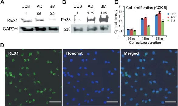
Figure 1. The expression levels of REX1, p38 MAPK and the cell proliferation of hMSCs.(A) The expression of REX1 in hUCB-MSCs, hAD-MSCs and hBM-hAD-MSCs. hUCB-hAD-MSCs and hAD-hAD-MSCs have strong REX1 expression, but hBM-hAD-MSCs have very weak REX1 expression. (B) The expression of p38 and phosphorylated p38 (Pp38) in hUCB-MSCs, hAD-MSCs and hBM-MSCs. The expression level of p38 was similar in the three types of hMSCs examined. hBM-MSCs have the strongest expression level of Pp38 among the three types of hMSCs. (C) Cell proliferation measured by CCK-8. The growth of hBM-MSCs was slowest among the three types of hMSCs. (D) REX1 primarily localized in the nucleus of hUCB-MSCs. Scale bars represent 100mm. Error bars represent the standard deviation from three independent experiments.
Results
The expression of REX1 is negatively correlated with the phosphorylation of p38 MAPK in hMSCs
REX1 is expressed in ESCs and MSCs. Among three well-characterized hMSCs, REX1 is strongly expressed in hUCB-MSCs and hAD-hUCB-MSCs; however, REX1 is weakly expressed in hBM-MSCs when grown under previously reported culture conditions [29–31]. In hUCB-MSCs, REX1 expression was nearly five-fold greater than in hBM-MSCs (Fig. 1A). However, in three types of hMSCs, the levels of phosphorylated p38 MAPK (Pp38) was in inverse proportion to the expression of REX1 (Fig. 1B). The level of Pp38 in hBM-MSCs was four-fold stronger than the level in hUCB-MSCs. The expression level of p38, itself, was similar between the three types of hMSCs (Fig. 1B). The cell proliferation rates of these three types of hMSCs were quite different. In repeated experiments, hUCB-MSCs and hAD-MSCs grew faster than hBM-MSCs (Fig. 1C). Therefore, the prolifer-ation rates of hMSCs positively correlate with the REX1 expression level but inversely correlate with the level of Pp38 expression. REX1 was expressed in the majority of hUCB-MSCs, and the localization of REX1 was primarily confined to the nucleus of hUCB-MSCs as shown by immunocytochemistry (Fig. 1D).
Knockdown of REX1 resulted in growth retardation of hUCB-MSCs
hUCB-MSCs grew faster than hBM-MSCs but at a similar pace as hAD-MSCs in in vitro culture. The discrepancy in the proliferation speed may be potentially due to the difference in REX1 expression in the different cell types. Therefore, a REX1 knockdown experiment was performed in hUCB-MSCs, which had the highest expression of REX1 among the three types of hMSCs. Four different shRNA producing lentiviruses that target different REX1-regions were used for the experiment. Two lentiviruses construct, REX1-2 (R2) and REX1-4 (R4), showed specific inhibition of REX1 after puromycin selection (Fig. 2A). After REX1 knockdown, cell growth severely decreased in hUCB-MSCs (Fig. 2B and 2C). For detailed analysis of cell cycle progression, FACS analysis was performed with the hUCB-MSCs (Fig. 2D). The composition of the G0/G1 phase significantly increased and the composition of the S phase significantly decreased in REX1 knocked-down hUCB-MSCs compared to vehicle control-infected hUCB-MSCs. Western blot analysis of cell cycle regulators was performed using vehicle control-infected and REX1 knocked-down hUCB-MSCs. After REX1 knockdown, the expression levels of CDK2, Cyclin B1, CDK4 and Cyclin D1 decreased compared to the levels of vehicle control-infected hUCB-MSCs. However, the expression of p27 did not change when REX1 was knocked-down in hUCB-MSCs compared to vehicle control-infected hUCB-MSCs (Fig. 2E).
Phosphorylation of p38 MAPK significantly increased following the knockdown of REX1 in hUCB-MSCs
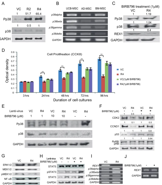
Among the detected MAPKs in hUCB-MSCs, p38 MAPK signaling significantly changed when REX1 was knocked-down. The expression level of p38 MAPK did not increase after REX1 knockdown; however, the level of Pp38 increased by more than fifty-fold after REX1 was knocked-down in hUCB-MSCs, which was verified in multiple experiments (Fig. 3A). SB203580 and SB202190 are potent inhibitors of the p38aand p38b isoforms, respectively, and all p38 MAPK isoforms are inhibited by BIRB796 [32]. The hUCB-MSCs express all four types of p38
isoforms (Fig. 3B); therefore, REX1 knocked-down hUCB-MSCs were treated with BIRB796. After p38 MAPK inhibitor treatment, the level of Pp38 in REX1 knocked-down hUCB-MSCs was not different compared to vehicle control-infected hUCB-MSCs (Fig. 3C and 3E). After BIRB796 treatment, the cell proliferation defect of REX1 knocked-down hUCB-MSCs recovered, and the proliferation rate was similar to the rate in vehicle control-infected hUCB-MSCs (Fig. 3D). Similarly, in hUCB-MSCs, 1mM or 10mM BIRB796 treatment repressed the phosphorylation of p38 MAPK (Fig. 3E). In REX1 knocked-down hUCB-MSCs, the suppressed expression levels of CDK2 and CCND1 recovered after BIRB796 treatment (Fig. 3F). The expression of p53 and hyper-phosphorylated RB (PpRB) did not change in REX1 knocked-down cells (Fig. 3F). In hUCB-MSCs, the expression levels of ERK1/2 and Mitogen-Activated Protein Kinase Kinase 1/2 (MAP2K1/2 or MEK1/2) did not change after the knockdown of REX1. The phosphorylation of MEK (pMEK1/2) and NF-kB expression were not significantly different after the knockdown of REX1 in hUCB-MSCs compared to vehicle control-infected hUCB-MSCs (Fig. 3G). REX1 inhibits signaling via the STAT pathway in F9 cells [12]; however, the expression of STAT5 did not significantly change after REX1 knockdown in hUCB-MSCs. The expression levels of STAT3 and phosphorylated STAT3 increased after REX1 knockdown without regard to p38 MAPK inhibitor treatment (Fig. 3H). After REX1 knockdown in hUCB-MSCs, the expression levels of the p38s did not significantly change Figure 2. REX1 knockdown resulted in the growth retardation of hUCB-MSCs. (A) The expression of REX1 decreased after REX1-knockdown lentivirus infection. (B) The cell proliferation of REX1 knocked-down hUCB-MSCs significantly decreased from two days after infection. (C) The morphology of cells three days after culturing. REX1 knocked-down cells are not growing at a rate similar to the control cells. (D) Cell cycle arrest was observed in REX1 knocked-down hUCB-MSCs using FACS analysis. (E) The expression levels of Cyclins, CDK and cell cycle inhibitors. The expression levels of CDK2, CCNB1, CDK4 and CCND1 decreased, but p27 did not change after REX1 knockdown. Error bars represent the standard deviation from three independent experiments. *, p,0.05; **, p,0.01.
Figure 3. The levels of p38 MAPK, Cyclins and cell cycle inhibitors changed after REX1 knockdown and p38 MAPK inhibitor treatment.(A) Significant phosphorylation of p38 MAPK (Pp38) was seen after REX1 knockdown in hUCB-MSCs. (B) hUCB-MSCs, hAD-MSCs and hBM-MSCs express all four types of p38 isoforms. (C) After p38 MAPK inhibitor treatment (1mM BIRB796), the phosphorylation of p38 MAPK was similar in REX1 knocked-down hUCB-MSCs compared to vehicle control-infected hUCB-MSCs. (D) Cell proliferation was measured with CCK-8 for four days. The cell proliferation of REX1 knocked-down hUCB-MSCs was similar with vehicle control-infected hUCB-MSCs after BIRB796 (1mM) treatment. Without BIRB796, the proliferation of REX1 knocked-down hUCB-MSCs significantly decreased compared with vehicle control-infected hUCB-MSCs. **, p,0.01. (E) The p38 MAPK of REX1 knocked-down hUCB-MSCs was not activated after 1mM or 10mM BIRB796 treatment compared to those of vehicle control-infected hUCB-MSCs. (F) Changes in the expression of CDK and cell cycle inhibitors. The decreased expression of CDK2 and CCND1 recovered after p38 MAPK inhibitor treatment. (G) The expression levels of ERK1/2, MEK, phospho-MEK (pMEK1/2) and NF-kB did not significantly change after REX1 knockdown in hUCB-MSCs. (H) The expression changes of STAT3 and STAT5. The expression levels of STAT3 and phospho-STAT3 were significantly increased after REX1 knockdown without regard to p38 MAPK inhibitor treatment. STAT5 expression did not change after the knockdown of REX1. (I) p38aand p38bwere up-regulated but p38cwas down-regulated after REX1 knockdown in hUCB-MSCs. (J)REX1expression of hUCB-MSCs did not change after BIRB796 (p38 MAPK inhibitor) treatment. Error bars represent the standard deviation from three independent experiments.
(Fig. 3I). In addition, BIRB 796 treatment did not influence REX1 expression in hUCB-MSCs (Fig. 3J).
MKK3 expression significantly increased after REX1 knockdown
In mammals, MKK3 and MKK6 are well known activators of p38 MAPK via the phosphorylation of p38 MAPK [33]. The expression ofMKK6did not significantly change after the knockdown of REX1, but the expression of MKK3 dramatically increased in REX1 knocked-down hUCB-MSCs (Fig. 4A). The MKK3 RNA level increased approximately seven-fold as confirmed by real-time RT-PCR. The level of MKK3 protein expression also increased 3.4-fold (Fig. 4B). In hMSCs, the expression of MKK3 was in inverse proportion to REX1 expression (Figs. 1A and 4C). The expression level of MKK3in hUCB-MSCs was 20-fold less than the level in hBM-MSCs. Therefore, the direct regulation ofMKK3by REX1 was investigated. The human genomic DNA sequences ofMKK3were analyzed for the presence of the REX1 binding motif. The DNA binding motif of REX1 contains the core sequences of GCAGCCAT
or GCCATTA [34]. In human genomic DNA, only three positions in MKK3contain consensus sequences for REX1 binding. These consensus sequences are located 1 Kb upstream of the first exon (Promoter region), inside of the first exon (Exon 1) and inside of the first intron (Intron 1) (Fig. 4D). A ChIP assay was performed to confirm the direct binding of REX1 toMKK3, and REX1 specifically binds only to the first exon ofMKK3(Fig. 4D). Other regions ofMKK3
did not contain REX1 binding motifs or a positive signal from the ChIP assay.
Alterations in the differentiation ability, and NOTCH and WNT signaling, of hUCB-MSCs following REX1
knockdown
The differentiation ability of hUCB-MSCs was investigated after the knockdown of REX1. The adipogenic potentiality in REX1 knocked-down hUCB-MSCs slightly increased or was similar to vehicle control-infected hUCB-MSCs after adipogenic differentiation as shown by Oil Red O staining. However, the osteogenic differentiation potential of REX1 knocked-down hUCB-MSCs visibly deteriorated after osteogenic induction as shown by Alizarin Red S staining (Fig. 5A).
Notch signaling is important for the differentiation of MSCs [35,36]. JAGGED1 (JAG1) is a ligand of the NOTCH receptor and plays key roles in cell differentiation and morphogenesis [37]. The expression ofJAG1was up-regulated in REX1 knocked-down hUCB-MSCs compared to vehicle control-infected hUCB-MSCs. NOTCH proteins are single-pass trans-membrane receptors that regulate cell fate decisions during development. The expression levels ofNOTCH1andNOTCH4also increased in REX1 knocked-down MSCs compared to vehicle control-infected hUCB-MSCs (Fig. 5B). As a consequence, the expression of HES1, a NOTCH target molecule, was up-regulated in REX1 knocked-down MSCs compared to vehicle control-infected hUCB-MSCs (Fig. 5B).
Canonical WNT/b-CATENIN signaling also plays an impor-tant role in regulating the differentiation of MSCs [38,39]. The expression levels ofb-CATENIN, GSK3-band phospho-GSK3-bat serine-9 decreased after REX1 inhibition in hUCB-MSCs (Fig. 5C). The expression of a WNT signaling related gene,
AXIN1, was not significantly different in REX1 knocked-down and vehicle control-infected hUCB-MSCs; however, the expres-sion levels of FZD2, LRP5 and DKK1 decreased in REX1 knocked-down hUCB-MSCs compared to vehicle control-infected hUCB-MSCs (Fig. 5D). The expression of ZNF281, a core transcription factor in stem cells that functions during hMSC osteogenesis (unpublished data), decreased in REX1 knocked-down hUCB-MSCs compared to the vehicle control-infected hUCB-MSCs (Fig. 5E). The expression of c-MYC, another core transcription factor of stem cells, did not change significantly after REX1 knockdown in hUCB-MSCs. In order to evaluate the changes in expression of other polycomb group genes, the expression levels of two polycomb group genes that are important to maintenance and specification of stem cells [40],SUZ12andBMI1, were measured. The expression levels of
SUZ12andBMI1decreased after REX1 knockdown in hUCB-MSCs (Fig. 5E).
REX1 inhibition did not affect apoptosis of hUCB-MSCs and Pp38 level was not significantly altered after REX1 knockdown in hBM-MSCs
Apoptosis is influenced by p38 MAPK, which is also important to cell growth. ANNEXIN V is used as a probe in the ANNEXIN V assay to detect cells that express phosphatidyl serine on the cell Figure 4. The expression of MKK3/6 in REX1 knocked-down
hUCB-MSCs and normally cultured hMSCs, and theMKK3ChIP assay in hUCB-MSCs.(A)MKK3expression significantly increased, but MKK6did not significantly change after REX1 knockdown as shown by RT-PCR. (B) MKK3 increased after REX1 knockdown at the protein expression level. (C) The expression level ofMKK3in hUCB-MSCs was 20-fold less than the level in hBM-MSCs under normal culture conditions as shown by real-time RT-PCR. (D) The ChIP assay for REX1. Three regions have REX1 consensus sequences in theMKK3genomic DNA. REX1 binds to the first exon region (MKK3Exon1) of MKK3. Abbreviations: MKK3Promt, MKK3 promoter region; MKK3Exon1, MKK3 exon 1 region; MKK3Intron1, MKK3 intron 1 region. Error bars represent the standard deviation from three independent experiments.
surface, a feature found in apoptosis as well as other forms of cell death [41,42]. ANNEXIN V staining was performed on three hUCB-MSCs clones derived from three different individuals (Fig. 6A). The number of apoptotic cells (Q2 and Q4 fraction) or necrotic cells (Q1 fraction) of REX1 knocked-down hUCB-MSCs did not significantly differ from the vehicle control-infected hUCB-MSCs (Fig. 6B). The expression of BAX, a marker protein of apoptosis, also did not increase in REX1 knocked-down hUCB-MSCs compared to vehicle control-infected hUCB-MSCs (Fig. 6C).
In hAD-MSCs, the level of Pp38 significantly increased after the knockdown of REX1 similar to hUCB-MSCs. However, in hBM-MSCs, the level of Pp38 increased slightly after REX1 knockdown (Fig. 6D). Basically, in hBM-MSCs, REX1 expression was very low (Fig. 1A). Also, under normal culture conditions, the intrinsic Pp38 and MKK3 expression levels were much higher in hBM-MSCs than in hUCB-hBM-MSCs or in hAD-hBM-MSCs (Figs. 1B, 4C and 6D). Therefore, the effect of the REX1 knockdown did not significantly influence Pp38 level in hBM-MSCs compared to other types of hMSCs.
Discussion
The p38 MAPK is important for cell growth, apoptosis and differentiation in mammalian cells. Under specific culture conditions, in hUCB-MSCs, which strongly express REX1, p38 MAPK is suppressed. However, hBM-MSCs, which have weak REX1 expression, have activated p38 MAPK and a high expression level of MKK3 under normal culture conditions. REX1 expression was inversely correlated with p38 MAPK activation and positively correlated with the proliferation rates of the three types of hMSCs examined.
REX1, a pluripotent marker gene, was expressed in some types of hMSCs and cancer cells as well as ESCs. In hUCB-MSCs, the knockdown of REX1 resulted in severe growth retardation. The functions of p38a have been previously reported and p38a is a major component of p38 MAPK in the majority of cells. p38acan negatively regulate cell cycle progression at both the G1/S and G2/M transitions by several mechanisms such as, the down-regulation of Cyclins and the up-down-regulation of CDK inhibitors [43]. In REX1 knocked-down hUCB-MSCs, the cell cycle was Figure 5. Differentiation study, NOTCH and WNT expression changes in REX1 knocked-down hUCB-MSCs.(A) After a three week induction, adipogenic and osteogenic differentiated hUCB-MSCs were stained with Oil Red O or Alizarin Red S. The number of adipogenic differentiated cells was similar to vehicle control-infected hUCB-MSCs or was slightly increased in REX1 knocked-down hUCB-MSCs. Osteogenesis of REX1 knocked-down hUCB-MSCs decreased compared to vehicle control-infected hUCB-MSCs. (B) The expression changes of NOTCH signaling genes after REX1 knockdown. The expression levels ofJAG1,NOTCH1andNOTCH4increased after REX1 knockdown. HES1 expression increased 1.2-fold after REX1 knockdown. (C) The expression levels of GSK3-b, phospho-GSK3-bat serine-9 (pGSK3b) andb-CATENIN decreased after REX1 knock-down in hUCB-MSCs. (D) The expression levels ofFZD2,LRP5andDKK1decreased after REX1 knockdown in hUCB-MSCs. (E) The expression levels of core transcription factors and polycomb group genes. The expression level ofZNF281 was down-regulated after REX1 knockdown in hUCB-MSCs. The expression levels ofSUZ12 andBMI1 also decreased after REX1 knockdown. The expression ofc-MYCdid not change after REX1 knockdown. Abbreviations: Adipo, adipogenic induction; Osteo, osteogenic induction. Error bars represent the standard deviation from three independent experiments. Scale bars represent 100mm.
arrested primarily in the G0/G1 stage, compared to vehicle control-infected hUCB-MSCs. Passage through the cell cycle requires the successive activation of different cyclin-dependent protein kinases (CDKs) and Cyclins [44,45]. Cyclin E, in association with CDK2, is required for the G1/S transition [46,47]. Both Cyclin A and the B-type Cyclins associate with CDC2 to promote entry into mitosis [48]. The expression levels of CDK2, CCND1 and Cyclin B1 decreased in REX1 knocked-down hUCB-MSCs compared with the levels in vehicle control-infected hUCB-MSCs. The expression levels of CDK2 and CCND1 recovered after p38 MAPK inhibitor treatment in REX1 knocked-down hUCB-MSCs. However, the expression
levels of CDK inhibitors, p53 and PpRB, did not change after p38 MAPK inhibitor treatment or REX1 knock-down in hUCB-MSCs. Although p38 MAPK was highly activated in REX1 knocked-down hUCB-MSCs, the cells grew slowly. Notch signaling, which increased in REX1 knocked-down hUCB-MSCs, also regulates stem cell number and has an opposite role of p38 MAPK [49]. Therefore, in REX1 knocked-down hUCB-MSCs, the growth inhibition signal by activated p38 MAPK was partially compensated by the cell proliferation signals from NOTCH activation.
Activation of p38 MAPK also contributes to chemically-induced cell death [50]. The possibility of increasing apoptosis in REX1 Figure 6. Apoptosis after REX1 knockdown in hUCB-MSCs and REX1 inhibition in hMSCs and the summary of the role of REX1 in stem cells.(A, B) Apoptotic cell death was not significantly different in REX1 knocked-down and vehicle control-infected hUCB-MSCs. (C) The expression of BAX did not change after REX1 knockdown in hUCB-MSCs. (D) Significant activation of p38 MAPK was observed after REX1 knockdown in hUCB-MSCs and hAD-MSCs but only slightly increased in hBM-MSCs. hBM-MSCs, which have low expression of REX1, have highly activated p38 MAPK (Pp38) in vehicle control-infected cells. (E) REX1 suppresses MKK3 expression, which activates p38 MAPK. REX1 also suppresses STAT3 expression and NOTCH signals. Error bars represent the standard deviation from three independent experiments.
knocked-down hUCB-MSCs was excluded because the results of the ANNEXIN V assay showed no significant difference between REX1 knocked-down and vehicle control-infected hUCB-MSCs. In addition, the expression of STAT3 increased in REX1 knocked-down hUCB-MSCs, which was also previously reported [12]. After p38 MAPK inhibitor treatment and REX1 knock-down, the STAT3 activation patterns did not change, suggesting that STAT3 suppression by REX1 is independent of p38 MAPK signaling.
Activation of p38 MAPK is important for the differentiation of mouse ESCs [51] and hMSCs [52]. The known up-stream regulators of p38 MAPK are MKK3 and MKK6, which are activated in response to many types of cell stresses [53,54]. However, in stem cells, the transcriptional regulation ofMKK3 and
MKK6was not previously reported. REX1 expression is specific to pluripotent stem cells, several types of hMSCs and cancer cells. REX1 is highly expressed in hUCB-MSCs, butMKK3expression is approximately 20-fold lower in hUCB-MSCs, compared to hBM-MSCs, where REX1 expression is low under normal culture conditions. REX1 suppresses the expression of MKK3 in hMSCs, which was mediated by REX1 binding directly to the first exon of
MKK3(Fig. 6E).
The Notch signaling pathway is implicated in the differentiation [55,56] and the immune-modulation of MSCs [57]. Notch signaling inhibits chondrogenesis of hMSCs [35]. Also, the expression of the NOTCH ligand and its receptors (JAG1, NOTCH1 and NOTCH 4) increased after the knockdown of REX1 in hUCB-MSCs. Therefore, osteogenic differentiation deformity was partially caused by NOTCH activation after REX1 knockdown in hUCB-MSCs. Notch signaling also has a role in adipogenesis, and in 3T3-L1 cells, the reduction of Hes-1 inhibits adipogenic differentiation [58]. The activation of p38 MAPK activates C/EBPb, a transcription factor that has a critical role in adipogenesis [59]. Therefore, after REX1 knockdown in hUCB-MSCs, increased adipogenesis was potentially caused by both NOTCH and p38 MAPK activation. The expression level of HES1, a target molecule of NOTCH signaling, increased in REX1 knocked-down hUCB-MSCs; therefore, REX1 may suppress the NOTCH signaling pathways and maintains the undifferentiated state of hUCB-MSCs (Fig. 6E).
In MSCs, Wnt/b-catenin (i.e., canonical) signaling is also important to maintain stemness and multipotency [60–62]. b -CATENIN decreased after REX1 knockdown in hUCB-MSCs. The function of GSK3-b, which inactivates b-CATENIN, is inhibited by the phosphorylation of serine-9 [63]. In REX1 knocked-down hUCB-MSCs, the expression level of GSK3-b
decreased, and the level of phosphorylated GSK3-b at serine-9 also decreased. p38 MAPK down-regulates WNT/b-CATENIN signaling via GSK3-b [64], which was confirmed by our results. Notch 1 over-expression inhibits osteoblastogenesis by suppressing Wnt/b-Catenin signaling [65], which correlated with osteogenic deformity in REX1 knocked-down hUCB-MSCs. The increased NOTCH signal also caused the suppression of WNT in REX1 knocked-down hUCB-MSCs. Several core transcription factors that have critical roles in ESCs were expressed in hUCB-MSCs. The effect of REX1 knockdown on these transcription factors was different in each gene expression. The expression of ZNF281
decreased in REX1 knocked-down hUCB-MSCs compared to vehicle control-infected hUCB-MSCs, but the expression of c-MYCdid not change when REX1 was knocked-down in hUCB-MSCs. REX1 is considered to be a member of the YY1 gene family which is a polycomb group gene. The expression levels of the polycomb group genes, SUZ12 and BMI1, were reduced in REX1 knocked-down hUCB-MSCs. Therefore, REX1 also has a role in modulating the chromatin structure of hUCB-MSCs, which needs to be further explored.
In conclusion, REX1 represses the expression of MKK3, which can activate p38 MAPK in hMSCs. The high expression level of REX1 in stem cells protects cell differentiation by suppressing p38 MAPK activation via MKK3 suppression and by suppressing NOTCH and STAT3 signaling. ASCs that highly express REX1 have more prominent cell-proliferation ability and multipotency than low REX1-expressing ASCs due to the p38 MAPK suppression in stem cells. Therefore, REX1 is a proper marker not only in ESCs but also in highly efficient multipotent human ASCs. Overall, the suppression of p38 MAPK is useful in the collection and culturing of multipotent human ASCs that do not have sufficient expression levels of REX1. This will be beneficial to the field of regenerative medicine.
Supporting Information
Table S1 PCR primers sequences used in experiment. Found at: doi:10.1371/journal.pone.0010493.s001 (0.12 MB DOC)
Author Contributions
Conceived and designed the experiments: KWS SKK KSK. Performed the experiments: DRB KWS KHR. Analyzed the data: DRB KWS JWJ. Contributed reagents/materials/analysis tools: KHR. Wrote the paper: KWS KSK.
References
1. Rossant J (2008) Stem cells and early lineage development. Cell 132: 527–531. 2. Takahashi K, Yamanaka S (2006) Induction of pluripotent stem cells from mouse
embryonic and adult fibroblast cultures by defined factors. Cell 126: 663–676. 3. Boyer LA, Lee TI, Cole MF, Johnstone SE, Levine SS, et al. (2005) Core
transcriptional regulatory circuitry in human embryonic stem cells. Cell 122: 947–956.
4. Wang J, Rao S, Chu J, Shen X, Levasseur DN, et al. (2006) A protein interaction network for pluripotency of embryonic stem cells. Nature 444: 364–368. 5. Boiani M, Eckardt S, Scholer HR, McLaughlin KJ (2002) Oct4 distribution and
level in mouse clones: consequences for pluripotency. Genes Dev 16: 1209–1219.
6. Ben-Shushan E, Thompson JR, Gudas LJ, Bergman Y (1998) Rex-1, a gene encoding a transcription factor expressed in the early embryo, is regulated via Oct-3/4 and Oct-6 binding to an octamer site and a novel protein, Rox-1, binding to an adjacent site. Mol Cell Biol 18: 1866–1878.
7. Niwa H, Miyazaki J, Smith AG (2000) Quantitative expression of Oct-3/4 defines differentiation, dedifferentiation or self-renewal of ES cells. Nat Genet 24: 372–376. 8. Hosler BA, LaRosa GJ, Grippo JF, Gudas LJ (1989) Expression of REX-1, a gene containing zinc finger motifs, is rapidly reduced by retinoic acid in F9 teratocarcinoma cells. Mol Cell Biol 9: 5623–5629.
9. Mongan NP, Martin KM, Gudas LJ (2006) The putative human stem cell marker, Rex-1 (Zfp42): structural classification and expression in normal human epithelial and carcinoma cell cultures. Mol Carcinog 45: 887–900.
10. Ramalho-Santos M, Yoon S, Matsuzaki Y, Mulligan RC, Melton DA (2002) ‘‘Stemness’’: transcriptional profiling of embryonic and adult stem cells. Science 298: 597–600.
11. Shi Y, Lee JS, Galvin KM (1997) Everything you have ever wanted to know about Yin Yang 1. Biochim Biophys Acta 1332: F49–66.
12. Xu J, Sylvester R, Tighe AP, Chen S, Gudas LJ (2008) Transcriptional activation of the suppressor of cytokine signaling-3 (SOCS-3) gene via STAT3 is increased in F9 REX1 (ZFP-42) knockout teratocarcinoma stem cells relative to wild-type cells. J Mol Biol 377: 28–46.
13. Masui S, Ohtsuka S, Yagi R, Takahashi K, Ko MS, et al. (2008) Rex1/Zfp42 is dispensable for pluripotency in mouse ES cells. BMC Dev Biol 8: 45. 14. Pearson G, Robinson F, Beers Gibson T, Xu BE, Karandikar M, et al. (2001)
Mitogen-activated protein (MAP) kinase pathways: regulation and physiological functions. Endocr Rev 22: 153–183.
16. Han J, Molkentin JD (2000) Regulation of MEF2 by p38 MAPK and its implication in cardiomyocyte biology. Trends Cardiovasc Med 10: 19–22. 17. Zarubin T, Han J (2005) Activation and signaling of the p38 MAP kinase
pathway. Cell Res 15: 11–18.
18. Schieven GL (2005) The biology of p38 kinase: a central role in inflammation. Curr Top Med Chem 5: 921–928.
19. Nebreda AR, Porras A (2000) p38 MAP kinases: beyond the stress response. Trends Biochem Sci 25: 257–260.
20. Campagnoli C, Roberts IA, Kumar S, Bennett PR, Bellantuono I, et al. (2001) Identification of mesenchymal stem/progenitor cells in human first-trimester fetal blood, liver, and bone marrow. Blood 98: 2396–2402.
21. Erices A, Conget P, Minguell JJ (2000) Mesenchymal progenitor cells in human umbilical cord blood. Br J Haematol 109: 235–242.
22. Gronthos S, Franklin DM, Leddy HA, Robey PG, Storms RW, et al. (2001) Surface protein characterization of human adipose tissue-derived stromal cells. J Cell Physiol 189: 54–63.
23. Igura K, Zhang X, Takahashi K, Mitsuru A, Yamaguchi S, et al. (2004) Isolation and characterization of mesenchymal progenitor cells from chorionic villi of human placenta. Cytotherapy 6: 543–553.
24. Tsai MS, Lee JL, Chang YJ, Hwang SM (2004) Isolation of human multipotent mesenchymal stem cells from second-trimester amniotic fluid using a novel two-stage culture protocol. Hum Reprod 19: 1450–1456.
25. Zvaifler NJ, Marinova-Mutafchieva L, Adams G, Edwards CJ, Moss J, et al. (2000) Mesenchymal precursor cells in the blood of normal individuals. Arthritis Res 2: 477–488.
26. Roche S, Richard MJ, Favrot MC (2007) Oct-4, Rex-1, and Gata-4 expression in human MSC increase the differentiation efficiency but not hTERT expression. J Cell Biochem 101: 271–280.
27. Guillot PV, Gotherstrom C, Chan J, Kurata H, Fisk NM (2007) Human first-trimester fetal MSC express pluripotency markers and grow faster and have longer telomeres than adult MSC. Stem Cells 25: 646–654.
28. Peng J, Li W, Li H, Jia Y, Liu Z (2009) Inhibition of p38 MAPK facilitates ex vivo expansion of skin epithelial progenitor cells. In Vitro Cell Dev Biol Anim 45: 558–565.
29. Seo KW, Lee SR, Bhandari DR, Roh KH, Park SB, et al. (2009) OCT4A contributes to the stemness and multi-potency of human umbilical cord blood-derived multipotent stem cells (hUCB-MSCs). Biochem Biophys Res Commun 384: 120–125.
30. Pal R, Venkataramana NK, Bansal A, Balaraju S, Jan M, et al. (2009) Ex vivo-expanded autologous bone marrow-derived mesenchymal stromal cells in human spinal cord injury/paraplegia: a pilot clinical study. Cytotherapy 11: 897–911.
31. Park JR, Jung JW, Lee YS, Kang KS (2008) The roles of Wnt antagonists Dkk1 and sFRP4 during adipogenesis of human adipose tissue-derived mesenchymal stem cells. Cell Prolif 41: 859–874.
32. Kuma Y, Sabio G, Bain J, Shpiro N, Marquez R, et al. (2005) BIRB796 inhibits all p38 MAPK isoforms in vitro and in vivo. J Biol Chem 280: 19472–19479. 33. Inoue T, Hammaker D, Boyle DL, Firestein GS (2005) Regulation of p38
MAPK by MAPK kinases 3 and 6 in fibroblast-like synoviocytes. J Immunol 174: 4301–4306.
34. Kim JD, Faulk C, Kim J (2007) Retroposition and evolution of the DNA-binding motifs of YY1, YY2 and REX1. Nucleic Acids Res 35: 3442–3452. 35. Grogan SP, Olee T, Hiraoka K, Lotz MK (2008) Repression of chondrogenesis
through binding of notch signaling proteins HES-1 and HEY-1 to N-box domains in the COL2A1 enhancer site. Arthritis Rheum 58: 2754–2763. 36. Otto WR, Rao J (2004) Tomorrow’s skeleton staff: mesenchymal stem cells and
the repair of bone and cartilage. Cell Prolif 37: 97–110.
37. Guarnaccia C, Pintar A, Pongor S (2004) Exon 6 of human Jagged-1 encodes an autonomously folding unit. FEBS Lett 574: 156–160.
38. Takada I, Kouzmenko AP, Kato S (2009) Wnt and PPARgamma signaling in osteoblastogenesis and adipogenesis. Nat Rev Rheumatol 5: 442–447. 39. Kawai M, Mushiake S, Bessho K, Murakami M, Namba N, et al. (2007) Wnt/
Lrp/beta-catenin signaling suppresses adipogenesis by inhibiting mutual activation of PPARgamma and C/EBPalpha. Biochem Biophys Res Commun 363: 276–282.
40. Rajasekhar VK, Begemann M (2007) Concise review: roles of polycomb group proteins in development and disease: a stem cell perspective. Stem Cells 25: 2498–2510.
41. Koopman G, Reutelingsperger CP, Kuijten GA, Keehnen RM, Pals ST, et al. (1994) Annexin V for flow cytometric detection of phosphatidylserine expression on B cells undergoing apoptosis. Blood 84: 1415–1420.
42. Vermes I, Haanen C, Steffens-Nakken H, Reutelingsperger C (1995) A novel assay for apoptosis. Flow cytometric detection of phosphatidylserine expression on early apoptotic cells using fluorescein labelled Annexin V. J Immunol Methods 184: 39–51.
43. Ambrosino C, Nebreda AR (2001) Cell cycle regulation by p38 MAP kinases. Biol Cell 93: 47–51.
44. Nigg EA (1995) Cyclin-dependent protein kinases: key regulators of the eukaryotic cell cycle. Bioessays 17: 471–480.
45. Murray AW (2004) Recycling the cell cycle: cyclins revisited. Cell 116: 221–234. 46. Knoblich JA, Sauer K, Jones L, Richardson H, Saint R, et al. (1994) Cyclin E controls S phase progression and its down-regulation during Drosophila embryogenesis is required for the arrest of cell proliferation. Cell 77: 107–120. 47. Ohtsubo M, Roberts JM (1993) Cyclin-dependent regulation of G1 in
mammalian fibroblasts. Science 259: 1908–1912.
48. King RW, Jackson PK, Kirschner MW (1994) Mitosis in transition. Cell 79: 563–571.
49. Androutsellis-Theotokis A, Leker RR, Soldner F, Hoeppner DJ, Ravin R, et al. (2006) Notch signalling regulates stem cell numbers in vitro and in vivo. Nature 442: 823–826.
50. Gills JJ, Castillo SS, Zhang C, Petukhov PA, Memmott RM, et al. (2007) Phosphatidylinositol ether lipid analogues that inhibit AKT also independently activate the stress kinase, p38alpha, through MKK3/6independent and -dependent mechanisms. J Biol Chem 282: 27020–27029.
51. Chakraborty S, Kang B, Huang F, Guo YL (2009) Mouse embryonic stem cells lacking p38alpha and p38delta can differentiate to endothelial cells, smooth muscle cells, and epithelial cells. Differentiation 78: 143–150.
52. Platt MO, Wilder CL, Wells A, Griffith LG, Lauffenburger DA (2009) Multipathway kinase signatures of multipotent stromal cells are predictive for osteogenic differentiation: tissue-specific stem cells. Stem Cells 27: 2804–2814. 53. Cuenda A, Rousseau S (2007) p38 MAP-kinases pathway regulation, function
and role in human diseases. Biochim Biophys Acta 1773: 1358–1375. 54. Mittelstadt PR, Salvador JM, Fornace AJ, Jr., Ashwell JD (2005) Activating p38
MAPK: new tricks for an old kinase. Cell Cycle 4: 1189–1192.
55. Li H, Yu B, Zhang Y, Pan Z, Xu W (2006) Jagged1 protein enhances the differentiation of mesenchymal stem cells into cardiomyocytes. Biochem Biophys Res Commun 341: 320–325.
56. Hiraoka K, Grogan S, Olee T, Lotz M (2006) Mesenchymal progenitor cells in adult human articular cartilage. Biorheology 43: 447–454.
57. Li YP, Paczesny S, Lauret E, Poirault S, Bordigoni P, et al. (2008) Human mesenchymal stem cells license adult CD34+hemopoietic progenitor cells to differentiate into regulatory dendritic cells through activation of the Notch pathway. J Immunol 180: 1598–1608.
58. Ross DA, Rao PK, Kadesch T (2004) Dual roles for the Notch target gene Hes-1 in the differentiation of 3T3-L1 preadipocytes. Mol Cell Biol 24: 3505–3513. 59. Ono K, Han J (2000) The p38 signal transduction pathway: activation and
function. Cell Signal 12: 1–13.
60. Etheridge SL, Spencer GJ, Heath DJ, Genever PG (2004) Expression profiling and functional analysis of wnt signaling mechanisms in mesenchymal stem cells. Stem Cells 22: 849–860.
61. Boland GM, Perkins G, Hall DJ, Tuan RS (2004) Wnt 3a promotes proliferation and suppresses osteogenic differentiation of adult human mesenchymal stem cells. J Cell Biochem 93: 1210–1230.
62. Bennett CN, Longo KA, Wright WS, Suva LJ, Lane TF, et al. (2005) Regulation of osteoblastogenesis and bone mass by Wnt10b. Proc Natl Acad Sci U S A 102: 3324–3329.
63. Stambolic V, Woodgett JR (1994) Mitogen inactivation of glycogen synthase kinase-3 beta in intact cells via serine 9 phosphorylation. Biochem J 303 (Pt 3): 701–704.
64. Bikkavilli RK, Feigin ME, Malbon CC (2008) p38 mitogen-activated protein kinase regulates canonical Wnt-beta-catenin signaling by inactivation of GSK3beta. J Cell Sci 121: 3598–3607.