T
ABSTRACT
BIOCOMPATIBILITY OF DIFFERENT INTRACANAL
MEDICATIONS IN RAT BUCCAL SUBMUCOSA TISSUE
Tereza Aparecida Delle Vedove SEMENOFF1, Alex SEMENOFF SEGUNDO2, José Antonio Poli de FIGUEIREDO3
1- DDS, MSc, PhD student in Oral Diagnosis, Araçatuba School of Dentistry, São Paulo State University, Araçatuba, SP, Brazil. 2- DDS, MSc, PhD in Periodontics, Araçatuba School of Dentistry, São Paulo State University, Araçatuba, SP, Brazil.
3- DD, MSc, PhD, Assistant Professor of Histology, Dental School, Federal University of Rio Grande do Sul, Porto Alegre, RS, Brazil.
Corresponding address: Tereza Aparecida Delle Vedove SEMENOFF - Rua da Aviação, 1800. Residencial Itamaracá, apartamento 11 bloco 11. Bairro Aviação, 16056-725, Araçatuba, SP, Brasil. [email protected]
Received: February 5, 2007 - Modification: July 5, 2007 - Accepted: October 24, 2007
he aim of this study was to analyze the buccal tissue responses of Wistar rats to 2% chlorhexidine solution, calcium hydroxide and the association of both products. For this purpose, 30 specimens were randomly implanted in the filtrum of the four upper and lower hemiarches with a polyethylene tube containing one of the following substances: 2% chlorhexidine solution, calcium hydroxide and 2% chlorhexidine solution (test groups); calcium hydroxide and distilled water and distilled water (control groups). Ten rats each were distributed according to time interval of evaluation at 7, 15 and 30 days. The histological sections were stained with Harris hematoxylin and eosin. Analysis was performed with an optical microscope at x100, x200 and x400 magnifications by an expert examiner blinded to the materials. The sections were classified by scores attributed to inflammatory events and by a ranking determined according to the severity of the inflammation. The results of the inflammatory events and severity ranking were submitted to the Kruskal-Wallis test at a 0.05 level of significance. No statistically significant difference occurred among the tested materials; however, all materials showed a decreased of severity with respect to longer time intervals.
Uniterms: Biocompatibility. Calcium hydroxide. Chlorhexidine. Endodontics. Rats.
INTRODUCTION
Chlorhexidine has been proposed in endodontic therapy in various concentrations as an irrigating solution, intracanal medication and in association with other substances, for cleansing of infected root canal systems (Barbosa, et al.11997;
Siqueira Jr. and Uzeda221997; Estrela, et al.4 2001; Schafer and
Bössmann20 2005).
This substance is a cationic detergent of the biguanide group, which is almost insoluble in water. For this reason, it is almost always prepared in salt form, chlorhexidine digluconate, which increases its solubility (Davies31973). It is considered
a wide spectrum antimicrobial substance because in addition to its efficacy against Gram-positive bacteria, it also acts against Gram-negative bacteria and has variable fungicidal action, depending on the specie. Furthermore, it has a very important property called substantivity, that is, residual effect (Rölla, et al.181970; Rosenthal, et al.19 2004), which means that
its presence is maintained in the blood and other bodily fluids. Although the use of chlorhexidine has been widely diffused in many dental fields, its efficacy in microbial control has been proven (Heling, et al.8,91992; Siqueira Jr. and Uzeda22
1997; Dametto, et al.22005) and the tolerance of oral tissues to
this substance has been shown (Löe e Schiott141970; Yates,
et al.241993), few studies have demonstrated the behavior of
conjunctive tissues in its presence (Fellippe, et al.51998).
When used as an intracanal medication, both in solution and in association with calcium hydroxide, chlorhexidine remains in intimate contact with the periapical tissues, usually for a prolonged period. Therefore, knowledge of tissue behavior in its presence is of paramount importance. Moreover, longitudinal observation of these reactions seems to be a relevant factor for elucidation of the type of response that host tissues can present in the presence of this substance. Thus, the purpose of this study was to evaluate the response of rat buccal submucosa tissue to the presence of distilled water, a mixture of calcium hydroxide (p.a.) and distilled water, a 2% chlorhexidine solution and a mixture of calcium hydroxide (p.a.) and 2% chlorhexidine solution.
MATERIAL AND METHODS
Thirty male Wistar rats weighing 220 g on average were selected. The animals were subjected to a light/dark cycle of 12 h, mean temperature of 22°C, 40-60% humidity, with free access to solid chow and water ad libitum, thus closely preserving their habitual situation. The rats were randomly separated and identified both as individuals and in groups.
For inoculation of the test materials, 120 cylinders measuring 5 mm in length and 1.3 mm internal diameter were obtained from polyethylene 19G tubes (Venescalp, Feira de Santana, BA, Brazil). The cylinders were divided equally according to experimental group.
Atoxic polyurethane sponges cut in sizes of approximately 6x2x1 mm were used for placement of 2% chlorhexidine solution and distilled water. As much as 0.02 mL of liquid was injected into the center of the sponges using a 1-mL insulin syringe to avoide liquid overflow at the edge of the cylinder. To obtain the pastes, calcium hydroxide powder (Iodontosul - Industrial Odontológica do Sul Ltda., Porto Alegre, RS, Brazil) was divided in two portions of approximately 4.6 g, weighed in an analytical balance. Concerning the quantity of liquid, whether using distilled water or 2% chlorhexidine, as much as 5.1 mL was stipulated, maintaining a proportion of 1 g of powder to 1.1 ml of liquid, resulting in a homogenous mixture with toothpaste consistency. The paste was injected into the cylinder using a 10 mL disposable syringe without needle.
All surgical procedures were performed under general anesthesia by intramuscular administration of 0.1 mL of ketamine hydrochloride (Dopalen, Agribrands. Saúde Animal, Paulínia, SP, Brazil) associated with 0.05 mL of xylazine hydrochloride (Rompun, Bayer, Saúde Animal, São Paulo, SP, Brazil)per 100 g of rat body weight.
For material insertion, four quadrants from each rat were used. With the aid of clinical tweezers, the rat’s mouth was opened and a 3-mm-long incision was made at the region of the buccal gingival sulcus with a #15 surgical scalpel (Med Blade, Medgoldman Ind. e Com. Ltda. Manaus, AM, Brazil), after which a randomly chosen cylinder was inserted in the submucosal tissue of the region. After implantation of each of the cylinders, the incision was closed using resorbable, #3 catgut sutures (Ethicon, Johnson & Johnson, São Paulo, SP, Brazil).
Following completion of each experimental time interval (7, 15 and 30 postoperative days), 10 randomly chosen rats were anesthetized as described above and sacrificed by cervical dislocation, the pieces being immediately immersed in 10% formalin.
Prior to histological processing, the cylinders were carefully located, removed from the histological samples, dehydrated in a gradual alcohol series and embedded in paraffin. The blocks were cut at the central portion, where 5 standardized semi-serial 5-µm-thick histological sections were obtained and stained with Harris hematoxylin and eosin. Five glass slides belonging to each histological block were examined with an optical microscope (Olympikus, Tokyo, Japan) by a single senior examiner who was blinded to the tested substances. In this initial stage, the whole extension of the glass slides was examined at x100, x200 and x400
magnification in order to visualize the tissue adjacent to at least one of the ends of the cylinder. This was established as an inclusion criterion to select the slide with the best quality staining and histological details (Figure 1). Regarding the region corresponding to the cylinder extremity, the area with the greatest degree of inflammation was chosen.
Once these selection criteria were established, a new phase began, in which the slides were analyzed qualitatively (Figueiredo, et al.62001), observing the presence of
neutrophils, lymphocytes, plasmocytes, eosinophils, macrophages, gigantocytes, condensation fibrosis and abscess. Based on these findings, the slides were then classified according to the following scores: 0 – Absence; 1 – Presence; 2 – Infiltrate.
For cell events, the following scores were considered: 0 -when inflammatory cells were absent or found only within the blood vessels; 1 - when inflammatory cells were sparsely present or in very small groups; and 2 - when inflammatory cells, in an area close to a tube extremity, were found in the entire field or were present in a large number, configuring great severity.
Condensation fibrosis received the following scores: 0 when absent; 1 when a fine layer involving the study material was formed; and 2 when a thick layer was formed.
Regarding abscess formation, the following scores were given: 0 - absent; 1 - the abscess was limited to the proximity of the cylinder containing the material; and 2 - the abscess reached more distant areas.
After qualitative analysis and attribution of scores to the observed events, the slides were scored according to inflammation severity. A ranking scale was established, which ordered the slides in increasing degree of inflammatory reaction, ranging from absent to highly inflammatory. All
analyzes were undertaken at x100, x200 and x400 magnification in the region close to a cylinder extremity, chosen as described above.
Statistical analysis
For comparison of the cell events observed using the tested materials at 7, 15 and 30 days, the data obtained were submitted to Kruskal-Wallis nonparametric test at 5% level of significance. The analysis of the degree of inflammation denoted in the ranking scale was also performed with
Kruskal-Wallis nonparametric test at 5% significance level. Initially, the data were submitted to statistical analysis of the possible influence of the quadrants used in the rat mouth in relation to the inflammatory response (Kruskal-Wallis; 5% significance level). As no statistically significant difference was observed, allp values were over 0.05, it was inferred that the factor quadrant exerted no influence on the results.
Neutrophils W 21 1.86
±
0.38 7 0.375 1.29±
0.49 7 0.495 0.57±
0.53 7 0.132C 22 1.43
±
0.53 7 1.43±
0.53 7 1.00±
0.53 8W+CH 22 1.67
±
0.50 9 1.67±
0.52 6 1.14±
0.38 7C+CH 21 1.75
±
0.46 8 1.25±
0.50 4 1.22±
0.67 9Eosinophils W 21 0.14
±
0.38 7 0.951 0.00±
0.00 7 0.488 0.14±
0.38 7 0.935C 22 0.14
±
0.38 7 0.14±
0.38 7 0.25±
0.46 8W+CH 22 0.22
±
0.44 9 0.00±
0.00 6 0.14±
0.38 7C+CH 21 0.13
±
0.35 8 0.00±
0.00 4 0.22±
0.44 9Lymphocytes W 21 1.86
±
0.38 7 0.860 1.86±
0.38 7 0.359 1.14±
0.38 7 0.672and C 22 1.71
±
0.49 7 1.57±
0.53 7 1.13±
0.25 8Plasmocytes W+CH 22 1.67
±
0.50 9 1.83±
0.41 6 1.14±
0.38 7C+CH 21 1.75
±
0.46 8 2.00±
0.00 4 1.33±
0.50 9Macrophages W 21 1.71
±
0.49 7 0.474 2.00±
0.00 7 0.392 1.43±
0.53 7 0.482and C 22 2.00
±
0.00 7 2.00±
0.00 7 1.75±
0.46 8Gigantocytes W+CH 22 1.89
±
033 9 1.83±
0.41 6 1.43±
0.53 7C+CH 21 1.88
±
0.35 8 2.00±
0.00 4 1.67±
0.50 9Condensation W 21 1.57
±
0.53 7 0.628 1.43±
0.53 7 0.357 0.86±
0.38 7 0.271Fibrosis C 22 1.43
±
0.53 7 1.43±
0.53 7 1.13±
0.35 8W+CH 22 1.33
±
0.50 9 1.17±
0.41 6 0.86±
0.38 7C+CH 21 1.25
±
0.46 8 1.00±
0.00 4 1.11±
0.33 9Abscess W 21 0.14
±
0.38 7 0.374 0.14±
0.38 7 0.359 0.00±
0.00 7 0.639C 22 0.00
±
0.00 7 0.43±
0.53 7 0.13±
0.35 8W+CH 22 0.44
±
0.73 9 0.17±
0.41 6 0.14±
0.38 7C+CH 21 0.25
±
0.46 8 0.00±
0.00 4 0.22±
0.44 9Time (days)
Variable Material n 7 15 30
X ± SD n P X ± SD n P X ± SD n P
RESULTS
The results are summarized in Table 1, which present the means and standard deviations of the cell events for the tested materials and the time interval.
No statistically significant differences were found in the analysis of cell events when comparing the tested materials (Figures 2 and 3). On the other hand, statistically significant differences were found while analyzing the tissue response caused by a single material according to the time interval: the severity of the inflammatory reaction to all substances decreased along the duration of the experiment (Figure 4).
Analyzing the results of the ranking scale, established according to the degree of general inflammation and with the cell events ordered gradually from those showing absence of inflammation to those of greatest inflammation, no significant differences were found regarding among the tested materials (Table 2). However, when testing the influence of the time interval on the degree of inflammation, the inflammatory
process clearly diminished as a result of a longer experimental time interval (Table 3).
Materials N Degree of Kruskal-Wallis
inflammation
mean p value
W 21 39.71 0.631
C 22 40.23
W + CH 22 46.68
C + CH 21 47.38
TABLE 2- Mean position of the materials of the entire sample in relation to the degree of inflammation - W – Distilled Water; C – Chlorhexidine; W+CH – Distilled Water associated with Calcium Hydroxide; C+CH – Chlorhexidine associated with Calcium Hydroxide. - Significance level 5% (p<0.05)
Time Intervals N Degree of Kruskal-Wallis
Inflammation
mean p value
7 days 31 53.81 0.005*
15 days 24 43.50
30 days 31 33.19
TABLE 3- Mean position of time intervals in relation to the degree of inflammation
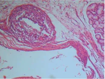
* Significant difference at a level of 5% FIGURE 2- Demonstrative image of tissue reaction,
considered severe, when using chlorhexidine associated with calcium hydroxide at 7 days. Abscess presence considered score 2 (arrow)
FIGURE 3- Demonstrative image of tissue reaction, considered severe, when using distilled water associated with calcium hydroxide at 7 days
DISCUSSION
The use of the submucosal region of rat buccal gingival sulcus was chosen due to the marked physiological similarity of this location with the periapical tooth region, rather than the dorsum of these animals, which is a commonly used area in several studies.
Figueiredo, et al.6(2001) used the submucosal tissue of
rabbits with success in a study that, aside from sample size, was similar to the present investigations in its methodological steps. The ease of execution of this technique, especially because it does not require trichotomy and tissue divulsion as necessary when using the dorsal region, should also be highlighted.
The stipulation of time intervals of 7, 15 and 30 days, contemplated the commonly used periods in Endodontics regarding the use of calcium hydroxide as an intracanal medication, permitting the observation of histological responses during short-, medium- and long-term periods, and was in agreement with the protocol of other studies that analyzed tissue response (Holland, et al.10 1999; Nelson Filho,
et al.15 1999; Holland, et al.11 2001).
The criteria for histological evaluation with 0-to-1 scores and the choice for neutrophils, eosinophils, lymphocytes/ plasmocytes and macrophages/giant cells, condensation fibrosis and abscess as markers of tissue reaction, were basically due to the direct relation of these elements with the inflammatory process (Figueiredo, et al.6 2001). An expert
examiner analyzed the inflammatory events because in addition to providing quantitative data using the above-mentioned scoring system, the examiner could present qualitative data on the inflammatory process, by interpreting tissue responses, such as the presence of abscess, condensation fibrosis and other events.
It is important to highlight that other methods of identifying inflammatory processes exist. Peters, et al.17(2002) suggested
quantification of angiogenesis using a specific software to provide a faster and more practical. However, it is our understanding that such a count is only quantitative, providing no information regarding the quality of the inflammatory process and allowing no data interpretation.
The use of a scoring system aimed to rank each slide according to the degree of inflammation present. Having observed that the quadrant position did not interfere with the inflammatory response, the scoring system was designed by mixing all materials and time intervals. The analysis of data referring to the measurement of the inflammatory events was performed (Kruskal-Wallis test; a=5%) at experimental periods of 7, 15 and 30 days.
No significant differences were found among the tested materials regarding the inflammatory reaction. However, some findings were interesting and should be addressed. In relation to the number of neutrophils, distilled water presented higher means than 2% chlorhexidine within a 7-day period. This is in agreement with the findings of Knuuttila et al.13 (1978) who,
using sponges impregnated with 0.01 to 0.2% chlorhexidine, observed that chlorhexidine demonstrated a certain influence on the diminished migration of leukocytes to the implantation
sites when compared to saline over 12-, 24- and 36-hour periods. Moreover, they reported that the greater the chlorhexidine concentration, the lower the number of inflammatory cells.
Using the same methodology, Paunio, et al.16(1978),
maintained the previously mentioned sponges for longer time intervals, between 3 and 23 days, obtaining results that indicated that chlorhexidine presented a clear delaying effect on the formation of granulation tissue.
Watts, et al.23(1989) affirmed that chlorhexidine presented a
mild chemotaxis over neutrophils at low concentrations; however, at high concentrations, this substance are able to immobilize cells. These results are in agreement with those of a previous study21, in which it was observed that, at low
concentrations, chlorhexidine caused a slight increase in the movement of neutrophils, while at higher concentrations, this substance determined a reduction in this movement. Perhaps for this reason, in the present study, a large number of neutrophils were verified in relation to the analysis of distilled water after the first time interval.
In contrast, other studies have demonstrated a more irritating capacity of chlorhexidine. Kenney, et al.12 (1972) analyzed the
effect of chlorhexidine on polymorphonuclear leukocytes and demonstrated that a concentration of 0.2% caused such a severe destruction of these cells that it was not possible to count them. Goldschmidt, et al.7(1977) concluded that chlorhexidine,
even at low concentrations, such as 0.001 and 0.2%, was shown to be cytotoxic.
Another event observed during the course of the study was the formation of abscesses throughout the experimental periods. To clarify the influence of the materials on the origin of these abscesses, a statistical analysis was performed (Table 1), which demonstrated that the materials did not influence abscess formation, since no differences were found between the tested materials.
In general, the cell events of this study resembled one another, denoting a certain similarity in the quality of inflammation.
Regarding the analysis of the influence of time interval on the tested substances, in accordance with the present methodology and the obtained results, it can be affirmed that all materials presented a statistically significant reduction in the inflammatory reaction at longer time intervals, which led us to infer that perhaps the initial inflammatory response was strongly influence by the aggression suffered during the surgical process and not only by the substances under analysis.
position of a tested substance within a scale.
The results presented hereby do not agree with those of Fellippe, et al.5(1998), who found differences between the
irritating potential of 0.12% chlorhexidine and saline for a 3-hour time interval. This different result may be attributed to methodological differences, especially because the present study used distilled water and longer time intervals.
This study did not intend to demonstrate that chlorhexidine solution and the association of this substance with calcium hydroxide are as biocompatible as distilled water and or the calcium hydroxide/distilled water pastes. On the contrary, we agree on the need for future works that may clarify the processes that take place in the periapical tissues when chlorhexidine is used as an intracanal medication.
CONCLUSIONS
Based on the methodology used in this study and according to the obtained results, it may be concluded that: 1. No differences in tissue response were verified regarding the use of 2% chlorhexidine solution, distilled water, the association of 2% chlorhexidine and calcium hydroxide and the association of distilled water and calcium hydroxide, at any of the experimental time intervals; 2. The severity of the inflammatory reaction caused by all substances decreased at the longer time intervals.
ACKNOWLEDGEMENTS
The authors are grateful to the CNPq (Brazilian National Council for Scientific and Technological Development) for financial support.
REFERENCES
1- Barbosa CA, Gonçalves RB, Siqueira JF Jr, De Uzeda M. Evaluation of the antibacterial activities of calcium hydroxide, chlorhexidine and camphorated paramonochlorophenol as Intracanal Medicament. A Clinical and Laboratory Study. J Endod. 1997;23(5):297-300.
2- Dametto FR, Ferraz CC, de Almeida Gomes SP, Zaia AA, Texeira FB, Souza-Filho FJ de. In vitro assessment of the immediate and prolonged antimicrobial action of chlorhexidine gels an endodontic irrigant against
Enterococcus faecalis. Oral Surg Oral Med Oral Pathol Oral Radiol Endod. 2005;99(6):768-72.
3- Davies A. The mode of action of chlorhexidine. J Periodontal Res. 1973;8(12):68-75.
4- Estrela C, Bammann LL, Pimenta FG, Pécora JD. Control of microorganisms in vitro by calcium hydroxide pastes. Int Endod J. 2001;34(5):341-5.
5- Fellippe WT, Bittencourt AZ, Mallimann J, Soares IJ. Evaluation of the Irritative Potential of the Chlorhexidine Solution (Vital Satin Exudation Technique). Braz Dent J. 1998;3(1):19-23.
6- Figueiredo JAP, Pesce HF, Gioso MA, Figueiredo MA. The histological effects of four endodontic sealers implanted in the oral mucosa: submucous injection versus implant in polyethylene tubes. Int Endod J. 2001;34(5):377-85.
7- Goldschmidt P, Cogen R, Taubman, S. Cytopathologic Effects of Chlorhexidine on Human Cells. J Periodontol. 1977;48(4):212-5.
8- Heling I, Steinberg D, Keing S, Gavrilovich I, Sela MN, Friedman M. Efficacy of a sustained-release device containing chlorhexidine and Ca(OH)2
in preventing secondary infection of dentinal tubules.Int Endod J.
1992;25(1)20-4.
9- Heling I, Sommer M, Steinberg D, Friedman M, Sela MN. Microbiological evaluation of the efficacy of the chlorhexidine in a sustained-release device for dentine sterilization. Int Endod J. 1992;25(1):15-9.
10- Holland R, de Souza V, Nery MJ, Otoboni Filho JA, Bernabé PF, Dezan E Jr. Reaction of rat connective tissue to implanted dentin tubes filled with Mineral Trioxide Aggregate or calcium hydroxide. J Endod. 1999;25(3):161-6.
11- Holland R, Souza V, Nery MJ, Faraco Júnior IM, Bernabé PF, Otoboni Filho JA, et al. Reaction of rat connective tissue to implanted dentin tube filled with Mineral Trioxide Aggregate, Portland cement or calcium hydroxide. Braz Dent J. 2001;12(1):3-8.
12- Kenney EB, Saxe SR, Bowles RD. Effect of chlorhexidine on human polymorphonuclear leucocytes. Arch Oral Biol. 1972;17(1):1633-6.
13- Knuuttila ML, Paunio KU, Mielityinen, H. Effect of chlorhexidine gluconate on acute nonmicrobial inflammation reaction. J Periodontol. 1978;49(2):96-101.
14- Löe H, Schiott CR. The effect of mouthrinses and topical application of chlorhexidine on the development of dental plaque and gingivitis in man. J. Periodontal Res. 1970;5(2):79-83.
15- Nelson P Filho, Silva LA, Leonardo MR, Utrilla LS, Figueiredo F. Connective tissue responses to calcium hydroxide-based root canal medicaments. Int Endod J. 1999;32(4):303-11.
16- Paunio KU, Knuuttila M, Mielityinen H. The effect of chlorhexidine gluconate on the formation of experimental granulation tissue. J Periodontol. 1978;49(2):92-5.
17- Peters K, Schmidt H, Unger RE, Otto M, Kamp G, Kirkpatrick CJ. Software-supported image quantification of angiogenesis in an in vitro culture system: application to studies of biocompatibility. Biomaterials. 2002;23(1):3413–9
18- Rölla G, Löe H, Schiott CR. The affinity of chlorhexidine for hydroxyapatite and salivary mucins. J. Periodontol Res. 1970;5(2):90-5.
19- Rosenthal S, Spangberg L, Safavi K. Chlorhexidine substantivity in root canal dentin. Oral Surg Oral Med Oral Pathol Oral Radiol Endod. 2004;98:488-92.
20- Schafer E, Bössmann K. Antimicrobial efficacy of chlorhexidine and two calcium hydroxide formulations against Enterococcus faecalis. J Endod. 2005;31(1):53-6.
21- Seymour KG, Watts TL, Addson IE, Johnson B. An in vivo study of neutrophil locomotion in relation to periodontal disease status and local chlorhexidine. Oral Microbiol Immunol.1990;5(2):95-7.
22- Siqueira-Júnior JF, Uzeda MD. Intracanal medicaments: evaluation of the antibacterial effects of chlorhexidine, metronidazole, and calcium hydroxide associated witht vehicles. J Endod. 1997;23(3):167-9.
23- Watts TLP, Addison IE, Johnson B. Effects of chlorhexidine solution on neutrophil locomotion. J Dent Res.1989;17(6):287-9.