De Novo
Assembly and Characterization of
Early Embryonic Transcriptome of the
Horseshoe Crab
Tachypleus tridentatus
Mingliang Chen1☯, Chenying Wang1☯, Wei Wang1☯, Gubiao Ji1, Bin Hu1, Mi Du1, Guosheng Liu1, Zengpeng Li1, Weiyi Wang1, Xiangzhi Lin2, Weibing Zheng2, Jianming Chen1*
1State Key Laboratory Breeding Base of Marine Genetic Resources, South China Sea Bio-Resource Exploitation and Utilization Collaborative Innovation Center, Third Institute of Oceanography, State Oceanic Administration, Xiamen, Fujian Province, China,2Center of Marine Biotechnology, Third Institute of Oceanography, State Oceanic Administration, Xiamen, Fujian Province, China
☯These authors contributed equally to this work. *[email protected]
Abstract
The horseshoe crabTachypleus tridentatusis a unique marine species and a potential model for marine invertebrate. Limited genomic and transcriptional data are currently avail-able to understand the molecular mechanisms underlying the embryonic development ofT.
tridentatus. Here, we reported for the first time thede novotranscriptome assembly forT. tri-dentatusat embryonic developmental stage using Illumina RNA-seq platform. Approximate 38 million reads were obtained and further assembled into 133,212 unigenes. Sequence homology analysis against public databases revealed that 33,796 unigenes could be anno-tated with gene descriptions. Of the annoanno-tated unigenes, we identified a number of key com-ponents of several conserved metazoan signaling pathways (Hedgehog, Wnt, TGF-beta and Notch pathways) and other important regulatory genes involved in embryonic develop-ment. Targeted searching ofPaxfamily genes which play critical roles in the formation of tis-sue and organ during embryonic development identified a complete set ofPaxfamily genes. Moreover, the full lengthT.tridentatus Pax1/9a(TtPax1/9a) andPax1/9b(TtPax1/ 9b) cDNA sequences were determined based on the transcriptome, demonstrating the immediate application of our database. Using quantitative real time PCR, we analyzed the expression patterns ofTtPax1/9aandTtPax1/9bin different tissues of horseshoe crab. Tak-ing advantage of Drosophila model, we further found thatTtPax1/9b, but notTtPax1/9a, can partly rescue the Drosophila homologPoxmdysfunction-caused lethality at the larval stage. Our study provides the embryonic transcriptome ofT.tridentatuswhich could be immedi-ately used for gene discovery and characterization, functional genomics studies inT. triden-tatus. This transcriptome database will also facilitate the investigations of molecular
mechanisms underlying embryonic development ofT.tridentatusand other marine arthro-pods as well.
OPEN ACCESS
Citation:Chen M, Wang C, Wang W, Ji G, Hu B, Du M, et al. (2016)De NovoAssembly and
Characterization of Early Embryonic Transcriptome of the Horseshoe CrabTachypleus tridentatus. PLoS ONE 11(1): e0145825. doi:10.1371/journal. pone.0145825
Editor:Utpal Pal, University of Maryland, College Park, UNITED STATES
Received:September 29, 2015
Accepted:November 2, 2015
Published:January 5, 2016
Copyright:© 2016 Chen et al. This is an open access article distributed under the terms of the
Creative Commons Attribution License, which permits unrestricted use, distribution, and reproduction in any medium, provided the original author and source are credited.
Data Availability Statement:The transcriptome sequencing data is available from the NCBI Short Read Archive database (accession number SRR946952).
Introduction
Emergence of new model organisms plays increasingly critical roles in embryogenesis and evo-lutionary developmental study. Until now, the most well-known model organism in arthro-pods is the fruit flyDrosophila melanogaster(belonging to Class Insecta), which is widely used in studies of genetics and embryogenesis [1,2]. The genome of water fleaDaphnia pulex (belonging to Class Crustacea) was also published recently, which facilitates the study of cellu-lar response to environmental challenges [3]. Nevertheless, due to lack of genomic information, understanding of developmental and molecular mechanisms of most arthropods is still lagged far behind that of the vertebrates, which consequentially hinders the appearance of new model organisms within arthropods. Fortunately, high throughputde novotranscriptome assembly has proven to be a valuable technology to obtain sequence information and expression level of large-scale target genes involved in a particular biological process without any prior knowledge of reference genome [4–7]. In fact, this technology has been applied to analyze transcriptomes from a variety of species in metazoan [8–12].
Horseshoe crabs (Family Limulidae, Order Xiphosura, Class Merostomata), which are an extremely ancient marine group, have emerged as a valuable laboratory animal model in devel-opmental study of marine invertebrate for decades [13–16]. They inhabit the areas around the shallow coastal seas and breed on intertidal shores. Even though horseshoe crabs show some common features of crustaceans (crab-like shell and claws), they are more closely related to arachnids, such as spiders and scorpions [17,18]. To date, only four extant horseshoe crab spe-cies have been discovered in two distinctly separate regions of the world, viz.Tachypleus triden-tatus,Tachypleus gigas,Carcinoscorpius rotundicauda(mainly found in Southeast Asia) and Limulus polyphemus(only found in western Atlantic coast of North America).T.tridentatus was once very common along the southeast coast of Mainland China. Unfortunately, its popu-lation has been declining for several decades due to excessive hunting by humans and environ-mental pollution. Fossil evidence revealed that the earliest horseshoe crab lived during the late Ordovician period, around 445 million years ago [19]. More strikingly, the horseshoe crab remains unchanged in overall morphology for over 200 million years, and therefore is consid-ered as a“living fossil”[20]. Besides their importance for the evolutionary studies and preserva-tion of ecological diversity, horseshoe crabs also serve as a multiple-use animal resource. For example, the Limulus Amebocyte Lysate test, which is widely applied in the detection and quantification of bacterial endotoxins, is based on an aqueous extract of amebocytes from horseshoe crab [21]. Moreover, it is believed that horseshoe crabs are essential for the mainte-nance of the ecology of estuarine and coastal communities [22].
It is reported that the horseshoe crabs take about 10–15 years to reach sexual maturity from fertilized eggs and more than ten molts occur during this period [23], which makes it difficult to record the whole growth process of horseshoe crab in natural habitat without interruption. Growth observation of horseshoe crabs from larvae to adulthood in laboratory also turned out to be a failure [24]. On the other hand, over the recent decades, the horseshoe crab has emerged as an experimental model for studying marine invertebrate embryology, structure-function relationship of the visual system and nervous system [15,25]. Morphological changes during early embryonic development of two horseshoe crab species, includingT.tridentatusandL. polyphemus, have been studied [14,23]. According to Sekiguchi’s classification [23], the embry-onic development of horseshoe crab could be roughly divided into the 21 stages, mainly includ-ing cleavage, blastula, gastrula, appearance of germ disc, formation of appendages and finally hatch-out to the first instar“trilobite larvae”. During hatch-out stage, the embryo grows remarkably and several organs further develop. For instance, the central eye becomes discern-able as a brownish spot. The appendages of the prosoma are further expanded [23]. All these
and analysis, decision to publish, or preparation of the manuscript.
changes enable the horseshoe crab to survive in the new challenging circumstance without the protection of chorion.
Although the study of horseshoe crab morphological changes during embryonic develop-ment has been described, the detailed molecular mechanism underlying this process remains unknown, which is largely due to the lack of genomic information. In the present study, we employed high-throughput Illumina Solexa sequencing and gene annotation to characterize the transcriptome ofT.tridentatusembryo at the hatch-out stage. We reported for the first time a comprehensive analysis of large-scale gene expression profile duringT.tridentatus embryonic development, which could be immediately used for further gene discovery and functional genomics study. Our data indicated that the major signaling pathways and key regu-latory factors involved in embryonic development were highly conserved betweenT. tridenta-tusand other metazoans, especiallyD.melanogaster. Molecular cloning and functional study of T.tridentatus Pax1/9aandPax1/9bwere also investigated in our study. Therefore, the tran-scriptome analysis reported here have important applications to the understanding of molecu-lar mechanisms underlyingT.tridentatusembryonic development.
Materials and Methods
Horseshoe crab maintenance and breeding
AdultT.tridentatuswere maintained in a 3 m×1 m×2 m tank containing natural seawater (temperature 25°C, salinity 30 ppt) and fed with bivalves. Fertilized eggs were obtained by nat-ural spawning and cultured in the laboratory with standard procedures [26]. The staging of embryos was according to Sekiguchi’s developmental tables [23]. The embryos at Stage 21 (the hatch-out stage) were collected for high throughput transcriptome sequencing.
RNA extraction and quality determination
Total RNA ofT.tridentatusembryos was isolated by TRIzol (Invitrogen, Carlsbad, CA, USA) according to the standard protocol. The RNA samples were treated with DNase I (TaKaRa, Japan) for 4 h. RNA was quantified by measuring the absorbance at 260 nm using a NanoDrop spectrophotometer (Thermo Fisher Scientific Inc., San Jose, CA, USA). The purity of RNA was assessed by the ratio of the absorbance at 260 and 280 nm. The integrity of the RNA samples was examined with an Agilent 2100 Bioanalyzer (Agilent Technologies, Santa Clara, CA, USA).
Cloning of full length
T
.
tridentatus Pax1/9
cDNAs
To obtain the full length cDNAs ofPax1/9genes, the BLAST search of humanPax1andPax9 genes were performed against theT.tridentatustranscriptome database, which resulted in two sequences with high homology. The 3’and 5’ends were obtained by rapid amplification of cDNA ends (RACE) approaches using 3’-Full RACE Core Set with PrimeScript™RTase and 5’ -Full RACE Kit with TAP (TaKaRa, Japan) following the manufacturer’s instructions. Primers for 3’-RACE and 5’-RACE were listed inS1 Table. The PCR products were ligated into pMD-19T vector (TaKaARa, Japan) and transformed into the competentE.coliTOP10 cells. Positive clones with the expected-size inserts were determined by colony PCR and DNA sequencing.
cDNA library preparation, Illumina sequencing and sequence assembly
cDNA synthesis using reverse transcriptase and random primers, followed by second strand cDNA synthesis using DNA polymerase I and RNase H. After second strain cDNA synthesis, fragments were treated with end repair, A-base tailing and adapter ligation consecutively. The sample was further treated by gel size fractionation and PCR amplification to create final cDNA library. The cDNA library was sequenced on the Illumina Cluster Station and Illumina Genome Analyzer system according to the manufacturer’s instructions. The Trinity method was used forde novoassembly of Illumina reads ofT.tridentatusembryos [27]. Briefly, the trinity usingde Bruijngraph algorithm was run on the paired-end sequences with the fixed defaultk-mer size of 25, minimum contig length of 200 and paired fragment length of 500.
Functional annotation
All possible coding sequences were predicted by GetORF model of EMBOSS (http://emboss. sourceforge.net/apps/cvs/emboss/apps/getorf.html) with default parameters. The longest ORF was considered as the candidate coding sequence (CDS). The assembled unigenes were anno-tated based on sequence similarity by sequential BLAST searches against National Center for Biotechnology Information (NCBI) non-redundant protein database (NR) and nucleotide sequences database (NT), the Swiss-Prot protein database, the Kyoto Encyclopedia of Genes and Genomes (KEGG) pathway database, the Cluster of Orthologous Groups (COG) database and the Translated EMBL Nucleotide Sequence Database (TrEMBL). The Blast2GO software was used for blasting and assigning associated gene ontology (GO) terms describing biological processes, molecular functions and cellular components.
Phylogenetic analysis
Related sequences fromhomo sapiensandDrosophila melanogasterwere included in the phylo-genetic analysis (S2 Table). The DNA binding domains of Pax family proteins was aligned manually and Mega 3 [28] was used to generate the phylogenetic trees. The neighbor joining method with 1000 bootstrap replications was used to calculate each tree.
Expression of TtPax1/9a and TtPax1/9b in T. tridentatus tissues
To assess the tissue distribution ofTtPax1/9aandTtPax1/9btranscripts, total RNA was ex-tracted from intestine, liver, yellow connective tissue, heart, stomach, muscle and gill, re-spectively. First strand cDNA synthesized from 2μg total RNA was used as templates for quantitative real time PCR using IQTM5 Multicolor Real-time PCR Detection System (Bio-Rad, Richmond, CA, USA). The TtPax1/9a specific primers are Pax1/9a-Fw (5’-AGCCGTT TACCTGAATCGAC-3’) and Pax1/9a-Rv (5’-AAATATTGTGCACTTGCTGGA-3’). The TtPax1/9b specific primers are Pax1/9b-Fw (5’-CCAGTGTCCATGCCATTAAG-3’) and Pax1/9b-Rv (5’-GTTTGCGGTGACACTGTTCT-3’). TheT.tridentatusGAPDH gene was used as the internal control. Specific primers forT.tridentatusGAPDH are GAPDH-Fw (5’-AT CATCAGCAATGCCTCTTG-3’) and GAPDH-Rv (5’-GCCTTAGAGCTTGGTCCATC-3’).
Generation of transgenic flies
orUAS-TtPax1/9btransgenic fly lines were crossed withpoxm8.4-Gal4fly to obtain theTtPax1/9a -andUAS-TtPax1/9b-expressing flies under the control ofpoxm8.4upstream region. The transgenic flies were imaged with Leica M205FA microscope (Leica, Wetzlar, German).
Results and Discussion
Sequencing and transcriptome assembly
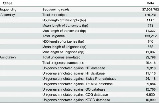
In this study, 10μg of total RNA isolated from theT.tridentatusembryos at hatch-out stage were used to prepare cDNA library for subsequent transcriptome sequencing using an Illumina HiSeq2000 sequencer. RNA was pooled from multipleT.tridentatusembryos to prepare one sample for sequencing. The summary of Illumima sequencing and annotation was shown in
Table 1. Sequencing of cDNA library generated a total of 37,902,792 paired end reads. These data are available from the NCBI Short Read Archive under accession number SRR946952. Transcrip-tome assembly was completed using RNAseqde novoassembler Trinity (k-mer length = 25). 4,864,778 contigs were identified with the nucleotide composition of A, T, C and G being 30.98%, 29.67%, 20.22% and 19.12% respectively, which gives rise to an overall GC content of 39.34% in the whole transcriptome. The contigs were further assembled into 176,231 transcripts falling into 133,212 unigenes (>200 bp) with a mean unigene length of 746 bp and an N50 of
568 bp (Table 1). The length distribution of transcripts and unigenes were shown inS1 Fig. Unigene annotation was achieved by BLASTx and BLASTn searches against NR, NT, Swis-sprot, and TrEMBL with an e-value less than 1×10⁻⁵. In total, there were 33,796 unigenes showing hits in one or more databases (Table 1), among which the NR database gave rise to the most annotations (29,918 hits). However, a large proportion of the sequences did not show sig-nificant blast hit. This was probably due to the lack of characterization of genes from closely related species, as the length distribution and the coverage of the annotated and unannotated unigenes were in a similar pattern (S2 Fig). The assembled sequences generated in our study are available from the authors upon request.
Table 1. Summary ofT.tridentatustranscriptome sequencing, assembly and annotation.
Stage Data
Sequencing Sequencing reads 37,902,792
Assembly Total transcripts 176,231
N50 length of transcripts (bp) 1147
Mean length of transcripts (bp) 713
Max length of transcripts (bp) 11,337
Total unigenes 133,212
N50 length of unigenes (bp) 746
Mean length of unigenes (bp) 568
Max length of unigenes (bp) 11,337
Annotation Total unigenes annotated 33,796
Total unigenes unannotated 99,416
Unigenes annotated against NR database 29,918 Unigenes annotated against NT database 11,116 Unigenes annotated against Swiss-Prot database 24,118 Unigenes annotated against TrEMBL database 29,884 Unigenes annotated against GO database 15,768 Unigenes annotated against COG database 6,920 Unigenes annotated against KEGG database 10,999
Functional annotation
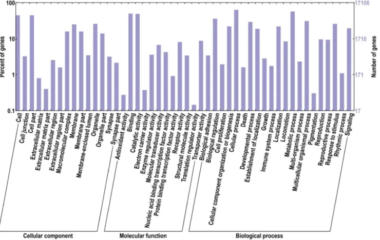
In order to predict the functions of these unigenes, all the sequences were analyzed according to gene ontology (GO) database and clusters of orthologous groups (COGs). 15,768 genes were suc-cessfully annotated with 95,305 GO terms and were separated into three categories: biological pro-cess, cellular component, and molecular function, which were further divided into 15, 12 and 21 functional groups, respectively (Fig 1). Among these GO terms, 49,724 unigene sequences were assigned to biological process, 27,948 to molecular function, and 17,363 to cellular component. Interestingly, the top 5 groups with most unigenes involved in the biological process were: cellular process, metabolic process, biological regulation, developmental process, and multicellular organ-ismal process. While the cell part and the cell functional groups were dominant in the cellular component category, and the binding and the catalytic activity were dominant in the molecular function category. The integral to membrane (GO: 0016021) contained 1572 unigenes, which belonged to the cellular component category. It had the largest number of unigenes among all the groups. In addition, the ATP binding (GO: 0005524) containing 1478 unigenes and the oxidation-reduction process (GO: 0055114) containing 619 unigenes were the most represented GO terms for the molecular function and biological process categories, respectively. A high percentage of unigenes involved in the following functional groups should also be noted: cellular process (GO: 0009987) with 561unigenes, protein phosphorylation (GO: 0006468) with 596 unigenes and regu-lation of transcription, DNA-dependent (GO: 0006355) with 556 unigenes. We were interested in the embryonic development process ofT.tridentatus, and thus investigated the unigenes with GO
Fig 1. GO distribution of the T. tridentatus unigenes.Go ontology distribution of the T. tridentatus unigenes were derived using BLAST2GO. The X-axis represents three main GO categories: cellular component, molecular function and biological process, which further separated into 15, 12 and 21 functional groups, respectively. The Y-axis represents percentages and numbers of unigenes mapping to the given functional GO group.
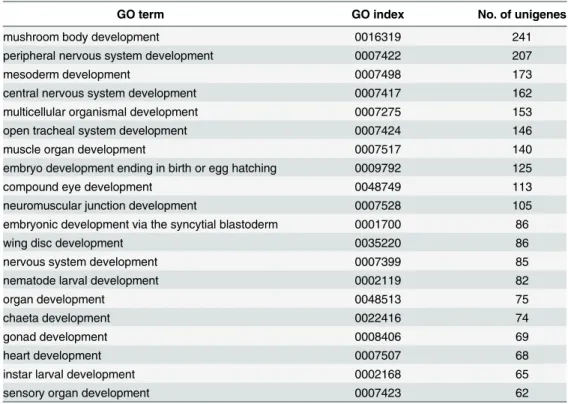
terms containing“embryo”or“development”. As a result, a total of 183 GO terms were found with unigene numbers ranging from 1 to 241. The top 20 terms with the most number of unigenes were listed inTable 2. This demonstrated that our transcriptome database contained a variety of unigenes related to the embryonic development, which provided an abundant resource for further investigation of regulatory mechanisms ofT.tridentatusembryonic development.
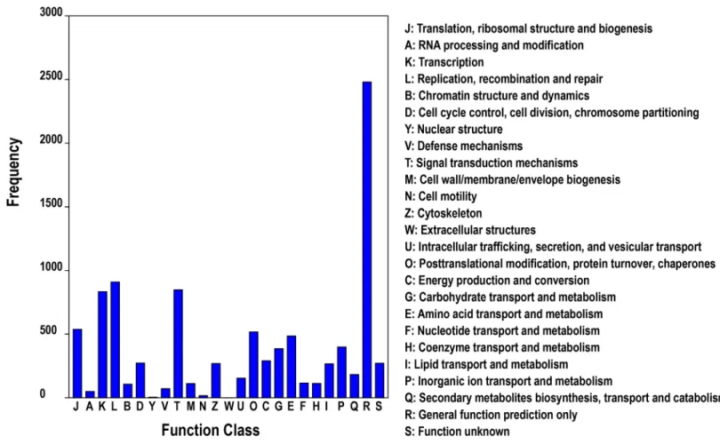
Meanwhile, COG annotation was used for phylogenetic classification of the putative pro-teins from theT.tridentatustranscriptome, in which a total of 6,920 proteins were assigned to 25 clusters (Fig 2). Among the COG library, the cluster of“General function prediction only”
consisted of the largest number of unigenes. The other five largest categories were:“replication, recombination and repair”(14.47%),“signal transduction mechanisms”(13.42%),“ transcrip-tion”(13.12%),“translation, ribosomal structure and biogenesis”(8.44%), and“ posttransla-tional modification, protein turnover, chaperones”(8.24%), suggesting the specific responses and molecular mechanisms ofT.tridentatusearly development to some extent.
To investigate the biological pathways which are possibly involved in early embryonic devel-opment process of T. tridentatus, we further mapped the unigenes to the reference canonical pathways in the Kyoto Encyclopedia of Genes and Genomes (KEGG) database. A total of 10,999 unigenes were assigned to 298 KEGG pathways. The pathways most represented by numbers of unigene sequences were“transcription factors”(865 unigenes),“chromosome”
(802 unigenes),“ubiquitin system”(798 unigenes),“protein kinases”(784 unigenes), and
“pathways in cancer”(518 unigenes).
Signaling pathways and regulatory genes involved in the development of
T
.
tridentatus
Formation of different cell types and patterns during development of multicellular organisms depends on a complex cascade of developmental decisions. An evolutionarily conserved
Table 2. Top 20 GO terms with most unigenes involved in embryonic development.
GO term GO index No. of unigenes
mushroom body development 0016319 241
peripheral nervous system development 0007422 207
mesoderm development 0007498 173
central nervous system development 0007417 162
multicellular organismal development 0007275 153
open tracheal system development 0007424 146
muscle organ development 0007517 140
embryo development ending in birth or egg hatching 0009792 125
compound eye development 0048749 113
neuromuscular junction development 0007528 105
embryonic development via the syncytial blastoderm 0001700 86
wing disc development 0035220 86
nervous system development 0007399 85
nematode larval development 0002119 82
organ development 0048513 75
chaeta development 0022416 74
gonad development 0008406 69
heart development 0007507 68
instar larval development 0002168 65
sensory organ development 0007423 62
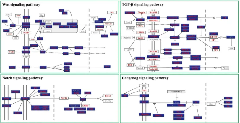
network of signaling pathways and regulatory genes plays key roles during this process [31]. Analyzing these pathways and factors involved in the early embryonic development ofT. tri-dentatusis of great importance for understanding of the developmental mechanisms of this ancient marine creature and may provide insights into the evolutional study of conserved sig-naling pathways and regulatory genes. In all the annotated unigenes ofT.tridentatus transcrip-tome, we identified a number of key components of four major signaling pathways (including Hedgehog, Wnt, transforming growth factor-βand Notch pathways) conserved in metazoan. As shown inFig 3, 98 possible homologues were identified from 126 key genes involved in these signaling pathways, representing 77.78% coverage of the total genes. The percentage of genes found in each pathway is 78.85% (Wnt), 77.27% (TGF-β), 77.78% (Hedgehog) and 87.5% (Notch) respectively. In case ofD.melanogaster-specific pathways, the percentage was much higher, about 91.57% (76/83) homologues could be found inT.tridentatus transcrip-tome, indicating the higher similarity ofT.tridentatuswithD.melanogasterthan other animal models in the aspect of signaling transduction pathways. It should also be noted that the sequence length of most gene fragments (>200 bp) is sufficient for functional studies of these
genes by modern molecular technology, such as real-time PCR quantification,in situ hybrid-ization and antibody preparation. Moreover, with the sequence information of these gene frag-ments, it would be more efficient and reliable to obtain the full length of desired genes
comparing with common degenerate PCR method. This is particularly helpful in case of gene discovery and functional study in the“non-model”organism horseshoe crab.
Fig 2. COG functional classification of theT.tridentatustranscriptome.
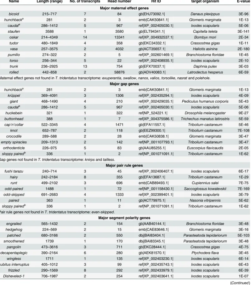
Regulatory genes are also key players in the network governing embryonic development [32].D.melanogasteris one of the best understood models of embryonic development, espe-cially pattern formation process, and thus we analyzed the regulatory genes potentially involved in theT.tridentatusaxis specification and patterning by usingD.melanogasteras a reference model. The genetic control of axis specification and patterning inD.melanogaster requires a cascade of gene regulation events before the onset of blastoderm stage. The major genes involved in this developmental process ofD.melanogastercan grouped into four catego-ries, viz. maternal effect genes, gap genes, pair rule genes and segment polarity genes [33,34]. This developmental process starts with the diffusion of the maternal effect genes which are responsible for setting the anterior-posterior polarity of the embryo. The gap genes, which are among the first genes transcribed in the embryo, participate in the establishment of the sub-domain of body plan under the control of maternal gradients. The pair rule genes subsequently divide the embryo into periodic units, whereas the segment polarity genes activated by pair rule genes further establish the periodicity of the embryo by dividing it into 14 segment-wide units [35,36]. Key regulatory genes of each category used in our study were selected according to S.F. Gilbert’s and R.M. Twyman’s classification [37,38]. Among all the 53 genes listed in
Table 3, we identified a total of 40 genes (75.47%) which had homologs inT.tridentatus, sug-gesting that functions of these genes conserved inT.tridentatus. It should be pointed out that the percentage of gene homologs identified in the maternal effect genes category was much lower (11/18, 61.11%) comparing to that of the other categories: gap genes (12/14, 85.71%), pair rule genes (7/8, 87.5%) and segment polarity genes (13/16, 81.25%). This is possibly
Fig 3. Major components from Conserved metazoan signaling pathways identified inT.tridentatus.The results were generated by BLASTing the unigenes ofT.tridentatusto known homologues with the e-value cutoff at 1e-10. Pathway schematics were adapted from KEGG pathway models (http:// www.genome.jp/kegg/).Drosophila melanogaster-specific pathways are colored blue. Genes identified and not identified inT.tridentatuswere marked with red font and grey font respectively.
Table 3. Representative regulatory genes involved in the early embryonic development ofT.tridentatus.
Name Length (range) No. of transcripts Read number Hit ID target organism E-value
Major maternal effect genes
bicoid 215–717 7 84 gb|EHJ73092.1| Danaus plexippus 3E-96
hunchbacka 281 2 3 emb|CAK50841.1| Glomeris marginata 1E-13
caudala 286
–1412 5 967 ref|XP_002405030.1| Ixodes scapularis 5E-06
staufen 3588 1 3580 gb|ELT94341.1| Capitella teleta 3E-141
oskar 214–4349 14 13341 ref|XP_004932537.1| Bombyx mori 2E-34
tudor 480–1849 4 358 gb|EKC34332.1| Crassostrea gigas 1E-11
vasa 257–3575 2 4032 gb|ACT35657.1| Haliotis asinina 2E-180
pumilio 274–322 2 5 ref|XP_002601469.1| Branchiostomafloridae 1E-45
torso 256–344 5 22 ref|XP_002408935.1| Ixodes scapularis 2E-10
trunk 236–2925 13 754 gb|EFX70037.1| Daphnia pulex 3E-03
rolled 442–858 2 58876 gb|ADV40083.1| Latrodectus hesperus 6E-59
Maternal effect genes not found inT.tridentatustranscriptome:exuperantia,swallow,nanos,valios,torsolike,nasratandpolehole.
Major gap genes
hunchbacka 281 2 3 emb|CAK50841.1| Glomeris marginata 1E-13
krüppel 369–4091 3 1306 ref|XP_002435294.1| Ixodes scapularis 4E-09
giant 468–1490 4 210 ref|XP_002429035.1| Pediculus humanus corporis 5E-43
caudala 286–1412 5 967 ref|XP_002405030.1| Ixodes scapularis 5E-06
huckebein 321 1 322 ref|NP_524221.1| Drosophila melanogaster 9E-27
buttonhead 388 1 7 ref|XP_004375586.1| Trichechus manatus latirostris 5E-59
cap'n'collar 522–3345 4 1471 gb|EFA11557.1| Tribolium castaneum 5E-46
knot 652–787 2 118 gb|EEZ99300.1| Tribolium castaneum 7E-108
crocodile 289–588 2 28 emb|CAK50838.1| Glomeris marginata 3E-47
empty spiracles 209–1313 2 142 ref|NP_001107793.1| Tribolium castaneum 3E-47
orthodenticle 226–975 5 83 gb|AAU85255.1| Euscorpiusflavicaudis 2E-05
sloppy pairedb 336 1 2 ref|NP_001071091.1| Tribolium castaneum 1E-62
Gap genes not found inT.tridentatustranscriptome:knirpsandtailless.
Major pair rule genes
fushi tarazu 240–714 3 45 ref|XP_002406407.1| Ixodes scapularis 6E-17
hairy 242–2184 8 355 gb|EFA13687.1| Tribolium castaneum 1E-29
runt 499–2102 3 696 emb|CAB89493.1| Cupiennius salei 7E-75
odd-paired 1488 1 72 ref|NP_001158430.1| Saccoglossus kowalevskii 7E-169
odd-skipped 691–2683 4 1333 ref|XP_002399401.1| Ixodes scapularis 3E-79
paired 363 1 11 gb|ACT79975.1| Nasonia vitripennis 5E-62
sloppy pairedb 336 1 2 ref|NP_001071091.1| Tribolium castaneum 1E-62
Pair rule genes not found inT.tridentatustranscriptome:even-skipped.
Major segment polarity genes
engrailed 565–1432 2 154 gb|AAB40144.1| Branchiostomafloridae 3E-48
hedgehog 224–569 2 15 emb|CAE83646.1| Glomeris marginata 3E-16
patched 680–3166 2 550 dbj|BAI83404.1| Parasteatoda tepidariorum 5E-103
smoothened 1739 1 170 dbj|BAI83345.1| Parasteatoda tepidariorum 3E-48
pangolin 473–3616 3 711 gb|EKC28444.1| Crassostrea gigas 4E-75
decapentaplegic 390–2164 6 280 gb|AEK81570.1| Ptychoderaflava 3E-45
wingless 1711 1 135 ref|XP_002403230.1| Ixodes scapularis 6E-14
cubitus interruptus 405–1012 3 99 ref|XP_002435743.1| Ixodes scapularis 6E-43
frizzled 290–1569 8 292 ref|XP_002433979.1| Ixodes scapularis 6E-39
Disheveled-1 706–1987 2 254 ref|XP_002403641.1| Ixodes scapularis 1E-07
because the mRNA products of the maternal gene homologs have been consumed at the hatch-out stage, the last stage ofT.tridentatusembryonic development. In addition, we also failed to identify some well-studied gene homologs (includingknirps,tailless,even-skipped,invected, fusedandcostal 2), which may be due to the extremely low expression of these gene homologs in our sample. Anyway, we identified the majority of the candidate regulatory genes that are potentially involved in the axis specification and patterning ofT.tridentatusembryo. Further study is required to characterize the function of these putative genes.
Case study: Detailed analysis of
Pax
family genes
In addition to transcriptome analysis of conserved signaling pathway components and regula-tory genes, we also tried to identify genes known to be important for embryogenesis based on our transcriptome data. Here, we focused specifically on thePaxfamily genes.Paxgenes, which are grouped into 4 subfamilies (Pax1/9,Pax2/5/8,Pax3/7andPax4/6), encode a group of transcription factors that have been conserved through millions of years of evolution and play roles in early development [39–41]. According to the homology within the highly con-served paired domain, the putative horseshoe crabPaxgenes were identified. A complete set of Paxfamily gene homologs was found inT.tridentatus. Phylogenetic tree was constructed by aligning the conserved paired domain with neighbor joining method usingPseudomonas trans-posaseas outgroup (Fig 4). Genes belonging toPax4/6subfamily branched out at the base of the tree. Genes fromPax4/6subfamily were divided into two clades. One includedPax4/6 genes fromHomo sapiens(HsPax4andHsPax6),D.melanogaster(DmtoyandDmey) andT. tridentatus(TtPax4/6aandTtPax4/6b). The second clade was composed of two sister groups with one containing the genesDmeygandDmtoefromD.melanogaster, and the other consist-ing ofTtPax4/6candTtPaX4/6dfromT.tridentatus. Interestingly, all theDmeyg,Dmtoe, TtPax4/6aandTtPaX4/6bhad a truncated paired domain. It is possible that the truncated paired domain already existed in the common ancestor of horseshoe crab and Drosophila. After split into two lineages, specific gene duplications occurred independently in horseshoe crab and Drosophila. The second clade includesPax2/5/8subfamily genes. All members of TtPax2/5/8genes were gathered closely, indicating that relatively recent duplications appear to have occurred in horseshoe crabs. The third clade was composed ofPax1/9subfamily genes. TtPax1/9abranched out at the base against all other genes in this clade, whereasTtPax1/9b gathered with the vertebratePax1and Pax9genes. The last clade was constituted of Pax3/7 subfamily. The horseshoe crabTtPax3/7awas clustered with DrosophilaDmgsbandDmgsbn genes.
While the majority of horseshoe crab genes of thePaxfamily show obvious homology to their respective subfamily, the placement ofTtPax3/7bin the phylogenetic tree is ambiguous. It occupied the most basal position of the tree without obvious orthologs. The homeodomain
Table 3. (Continued)
Name Length (range) No. of transcripts Read number Hit ID target organism E-value
shaggy 526 1 15 gb|EFN78376.1| Harpegnathos saltator 1E-08
armadillo 350–657 5 75 gb|EKC38770.1| Crassostrea gigas 1E-73
gooseberry 771 1 23 gb|EHJ65894.1| Danaus plexippus 2E-103
Segment polarity genes not found inT.tridentatustranscriptome:invected,fusedandcostal 2.
agene grouped in both maternal effect genes and gap genes bgene grouped in both gap genes and segment polarity genes
Fig 4. Phylogenetic analysis of paired domains of T. tridentatus Pax family proteins.Neighbour joining tree of the paired domains of Pax proteins with paired deletion option. Since TtPax4/6 has only partial paired domain available, the phylogeny of Pax4/6 subfamily was constructed with complete deletion option. A pseudomonas transposase sequence (AF169828) serves as outgroup. Numbers above branches are the percentage of the trees in which the topology appears. Paired domain sequences of Pax proteins were shown inS2 Table. Tt:Tachypleus tridentatus. Hs:homo sapiens. Dm:Drosophila melanogaster.
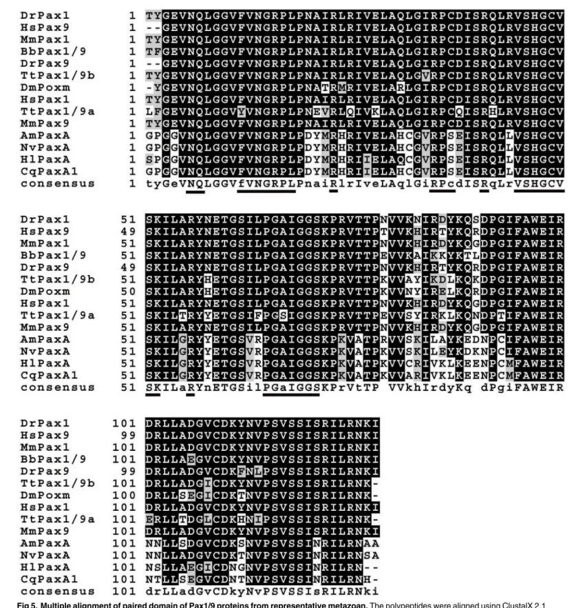
Fig 5. Multiple alignment of paired domain of Pax1/9 proteins from representative metazoan.The polypeptides were aligned using ClustalX 2.1 program and Boxshade online software (http://www.ch.embnet.org/software/BOX_form.html) were employed to highlight conserved amino acids. Conserved DNA binding motifs were underlined. The sequences used in the alignment are as follows: AmPaxA:Acropora milleporaPaxA [GenBank: AAC15713.2], BbPax1/9:Branchiostoma belcheriPax1/9 [GenBank: ABK54274.1], CqPaxA1:Chrysaora quinquecirrhaPaxA1 [GenBank: AAB58292.1], DmPoxm:
Drosophila melanogasterPoxm [GenBank: ABE69189.1], HlPaxA:Hydra littoralisPaxA [GenBank: AAB58290.1], DrPax1:Danio rerioPax1 [GenBank: NP_001074061.1], DrPax9:Danio rerioPax9 [GenBank: NP_571373.1], HsPax1:Homo sapiensPax1 [GenBank: NP_006183.2], HsPax9:Homo sapiens
sequence ofTtPax3/7bwas blasted against the GenBank database. Sequence comparison result showed that all the genes highly similar (over 78%) withTtPax3/7bhomeodomain belonged to the Pax3/7 subfamily. Therefore, it is possible thatTtPax3/7bgenes belong to the Pax3/7 sub-family, but have been evolving at a rate that obscures its orthology.
To further test the reliability of our database in gene discovery, the full length cDNAs encoding TtPax1/9a and TtPax1/9b proteins were cloned based on the partial sequences obtained from transcriptome assembly. The completeTtPax1/9acDNA contained 258 bp of 5’
untranslated region (UTR), 681 bp of open reading frame (ORF) and 150 bp of 3’UTR. The TtPax1/9aORF can be translated into a polypeptide of 226 amino acids (aa) with a predicted molecular weight (MW) of 25.4 KDa. A conserved paired domain of 127 aa was included in the predicted protein sequence (S3 Fig). The full lengthTtPax1/9bcDNA contains 1893 bp, while the ORF is of 912 bp encoding 303 aa with a predicted MW of 33.2 KDa. A conserved paired domain with 127 aa in length is also present in theTtPax1/9bprotein sequence (S4 Fig). A mul-tiple alignment of the paired domain sequences ofPax1/9was performed inT.tridentatusand other representative metazoan species. As shown inFig 5, thePax1andPax9showed high sim-ilarity among all the species examined. Interestingly, although bothT.tridentatusandD. mela-nogasterbelong to the Arthropoda,TtPax1/9aandTtPax1/9bshowed higher protein identity to zebrafish (Danio renio), human (Homo sapiens) and mouse (Mus musculus) which belong to Chordata, comparing toD.melanogaster. Whereas, polypeptide identities of Pax paired domain betweenT.tridentatusand the Coelenterata species, includingAcropora millepora, Chrysaora quinquecirrha,Hydra littoralisandNematostella vectensis, were relatively low. In summary, our data demonstrated the immediate application of the transcriptome data for gene discovery in the“non-model”organismT.tridentatus.
Tissue distribution of Pax1/9a and Pax1/9b
Expression patterns ofTtPax1/9aandTtPax1/9bgenes in different tissues of horseshoe crab were analyzed by quantitative RT-PCR. The highest expression level ofTtPax1/9awas found in the heart (Fig 6).TtPax1/9awas also abundantly expressed in the muscle, liver and intestine. On the other hand, the expression level ofTtPax1/9ain the stomach, yellow connective tissue
Fig 6. Tissue distribution ofTtPax1/9aandTtPax1/9bdetermined by quantative real-time PCR.TheT.
tridentatus GAPDHtranscript was used as internal standard. I, intestine; L: liver; Y, yellow connective tissue; H, heart; S: stomach; M, muscle; G, gill.
and gill was significantly lower. Expression pattern ofTtPax1/9bis similar to that ofTtPax1/ 9a, albeit at much lower levels.
TtPax1/9b
partly rescues Poxm dysfunction-caused larval lethality of
Drosophila
Due to lack of genetic manipulation tools for the gene function study inT.tridentatus, we fur-ther explore the function ofTtPax1/9aandTtPax1/9bgenes by taking advantage of Drosophila model. Drosophila Pax genePox meso(Poxm) plays a crucial role in the early development. Our previous study showed that the loss-of-function mutant inpoxm,PoxmR361, causes the Drosophila death [29]. According to our protein sequence alignment data,TtPax1/9aand TtPax1/9bgenes are homologs of the Drosophilapoxmgene. Therefore in this experiment, we investigated whether theTtPax1/9aandTtPax1/9bgenes rescuePoxmR361-caused embryonic lethality of Drosophila. TheTtPax1/9aandTtPax1/9bgenes were expressed in the Drosophila under the control of the 8.4 kbpoxmupstream region using GAL4/UAS system [29]. The
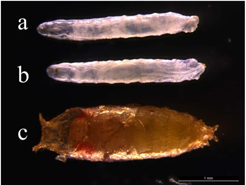
Fig 7. Rescue study of PoxmR361 mutant by the expression ofTtPax1/9aandTtPax1/9bgenes.(a)PoxmR361mutant. (b)PoxmR361mutant with
TtPax1/9agene expression. (c)PoxmR361mutant withTtPax1/9bgene expression. Anterior is to the left.
PoxmR361mutant alone (Fig 7A) and thePoxmR361mutant withTtPax1/9agene expression (Fig 7B) resulted in the Drosophila death at the larval stage. Surprisingly, ThePoxmR361mutant in the presence ofTtPax1/9bsurvived at the pupal stage (Fig 7C). These results imply that the TtPax1/9bcan at least partly rescue Poxm dysfunction-caused larval lethality of Drosophila.
Conclusions
In this study, we, for the first time, performedde novotranscriptome sequencing ofT. tridenta-tusembryos in the absence of a reference genome using Illumnia Solexa platform. Of 133,212 unigenes obtained, 33,796 were annotated by BLAST with NR, NT, Swiss-Prot, GO, COG and KEGG databases. We further identified a number of candidate genes potentially involved in embryonic development ofT.tridentatus, shedding light on future study on the characteriza-tion and funccharacteriza-tion of genes of interest and detailed molecular mechanisms underlying embry-onic development ofT.tridentatus. Moreover, the cloning and phylogenetic analysis ofPax family genes were performed, demonstrating that the transcriptome sequencing is a fast and reliable technology for high-throughput gene discovery in“non-model”organisms and for evo-lutionary developmental biology study as well. Rescue study further indicated thatTtPax1/9b gene is functionally relative to DrosophilaPoxmgene.
Supporting Information
S1 Fig. Sequence length distribution of transcripts and unigenes assembled from Illumina reads.All Illumina transcripts and unigenes with length over 200 bp were analyzed.
(TIF)
S2 Fig. Contour plot of length and coverage distribution of annotated and unannotated unigenes.Transcripts were annotated using Blast2GO software. The Burrows-Wheeler Aligner (BWA) program was used for reads mapping. The color bar indicates log10 transformed count values.
(TIF)
S3 Fig. Nucleotide and deduced amino acid sequence ofT.tridentatus Pax1/9a.Numbers
on the left indicate numbers of nucleotides or amino acids. Boxing indicates the conserved paired domain andstands for putative stop codon. The polyadenylation signal is underlined.
(TIF)
S4 Fig. Nucleotide and deduced amino acid sequence ofT.tridentatus Pax1/9b.Numbers
on the left indicate numbers of nucleotides or amino acids. Boxing indicates the conserved paired domain andstands for putative stop codon.
(TIF)
S1 Table. List of primer sequences used in this study.
(DOCX)
S2 Table. Paired domain amino acid sequences used in the phylogenetic analysis.Tt: Tachy-pleus tridentatus, Hs:homo sapiens, Dm:Drosophila melanogaster.
(DOCX)
Acknowledgments
Author Contributions
Conceived and designed the experiments: MC JC. Performed the experiments: MC CW Wei Wang. Analyzed the data: MC GJ BH MD ZL. Contributed reagents/materials/analysis tools: GL XL WZ Weiyi Wang. Wrote the paper: MC GJ JC.
References
1. Lawson D, Arensburger P, Atkinson P, Besansky NJ, Bruggner RV, Butler R, et al. VectorBase: a data resource for invertebrate vector genomics. Nucleic Acids Res. 2009; 37: D583–587. doi:10.1093/nar/
gkn857PMID:19028744
2. Arias AM. Drosophila melanogaster and the development of biology in the 20th century. Methods Mol Biol. 2008; 420: 1–25. doi:10.1007/978-1-59745-583-1_1PMID:18641938
3. Colbourne JK, Pfrender ME, Gilbert D, Thomas WK, Tucker A, Oakley TH, et al. The ecoresponsive genome ofDaphnia pulex. Science. 2011; 331: 555–561. doi:10.1126/science.1197761PMID:
21292972
4. Cahais V, Gayral P, Tsagkogeorga G, Melo-Ferreira J, Ballenghien M, Weinert L, et al. Reference-free transcriptome assembly in non-model animals from next-generation sequencing data. Mol Ecol Resour. 2012; 12(5): 834–845. doi:10.1111/j.1755-0998.2012.03148.xPMID:22540679
5. Martin JA, Wang Z. Next-generation transcriptome assembly. Nat Rev Genet. 2011; 12(10): 671–682.
doi:10.1038/nrg3068PMID:21897427
6. Surget-Groba Y, Montoya-Burgos JI. Optimization of de novo transcriptome assembly from next-gener-ation sequencing data. Genome Res. 2010; 20(10): 1432–1440. doi:10.1101/gr.103846.109PMID:
20693479
7. Grabherr MG, Haas BJ, Yassour M, Levin JZ, Thompson DA, Amit I, et al. Full-length transcriptome assembly from RNA-Seq data without a reference genome. Nat Biotechnol. 2011; 29(7): 644–652. doi:
10.1038/nbt.1883PMID:21572440
8. Micallef G, Bicherdike R, Reiff C, Fernandes JM, Bowman AS, Martin SA. Exploring the transcriptome of Atlantic Salmon (Salmo salar) skin, a mjor defense organ. Mar Biotechno (NY). 2012; 14(5): 559–
569.
9. Zhao X, Wang Q, Jiao Y, Huang R, Deng Y, Wang H, et al. Identification of genes potentially related to biomineralization and immunity by transcriptome analysis of pearl sac in pearl oysterPinctada marten-sii. Mar Biotechnol (NY). 2012; 14(6): 730–739.
10. Wang H, Jiang J, Chen S, Qi X, Peng H, Li P, et al. Next-generation sequencing of theChrysanthemum nankingense(Asteraceae) transcriptome permits large-scale unigene assembly and SSR marker dis-covery. PLoS One. 2013; 8(4): e62293. doi:10.1371/journal.pone.0062293PMID:23626799
11. Vidotto M, Grapputo A, Boscari E, Barbisan F, Coppe A, Grandi G, et al. Transcriptome sequencing and de novo annotation of the critically endangered Adriatic sturgeon. BMC Genomics. 2013; 14(1): 407.
12. Meng XL, Liu M, Jiang KY, Wang BJ, Tian X, Sun SJ, et al.De NovoCharacterization of Japanese Scal-lopMizuhopecten yessoensisTranscriptome and Analysis of Its Gene Expression following Cadmium Exposure. PLoS One. 2013; 8(5): e64485. doi:10.1371/journal.pone.0064485PMID:23741332
13. Sugita H, Sekiguchi K. Horseshoe crab developmental studies II. Physiological adaptation of horse-shoe crab embryos to the environment during embryonic development. Prog Clin Biol Res. 1982; 81: 75–82. PMID:7122560
14. Sekiguchi K, Yamamichi Y, Costlow JD. Horseshoe crab developmental studies I. Normal embryonic development ofLimulus polyphemuscompared withTachypleus tridentatus. Prog Clin Biol Res. 1982; 81: 53–73. PMID:7122559
15. Harzsch S, Vilpoux K, Blackburn DC, Platchetzki D, Brown NL, Melzer R, et al. Evolution of arthropod visual systems: development of the eyes and central visual pathways in the horseshoe crabLimulus polyphemus Linnaeus,1758(Chelicerata, Xiphosura). Dev Dyn. 2006; 235 (10): 2641–2655. PMID:
16788994
16. Mittmann B. Early neurogenesis in the horseshoe crab Limulus polyphemus and its implication for arthropod relationships. Biol Bull. 2002; 203(2): 221–222. PMID:12414588
17. Walls EA, Berkson J, Smith SA. The horseshoe crab,Limulus polyphemus: 200 million years of exis-tence, 100 years of study. Rev Fish Sci. 2002; 10(1): 39–73.
19. Rudkin DM, Young GA, Nowlan GS. The oldest horseshoe crab: a new xiphosurid from Late Ordovician Konservat-Lagerstätten deposits, Manitoba, Canada. Palaeontology. 2008; 51(1): 1–9.
20. Fisher DC. The Xiphosurida: archetypes of bradytely? In: Eldredge N, Stanley SM editors. Living Fos-sils. New York: Springer; 1984. pp. 196–213.
21. Cooper JF, Pearson SM. Detection of endotoxin in biological products by the limulus test. Dev Biol Stand. 1997; 34: 7–13.
22. Botton ML. The Ecological Importance of Horseshoe Crabs in Estuarine and Coastal Communities: A Review and Speculative Summary. In: Tanacredi JT, Botton ML, Smith DR, editors. Biology and Con-servation of Horseshoe Crabs. New York: Springer; 2009. pp. 45–64.
23. Sekiguchi K. Biology of horseshoe crabs. Portland: International Specialized Book Service Incorpo-rated; 1988.
24. Sekiguchi K, Seshimo H, Sugita H. Post-embryonic development of the horseshoe crab. Biological Bul-letin. 1988; 174(3): 337–345.
25. Mittmann B, Scholtz G. Development of the nervous system in the "head" ofLimulus polyphemus (Che-licerata: Xiphosura): morphological evidence for a correspondence between the segments of the chelic-erae and of the (first) antennae of Mandibulata. Dev Genes Evol. 2003; 213(1): 9–17. PMID:12590348 26. Smith SA, Berkson J. Laboratory culture and maintenance of the horseshoe crab (Limulus
polyphe-mus). Lab Anim (NY). 2005; 34(7): 27–34.
27. Grabherr MG, Haas BJ, Yassour M, Levin JZ, Thompson DA, Amit I, et al. Full-length transcriptome assembly from RNA-Seq data without a reference genome. Nat Biotechnol. 2011; 29(7): 644–652. doi:
10.1038/nbt.1883PMID:21572440
28. Kumar S, Tamura K, Nei M. MEGA3: Integrated software for Molecular Evolutionary Genetics Analysis and sequence alignment. Brief Bioinform. 2004; 5(2): 150–163. PMID:15260895
29. Duan H, Zhang C, Chen J, Sink H, Frei E, Noll M. A key role ofPox mesoin somatic myogenesis of Dro-sophila. Development. 2007; 134: 3985–3997. PMID:17942482
30. Del Valle Rodriguez A, Didiano D, Desplan C. Power tools for gene expression and clonal analysis in Drosophila. Nat Methods. 2011; 9(1): 47–55. doi:10.1038/nmeth.1800PMID:22205518
31. Pires-daSilva A, Sommer RJ. The evolution of signalling pathways in animal development. Nat Rev Genet. 2003; 4(1): 39–49. PMID:12509752
32. Davidson EH, Levine MS. Properties of developmental gene regulatory networks. Proc Natl Acad Sci U S A. 2008; 105(51): 20063–20066. doi:10.1073/pnas.0806007105PMID:19104053
33. Jaeger J, Surkova S, Blagov M, Janssens H, Kosman D, Kozlov KN, et al. Dynamic control of positional information in the early Drosophila embryo. Nature. 2004; 430(6997): 368–371. PMID:15254541 34. Wunderlich Z, DePace AH. Modeling transcriptional networks in Drosophila development at multiple
scales. Curr Opin Genet Dev. 2011; 21(6): 711–718. doi:10.1016/j.gde.2011.07.005PMID:21889888 35. Jaeger J. The gap gene network. Cell Mol Life Sci. 2011; 68(2): 243–274. doi:
10.1007/s00018-010-0536-yPMID:20927566
36. Schroeder MD, Pearce M, Fak J, Fan H, Unnerstall U, Emberly E, et al. Transcriptional control in the segmentation gene network of Drosophila. PLoS Biol. 2004; 2(9): E271. PMID:15340490
37. Gilbert SF. Developmental biology. 8th ed. Oxford, UK: Sinauer Associates Inc; 2006.
38. Twyman R. BIOS Instant Notes in Developmental Biology. Oxford, UK: Taylor & Francis Group; 2001.
39. Blake JA, Ziman MR. Pax genes: regulators of lineage specification and progenitor cell maintenance. Development. 2014; 141(4): 737–751. doi:10.1242/dev.091785PMID:24496612
40. Lang D, Powell SK, Plummer RS, Young KF, Ruggeri BA. PAX gene: role in development, pathophysi-ology, and cancer. Biochem Parmacol. 2007; 73(1): 1–14.