CASE REPORT
J of Evidence Based Med & Hlthcare, pISSN- 2349-2562, eISSN- 2349-2570/ Vol. 2/Issue 15/Apr 13, 2015 Page 2328
SMALL CELL VARIANT OF OSTEOSARCOMA AT DIAPHYSIS OF
TIBIA: A RARE CASE REPORT WITH REVIEW OF LITERATURE
A. Venkatalakshmi1, S. Jagdeeswari2, A. Bhagyalakshmi3, S. Satish Kumar4HOW TO CITE THIS ARTICLE:
A. Venkatalakshmi, S. Jagdeeswari, A. Bhagyalakshmi, S. Satish Kumar. ”Small Cell Variant of Osteosarcoma at Diaphysis of Tibia: A Rare Case Report with Review of Literature”. Journal of Evidence based Medicine and Healthcare; Volume 2, Issue 15, April 13, 2015; Page: 2328-2332.
ABSTRACT: Osteosarcoma is the most common primary malignant tumor of bone involving predominantly metaphysis of the long bones. It accounts for 20% of primary bone cancers. Diaphyseal osteosarcoma is a rare form which accounts for approximately 10% of all cases of osteosarcomas. We present a case of Small cell variant of osteosarcoma in a 25 year old female presented in the diaphysis of left tibia.
KEYWORDS: Osteosarcoma, small cell variant, diaphysis.
INTRODUCTION: Osteosarcoma is a high grade malignant mesenchymal tumor affecting bone. The world health organization recognized several variants which differ in location, clinical behavior, degree of cellular atypia.1 This is a rare case of Small cell variant of osteosarcoma in a 25 year old female presented in the diaphysis of left tibia.
CASE REPORT: A 25 year old female presented with history of fall from height and pain since 6 months and had pathological fracture and swelling since 2 months at lower end of left leg.
Clinical examination revealed a single firm to hard swelling of 10x7 cm occupying the anterior and medial aspect of the left leg, middle and lower third approximately 10 cm from medial malleolus.
Radiological examination showed an intramedullary cystic lesion of the diaphyseal lower end of tibia with periosteal reaction and soft tissue extension with Moth eaten appearance.
Ultrasound abdomen and Computer tomography of chest, X-ray spine, skull, pelvis showed normal study.
Grossly, received an above knee amputation of leg, with swelling in middle of leg measuring 15x10x4 cm and showing thinning of skin. Cut section was grey white, glistening and gritty to cut.
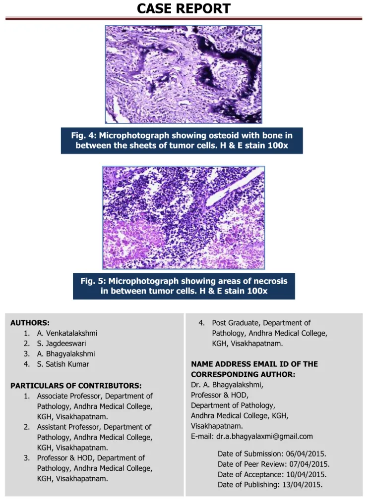
Microscopic examination revealed round to medium sized pleomorphic tumor cells arranged in loose sheets separated by connective tissue stroma (Fig. 1). The cells have scanty to moderate amount of eosinophilic cytoplasm with hyperchromatic nuclei (Fig. 2). Tumor osteoid punctuated by malignant tumor cells with bone formation and cartilaginous areas are seen (Fig. 3 & 4). Mitotic activity is increased and tumor giant cells are seen. There are areas of necrosis and hemorrhages (Fig. 5). Features are suggestive of small cell variant of osteosarcoma.
CASE REPORT
J of Evidence Based Med & Hlthcare, pISSN- 2349-2562, eISSN- 2349-2570/ Vol. 2/Issue 15/Apr 13, 2015 Page 2329
subtypes. 2) Intramedullary well-differentiated osteosarcoma and 3) Surface osteosarcoma. 60% of osteosarcoma is common in patients younger than 25 years and males are affected more frequently than females at a ratio of 1.3-1. 6:1. Osteosarcoma usually arises from the metaphysis of the long bones and uncommon in diaphysis.
Microscopically Osteosarcoma is again sub classified into Osteoblastic, Chondroblastic, Fibroblastic, Telangiectatic, Small cell, Giant cell and Epitheloid variants. In small cell variant of osteosarcoma, the cells are round to oval, with indistinct cell borders and have hyperchromatic nuclei, arranged in sheets with neoplastic osteoid and bone formation. There is another tumor known as E wing/ primitive neuroectodermal tumor is also a primary small round cell tumor of bone and soft tissue tumor and have similar picture except osteoid and bone formation. It is common in males in the first and second decade of life. Microscopically if there is no osteiod and bone among the tumor components then IHC markers help in confirmation of the diagnosis.
Small cell osteosarcoma is a rare but distinct variant of Osteosarcoma. Although Hultes et al2 in 1966 & Jacobson in 1977 described small cell tumor of bone capable of differentiating in to bone and cartilage, Sim et al3 reporting in 1979 on 24 patients at the Mayo clinic was the first to delineate the clinico pathological features of this entity. Further classification was given by other investigations, but few large series of patients with the lesions have been studied & there have been few case reports of these lesions. The tumor usually arises from metaphysis but rarely from diaphysis.4 Diaphysial osteosarcoma is a rare form which accounts for approximately 10% of all osteosarcomas.5 Although osteosarcoma usually arises in the medullary cavity of the metaphysis of a growing long bone, it also may arise on the surface of bone, it may be confined to the cortex or it even may arise in an extra skeletal site.6
Small cell osteosarcoma constitutes between 1.3% of all Osteosarcomas7 arising from bones. The osteoid production is a typical characteristic of this tumor and alters treatment strategy.8 Presence of Osteoid is a pre- requisite for differentiating Small cell osteosarcoma from
Ewing’s sarcoma. Although even in Ewing's sarcoma reactive bone sclerosis and soft tissue mineralization can be seen in the form of periosteal laminated bone, but in small cell osteosarcoma mineralized tumor matrix is usually noted. However, the diagnosis of small cell osteosarcoma depends on the identification of produced osteoid, which again can be quite variable. The problem can be in the absence of mineralization or to differentiate hyalinized collagen from osteoid or even sampling error could influence the diagnosis. The defining feature present in small cell osteosarcoma is mineralized matrix and in the absence of identifiable mineralized matrix, it is difficult to differentiate fibrin deposit found between individual cells of
Ewing’s sarcoma from osteoid.9 Nakajima et al. stated that if in doubt the diagnosis of Ewing’s sarcoma should be made.7 The other small cell tumors including Ewing’s sarcoma should be ruled out using immune histo chemistry. CD-99 Positivity has been noted in small cell osteosarcoma. Positive reaction for either of these LCA, S-100, EMA, SMA, factor VIII, smooth muscle acting, Neuron specific enolase, synoptophysin, etc., would favor the exclusion of small cell osteosarcoma.10 Most small cell osteosarcoma show vimentin positivity and occasional minority may be muscle specific actin (HHHF-35) positive.
CASE REPORT
J of Evidence Based Med & Hlthcare, pISSN- 2349-2562, eISSN- 2349-2570/ Vol. 2/Issue 15/Apr 13, 2015 Page 2330
insensitive to that approach.6 Pre-operative chemotherapy is of no value of prognostic significance.11 Post-operative chemotherapy and radio therapy is administered. The mainly used chemotherapeutic agents are vincristin, adriamycin, actinomycin D and cyclophosphamide6. But chemotherapy together is not necessary without evidence of any malignant cells on the surgical margins or the presence of distant metastasis.8 The 5 years survival rate for the classic osteosarcoma is 77%, whereas it is 28% for small cell osteosarcoma.8 Overall survival rate depends upon prognostic factors including tumor size, location, and histologic grade. The prognosis of small cell osteosarcoma was considered to be worse than conventional osteosarcoma and Ewing’s sarcoma.9
CONCLUSION: A rare histological type of small cell variant of osteosarcoma to be considered in the differential diagnosis of small cell lesions (round blue cell tumors) of the bone at the diaphysis which is useful to the clinician for the planning of the therapy.
REFERENCES:
1. Schajowicz F, Sissons HA, Sobin LH. The World Health Organization Histological Classification of bone tumors. A comment on the second edition CANCER 1995; 75; 1208-14.
2. Hulter, RUP, Foot, FW (1966) Francis KC, Sherman RS. Primitive multipotential primary sarcoma of bone. CANCER 19: PP 1-25.
3. Sim FH, Unni KK, Beabout JW, Dahlin DC. Osteosarcoma with small cells simulating Ewings tumor. J Bone Joint Surg Am. 1979; 61; 207-15.
4. Juan Rosai, Bone and joints. In, Juan Rosai (ed). Rosai and Ackerman’s Surgical Pathology, 10th edition. Edinburgh, Mosby, 2011; 2024-2032.
5. Harsh Kumar, Archana C. Buch, Vinay M. Sawlani, Shirish S. Chandanwale. Diaphysial osteosarcoma with varying histomorphologic patterns. Adv Biomed Res. 2014; 3: 33. Published online 2014 January 9. doi: 10.4103/2277-9175.124685
6. Michael J. Klein, MD, and Gene P. Siegal, MD, PhD Anatomic & Histologic Variants of Oseosarcoma. American Journal of clinical pathology 2006; 125; 555-581.
7. Nikajima H, Sim FH, Bond JR, et al Small cell osteosarcoma of bone; review of 72 cases, Cancer 1997; 79; 2095-2106.
8. Gokturk Findik, Ersin Gunay, Yetkin Agackiran, Koray Aydogdu, Ertan aydin, Sadikaya et al. Small cell osteosarcoma of rib; Diagnosis and treatment of the rare case. Tuberkuloz ve toraks 06/2012; 60(2): 172-5.
9. Amit Sethi 1, Shweta Rehani 2, Kundendu Arya 3. Small cell osteosarcoma of Mandible; A Rare case report and review of literature. J Clin Exp Dent. 2010; 2(2): e96-9.
10.Meera Hameed et al, Small round cell tumors of Bone. Arch pathol Lab Med -Vol 131, February 2007.
CASE REPORT
J of Evidence Based Med & Hlthcare, pISSN- 2349-2562, eISSN- 2349-2570/ Vol. 2/Issue 15/Apr 13, 2015 Page 2331
Fig. 1: Microphotograph showing sheets of round to oval pleomorphic cells separated by connective tissue stroma. H & E stain 100x
Fig. 2: Higher magnification of tumor cells having scanty cytoplasm and hyperchromatic nuclei. H & E stain 400x
CASE REPORT
J of Evidence Based Med & Hlthcare, pISSN- 2349-2562, eISSN- 2349-2570/ Vol. 2/Issue 15/Apr 13, 2015 Page 2332 4. Post Graduate, Department of
Pathology, Andhra Medical College, KGH, Visakhapatnam.
NAME ADDRESS EMAIL ID OF THE CORRESPONDING AUTHOR: Dr. A. Bhagyalakshmi,
Professor & HOD,
Department of Pathology, Andhra Medical College, KGH, Visakhapatnam.
E-mail: [email protected]
Date of Submission: 06/04/2015. Date of Peer Review: 07/04/2015. Date of Acceptance: 10/04/2015. Date of Publishing: 13/04/2015.
AUTHORS:
1. A. Venkatalakshmi 2. S. Jagdeeswari 3. A. Bhagyalakshmi 4. S. Satish Kumar
PARTICULARS OF CONTRIBUTORS: 1. Associate Professor, Department of
Pathology, Andhra Medical College, KGH, Visakhapatnam.
2. Assistant Professor, Department of Pathology, Andhra Medical College, KGH, Visakhapatnam.
3. Professor & HOD, Department of Pathology, Andhra Medical College, KGH, Visakhapatnam.
Fig. 4: Microphotograph showing osteoid with bone in between the sheets of tumor cells. H & E stain 100x