RECUPERAÇÃO TARDIA DA
TRABECULECTOMIA ATRAVÉS
DO AGULHAMENTO COM
MITOMICINA C
Orientador: Prof. Dr. Sebastião Cronemberger
Faculdade de Medicina
Universidade Federal de Minas Gerais
Belo Horizonte
RECUPERAÇÃO TARDIA DA
TRABECULECTOMIA ATRAVÉS
DO AGULHAMENTO COM
MITOMICINA C
Tese apresentada ao Programa de Pós-Graduação em Ciências Aplicadas à Cirurgia e à Oftalmologia da Faculdade de Medicina da Universidade Federal de Minas Gerais, como requisito parcial para obtenção do título de Doutor em Medicina.
Área de concentração: Oftalmologia
Orientador: Prof. Dr. Sebastião Cronemberger Universidade Federal de Minas Gerais
Faculdade de Medicina
Universidade Federal de Minas Gerais
Belo Horizonte
Maestrini, Heloisa Andrade.
M186r Recuperação tardia da trabeculectomia através do agulhamento com Mitomicina C [manuscrito]. / Heloisa Andrade Maestrini. - - Belo Horizonte: 2009.
120f.: il.
Orientador: Sebastião Cronemberger
Área de concentração: Ciências Aplicadas à Cirurgia e à Oftalmologia. Tese (doutorado): Universidade Federal de Minas Gerais, Faculdade de Medicina.
1. Trabeculectomia. 2. Glaucoma. 3. Epitélio Posterior. 4. Mitomicina C/uso terapêutico. 5. Mitomicina C/administração & dosagem. 6.
Reoperação. 7. Dissertações Acadêmicas. I. Cronemberger, Sebastião. II. Universidade Federal de Minas Gerais, Faculdade de Medicina. III. Título.
II
UNIVERSIDADE FEDERAL DE MINAS GERAIS
Magnífico Reitor
Prof. Ronaldo Tadêu Pena
Pró-Reitora de Pós-Graduação
Profa. Elizabeth Ribeiro da Silva
Pró-Reitor de Pesquisa
Prof. Carlos Alberto Pereira Tavares
Diretor da Faculdade de Medicina
Prof. Francisco José Penna
Coordenador do Centro de Pós-Graduação da Faculdade de Medicina
Prof. Carlos Faria Santos Amaral
Coordenador do Programa de Pós-Graduação em Ciências Aplicadas à
Cirurgia e à Oftalmologia
Prof. Edson Samesina Tatsuo
Chefe do Departamento de Oftalmologia e Otorrinolaringologia
Profa. Ana Rosa Pimentel de Figueiredo
Membros do Colegiado do Programa de Pós-Graduação em Ciências Aplicadas
à Cirurgia e à Oftalmologia
Prof. Edson Samesina Tatsuo Prof. Marcelo Dias Sanches Prof. Alcino Lázaro da Silva Prof. Márcio Bittar Nehemy Prof. Marco Aurélio Lana Peixoto Prof. Tarcizo Afonso Nunes
III
A comissão organizadora que assina abaixo _______________ a tese de doutorado intitulada “RECUPERAÇÃO TARDIA DA TRABECULECTOMIA ATRAVÉS DO AGULHAMENTO COM MITOMICINA C”, apresentada e defendida, em sessão pública, por HELOISA ANDRADE MAESTRINI, para a obtenção do título de Doutor em Medicina, pelo Programa de Pós-Graduação em Ciências Aplicadas à Cirurgia e à Oftalmologia da Faculdade de Medicina da Universidade Federal de Minas Gerais.
Belo Horizonte, _____ de ___________________ de _________
___________________________________________ Prof. Dr. Sebastião Cronemberger
Orientador
Universidade Federal de Minas Gerais
___________________________________________ Prof. Dr. Nassim da Silveira Calixto
Universidade Federal de Minas Gerais
___________________________________________ Prof. Dr. Homero Gusmão de Almeida
Universidade Federal de Minas Gerais
___________________________________________ Prof. Dr. Angelo Ferreira Passos
Universidade Federal do Espírito Santo
___________________________________________ Prof. Dr. Carlos Akira Omi
IV
Suplentes:
___________________________________________ Dr. Galton Carvalho Vasconcelos
Universidade Federal de Minas Gerais
___________________________________________ Dra. Ana Cláudia Alves Pereira
V
A meus pais, Angelo e Maria Célia, pelo contínuo apoio e pelo
maravilhoso exemplo de vida.
A meu marido Wladmir, pelo amor, carinho, suporte e bom humor
diante das dificuldades.
A minha filha Júlia, pelas surpresas e alegrias infinitas.
A minha irmã Angela, pelo exemplo e pelo apoio incondicional de
todas as horas.
A meus irmãos Cid e Marco, pela convivência carinhosa e divertida.
VI
AGRADECIMENTOS
Ao meu orientador, Prof. Dr. Sebastião Cronemberger, pela amizade, pelo estímulo e por saber semear seu espírito investigativo entre todos nós.
Ao Prof. Dr. Nassim Calixto, pela honra de sua convivência, pelo maravilhoso exemplo e pelo incansável desejo de ensinar.
Ao Prof. Dr. Angelo Ferreira Passos, inspirador do tema desta tese, pelos ensinamentos e pela contínua disponibilidade em ajudar nos casos difíceis.
À Dra. Hérika Danielle de Miranda Santos Matoso, pela ajuda inestimável durante toda a condução desta pesquisa.
Ao Dr. Flávio Marigo, excelente mestre e amigo, por ter compartilhado tantas idéias e ensinamentos.
Ao Dr. Galton Carvalho Vasconcelos, amigo irmão de tantos anos, pelo constante interesse e apoio.
À querida amiga Núbia Vanessa dos Anjos Lima Henrique de Faria, companheira, desde os primeiros passos, nesta longa caminhada pelos tortuosos caminhos do glaucoma.
Aos colegas Dr. José Roberto Costa Reis, Dr. Rafael Vidal Mérula, Dr. Alberto Diniz Filho, Emília Sakurai e Graziele Alves Ferreira, co-autores dos trabalhos, pelo entusiasmo com que participaram desta pesquisa.
Aos funcionários do Serviço de Glaucoma do Hospital São Geraldo, em especial ao Sr. José Francisco do Nascimento, Sra. Maria Lúcia dos Santos e Sra. Maria dos Anjos Alves Gomes, pela amizade e pelo convívio de tantos anos.
Às funcionárias do Departamento de Oftalmologia e Otorrinolaringologia da Faculdade de Medicina da UFMG, em especial à Srta. Rosemary Rodrigues Silva. Aos funcionários do Centro de Pós-Graduação da Faculdade de Medicina da UFMG, especialmente à Maricrislei Rocha Torres, pelo apoio e pela atenção prestada.
Às amigas Dra. Silvia Mandello Carvalhaes e Dra. Lílian Paula de Souza, pela convivência carinhosa e pela presença constante em todos os momentos da minha vida.
Ao meu marido Wladmir, companheiro inseparável, por saber compreender os inevitáveis momentos de ausência dedicados a esta tese.
À minha família, pelo suporte imprescindível e inabalável.
VII
“All our science, measured against reality, is primitive and
childlike – and yet it is the most precious thing we have.”
Albert Einstein
VIII
LISTA DE ABREVIATURAS
5-FU 5-fluorouracil
CCT central corneal thickness DNA deoxyribonucleic acid ECC espessura corneana central et al. et alii (e outros)
Fig. Figura IL Illinois Inc. Incorporation
IOP intraocular pressure MD Medical Doctor mg miligrama ml mililitro mm milímetro
mm2 milímetro quadrado mmHg milímetro de mercúrio MMC mitomicina C
µg micrograma µm micrômetro
n tamanho da amostra No. number
P p-valor
PA Pensilvânia PhD Doctor of Philosophy PIO pressão intraocular postop postoperative RNA ribonucleic acid SD standard deviation
SPSS Statistical Package for the Social Sciences Trab trabeculectomy
USA United States of America vs. versus
χ2
IX
LISTA DE TABELAS
PRIMEIRO TRABALHO
TABELA 1 – Características da amostra estudada ... 33
TABELA 2 – Pressão intraocular e medicações hipotensoras ... 37
TABELA 3 – Complicações pós-operatórias ... 39
SEGUNDO TRABALHO TABELA 1 – Características da amostra estudada ... 58
TABELA 2 – Cirurgias prévias e exposição a anti-metabólitos ... 58
TABELA 3 – Variáveis contínuas nos grupos de sucesso e fracasso ... 63
TABELA 4 – Variáveis categóricas e taxas de sucesso ... 63
TERCEIRO TRABALHO TABELA 1 – Características da amostra estudada ... 83
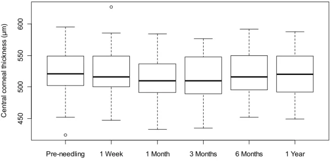
TABELA 2 – Espessura corneana central ao longo do tempo ... 86
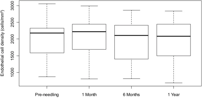
TABELA 3 – Contagem endotelial ao longo do tempo ...86
X
LISTA DE FIGURAS
PRIMEIRO TRABALHO
FIGURA 1 – Mudança na pressão intraocular (PIO) após todos os agulhamentos. Pontos à direita da linha de equivalência indicam melhora da PIO... 37 FIGURA 2 – Pressão intraocular média no pós-operatório ... 38 FIGURA 3 – Curvas de Kaplan-Meier comparando o primeiro agulhamento a todos os agulhamentos. Critério de sucesso: PIO ≤ 16 mmHg ... 40 FIGURA 4 – Aparência da bolsa fistulante antes e depois do agulhamento ... 42
TERCEIRO TRABALHO
XI
RESUMO
TÍTULO: Recuperação Tardia da Trabeculectomia através do Agulhamento com
Mitomicina C.
OBJETIVOS: Avaliar a eficácia e a segurança do agulhamento com mitomicina C
(MMC) na recuperação tardia da trabeculectomia, identificar os fatores associados a seu sucesso e estudar seus efeitos sobre o endotélio corneano.
MATERIAL E MÉTODOS: Esta pesquisa consta de três trabalhos prospectivos. Para
o primeiro e o segundo trabalho foram selecionados 125 olhos de 98 pacientes portadores de glaucoma sem controle adequado. Todos haviam sido submetidos a pelo menos uma trabeculectomia e apresentavam a bolsa fistulante plana e o óstio interno pérvio à gonioscopia. O agulhamento associado à injeção subconjuntival de 8 µg de mitomicina C foi realizado no bloco cirúrgico, pela mesma cirurgiã, e repetido quando necessário. Para o terceiro trabalho, foram selecionados 42 olhos de 36 pacientes para que tivessem o endotélio corneano estudado e monitorizado antes e depois do agulhamento. A espessura corneana central (ECC) foi avaliada através da paquimetria ultra-sônica antes do agulhamento e após uma semana, 1, 3, 6 e 12 meses. A contagem endotelial foi realizada com microscópio especular de não contato antes do agulhamento e após 1, 6 e 12 meses.
RESULTADOS: Foram realizados 186 agulhamentos nos 125 olhos (média de 1,49
XII
medicações hipotensoras por olho caiu de 2,35 ± 1,14 antes do agulhamento para 0,78 ± 1,30 (p < 0,001) na última consulta. A taxa global de sucesso (PIO ≤ 16 mmHg) foi de 76% (58,4% sem medicação e 17,6% com o auxílio de medicações hipotensoras). As complicações incluíram hifema discreto (25,8%), câmara anterior rasa (18,3%), descolamento seroso da coróide (15,6%), extravasamento de humor aquoso pelo orifício de entrada da agulha (8,6%) e bolsa encapsulada (7,5%). A maioria das complicações foi leve e transitória, sem necessidade de tratamento. A principal variável associada ao sucesso foi a PIO baixa antes do agulhamento (p < 0,001). O sucesso também foi correlacionado a uma PIO baixa no primeiro dia após o agulhamento (p = 0,005), um maior intervalo de tempo entre a trabeculectomia e o agulhamento (p = 0,030) e à idade (p = 0,050; significância limítrofe), sendo que quanto mais idoso o paciente, maior sua chance de sucesso. Observou-se maior tendência ao sucesso também nos pacientes brancos, em olhos pseudofácicos e em olhos com trabeculectomias de base fórnice. Não houve diferença estatisticamente significativa entre as medidas da ECC e da contagem endotelial antes e após o agulhamento, durante todo o primeiro ano de acompanhamento.
CONCLUSÕES: O agulhamento com MMC é eficaz na recuperação de bolsas
fistulantes falidas e planas, proporcionando um bom controle da PIO, mesmo quando realizado anos após a trabeculectomia. Com relação às complicações, o procedimento é relativamente seguro e parece não afetar o endotélio corneano. Maiores taxas de sucesso foram alcançadas em olhos com menor PIO pré-operatória, menor PIO no primeiro dia após o agulhamento, maior intervalo de tempo entre a trabeculectomia e o agulhamento e em pacientes mais idosos.
PALAVRAS-CHAVE: Trabeculectomia, Glaucoma, Epitélio Posterior, Mitomicina
XIII
ABSTRACT
TITLE: Late Needling of Filtering Blebs with Adjunctive Mitomycin C.
OBJECTIVES: To asses the efficacy and safety of needle revision with mitomycin C
(MMC) in reviving failed filtering blebs during the late postoperative period, to identify factors associated with success, and to study its effect on the corneal endothelium.
MATERIAL AND METHODS: This research consists of three prospective studies.
The first and second studies investigated 125 eyes from 98 patients with uncontrolled glaucoma. All had at least one failed trabeculectomy, a flat filtering bleb, and a patent internal ostium on gonioscopy. Needle revision with subconjunctival injection of 8 µg of MMC was performed in the operating room, by a single surgeon, and repeated if necessary. The third paper included 42 eyes of 36 patients to study the corneal endothelium before and after needle revision. Central corneal thickness (CCT) was measured by ultrasonic pachymetry preoperatively and 1 week and 1, 3, 6, and 12 months after revision. Corneal endothelial cell density was measured with a non-contact specular microscope preoperatively and after 1, 6, and 12 months.
RESULTS: Overall, 186 needling procedures were performed on 125 eyes (mean
XIV
anterior chamber (18.3%), serous choroidal detachment (15.6%), bleb leakage (8.6%), and encapsulated bleb (7.5%). Most complications were minor, transient, and required no treatment. The most important variable associated with success was a lower pre-needling IOP (P < 0.001). Successful outcomes also correlated significantly with a lower IOP on the first postoperative day (P = 0.005), a longer time between trabeculectomy and needling (P = 0.030), and older age (P = 0.050; borderline significance). The success rates tended to be greater in whites, pseudophakic eyes, and in eyes with a previous fornix-based trabeculectomy. There was no statistically significant difference between preoperative and postoperative CCT and endothelial cell density during the first year of follow-up.
CONCLUSIONS: Needle revision with adjunctive MMC is effective for reviving flat
filtering blebs and controlling IOP, even several years after the original trabeculectomy, and seems to be safe for the corneal endothelium. Complications were minor and transient. Higher success rates were achieved in eyes with lower pre-needling IOP, lower IOP on the first postoperative day, longer interval between trabeculectomy and needling, and in older patients.
KEY-WORDS: Trabeculectomy, Glaucoma, Corneal Endothelium, Mitomycin C/
XV
SUMÁRIO
INTRODUÇÃO ... 17
OBJETIVOSDA TESE... 22
ANÁLISE DOS TRABALHOS ... 23
PRIMEIRO TRABALHO ... 24
OBJETIVOS ……… 25
RESUMO ………...…. 26
“Late Needling of Flat Filtering Blebs with Adjunctive Mitomycin C: A Prospective Study” ……….……… 28
Abstract ………. 29
Introduction ... 31
Methods ... 32
Results ... 36
Discussion ... 40
References ... 46
CONCLUSÃO DO PRIMEIRO TRABALHO, FORMULAÇÃO DE NOVAS HIPÓTESES E OBJETIVOS DO SEGUNDO TRABALHO ... 50
SEGUNDO TRABALHO ... 51
RESUMO ... 52
“Predictors of Success in Needle Revision of Filtering Blebs with Mitomycin C: A Prospective Study” ..………..…….…………. 54
Abstract ………. 55
Introduction ………... 57
Patients and Methods ... 57
Results ………... 61
Discussion ……….…… 64
XVI
CONCLUSÃO DO SEGUNDO TRABALHO, FORMULAÇÃO DE NOVAS
HIPÓTESES E OBJETIVOS DO TERCEIRO TRABALHO ... 73
TERCEIRO TRABALHO ... 75
RESUMO ……….... 76
“Corneal Thickness and Endothelial Density before and after Needling with Mitomycin C: A Prospective Study”………..……….... 78
Abstract ………. 79
Introduction ………... 81
Methods ... 82
Results ... 85
Discussion ... 88
References ... 93
CONCLUSÃO DO TERCEIRO TRABALHO ... 98
DISCUSSÃO ... 99
CONCLUSÕES ... 104
REFERÊNCIAS ... 106
INTRODUÇÃO
A trabeculectomia ainda é a técnica padrão para o tratamento cirúrgico do glaucoma. Apesar de ser altamente eficaz, ela possui elevada taxa de falência, tanto precoce quanto tardia. Estudos mostram taxas de falência em uma primeira trabeculectomia em torno de 20% no primeiro ano e até 52% após 5 anos (The Fluorouracil Filtering Surgery Study Group, 1996). O risco de falência é ainda maior após uma segunda trabeculectomia, podendo variar de 36 a 64%, dependendo do estudo (INABA, 1982; MIETZ; RASCHKA; KRIEGLSTEIN, 1999; YOU et al., 2002).
A resistência ao fluxo do humor aquoso pode ocorrer em qualquer local ao longo da via de filtração, ou seja, no óstio interno, na altura do retalho escleral ou na interface episclera-tenon-conjuntiva. No entanto, na maioria dos casos, a falência está relacionada à excessiva proliferação de fibroblastos e à fibrose subconjuntival (SKUTA; PARRISH II, 1987). Nestes casos, podemos observar dois tipos de bolsa: o primeiro seria a bolsa encapsulada ou cisto de Tenon. O segundo tipo e mais comum é a bolsa plana ou ausente, geralmente com a conjuntiva firmemente aderida à esclera.
A eficácia do agulhamento já está bem estabelecida para a recuperação da trabeculectomia no pós-operatório precoce, ou seja, nos primeiros meses (CHANG; HOU, 2002; FAGERLI; LOFORS; ELSAS, 2003; GREENFIELD et al., 1996; GUTIERREZ-ORTIZ; CABARGA; TEUS, 2006; OPHIR; WASSERMAN, 2002; ROTCHFORD; KING, 2008). No entanto, ainda não existe aceitação a respeito de sua eficácia na recuperação das cirurgias falidas há mais tempo (vários meses ou até mesmo anos), nas quais já nem se consegue distinguir o retalho escleral sob a conjuntiva. Portanto, muitos profissionais interpretam estas cirurgias como definitivamente perdidas e reintroduzem o tratamento clínico ou partem para uma nova cirurgia, acreditando não ser possível a recuperação da primeira.
Em 1996, Mardelli et al. foram os primeiros a descrever a recuperação tardia de trabeculectomias através do agulhamento realizado à lâmpada de fenda, associado à injeção subconjuntival de mitomicina C. Outros poucos trabalhos foram publicados sobre o tema (KAPASI; BIRT, 2009; PARIS; ZHAO; SPONSEL, 2004; PASSOS et al., 2002; UNG; VON LANY; CLARIDGE, 2003), porém vemos que este procedimento ainda é pouco aceito e divulgado. Alguns autores chegam até mesmo a acreditar que só são passíveis de recuperação aquelas cirurgias em que existe alguma evidência de funcionamento ou nas quais se pode distinguir o retalho escleral sob a conjuntiva (EWING; STAMPER, 1990).
1993) e a existência de cirurgia conjuntival prévia (BROADWAY; GRIERSON; HITCHINGS, 1998; GREENFIELD et al., 1996; STURMER; BROADWAY; HITCHINGS, 1993), uveíte (MIETZ; RASCHKA; KRIEGLSTEIN, 1994), glaucoma neovascular (MIETZ; RASCHKA; KRIEGLSTEIN, 1999) e afacia (HEUER et al., 1984). Outros fatores, estes já específicos do agulhamento, também já foram associados ao fracasso do procedimento, como o intervalo entre a trabeculectomia e o agulhamento (GUTIERREZ-ORTIZ; CABARGA; TEUS, 2006; MARDELLI et al., 1996; PASSOS et al., 2002; SHETTY; WARTLUFT; MOSTER, 2005), a PIO elevada antes do agulhamento (BROADWAY et al., 2004; GREENFIELD et al., 1996; KAPASI; BIRT, 2009; SHIN et al., 2001), múltiplos agulhamentos (GREENFIELD et al., 1996; NASCIMENTO et al., 2007; ROTCHFORD; KING, 2008) e a obtenção de uma PIO elevada logo após o procedimento (ANAND; KHAN, 2009; BROADWAY et al., 2004; KAPASI; BIRT, 2009; ROTCHFORD; KING, 2008; SHIN et al., 2001).
é tóxico para a superfície ocular, causando quadros de desepitelizações conjuntivais e corneanas desconfortáveis e de lenta recuperação, a MMC é altamente tóxica para o endotélio corneano (HERNANDEZ-GALILEA et al., 2000; ROH et al., 2008; SILVA; GREGÓRIO, 2009; WU et al.; 1999; WU; WANG; HONG, 2008) e o corpo ciliar (MIETZ et al., 1994; SARI et al., 2005; SCHRAERMEYER et al., 1999), além de já terem sido relatados afilamentos conjuntivais e esclerais após seu uso (SAIFUDDIN; ZAWAWI, 1995; YAMANOUCHI, 1983). Por isso, uma das maiores preocupações quanto ao uso da MMC é quanto a sua segurança, principalmente para o endotélio corneano, devido a sua incapacidade de regeneração. McDermott et al. (1994) realizaram um estudo no qual o endotélio corneano humano foi exposto a duas concentrações de MMC. Na concentração mais baixa (20 µg/ml) não se observaram alterações significativas. No entanto, a exposição a uma concentração 10 vezes maior (200 µg/ml) resultou em imediata destruição do endotélio corneano.
OBJETIVOS DA TESE
A presente pesquisa foi idealizada e planejada para responder às seguintes perguntas:
1) O agulhamento é eficaz para recuperar bolsas fistulantes falidas em uma fase tardia do pós-operatório, ou seja, meses ou anos após a trabeculectomia? 2) O agulhamento é eficaz no caso de bolsas totalmente planas, ou seja, na
ausência de bolsa fistulante?
3) Quais são os fatores que podem influenciar a taxa de sucesso do agulhamento com MMC?
4) O agulhamento com MMC é seguro no que diz respeito a suas complicações? 5) O agulhamento com MMC é seguro para o endotélio corneano?
Para responder a essas perguntas foram conduzidos os seguintes trabalhos: 1) Agulhamento tardio de bolsas fistulantes planas com mitomicina C: estudo
prospectivo;
2) Fatores preditivos para o sucesso do agulhamento com mitomicina C: estudo prospectivo;
3) Espessura corneana e densidade endotelial antes e depois do agulhamento com mitomicina C: estudo prospectivo.
PRIMEIRO TRABALHO
“Late Needling of Flat Filtering Blebs with Adjunctive
Mitomycin C: A Prospective Study”
“Agulhamento Tardio de Bolsas Fistulantes Planas com
OBJETIVOS DO PRIMEIRO TRABALHO
Os objetivos principais do primeiro trabalho foram:
1) Determinar a eficácia do agulhamento episcleral com MMC na recuperação de fístulas antiglaucomatosas falidas, quando realizado no pós-operatório tardio (mínimo de 6 meses após a trabeculectomia).
2) Avaliar especificamente sua eficácia no caso de bolsas planas (ausência de bolsa fistulante).
3) Avaliar a segurança do agulhamento com MMC através do estudo de suas complicações.
RESUMO DO PRIMEIRO TRABALHO
OBJETIVO: Avaliar a eficácia e a segurança do agulhamento com mitomicina C
(MMC) na recuperação tardia de trabeculectomias com bolsas fistulantes planas.
MÉTODO: Neste estudo prospectivo, foram selecionados 125 olhos de 98 pacientes
portadores de glaucoma sem controle adequado. Todos haviam sido submetidos a pelo menos uma trabeculectomia e apresentavam a bolsa fistulante plana e o óstio interno pérvio à gonioscopia. O intervalo de tempo médio entre a trabeculectomia e o agulhamento foi de 5,31 ± 5,29 anos (mínimo de 6 meses e máximo de 30 anos). O agulhamento associado à injeção subconjuntival de 8 µg de mitomicina C foi realizado no bloco cirúrgico, pela mesma cirurgiã e repetido, quando necessário.
RESULTADOS: Foram realizados 186 agulhamentos nos 125 olhos (média de 1,49
Late Needling of Flat Filtering Blebs with Adjunctive Mitomycin C: A
Prospective Study.
AUTHORS
1) Heloisa A. Maestrini1, MD
2) Sebastião Cronemberger1, MD, PhD 3) Hérika Danielle S. Matoso1, MD 4) Flávio A. Marigo1, MD, PhD 5) Emília Sakurai2, PhD
Financial Support: None.
None of the authors have any financial/conflicting interests to disclose.
Running head: Late Needling of Flat Filtering Blebs with MMC
Reprint requests to Heloisa A. Maestrini, MD
Department of Ophthalmology, Federal University of Minas Gerais.
Av. Alfredo Balena, 190. ZIP Code: 30.130-100. Belo Horizonte, MG, Brazil. e-mail: [email protected]
1
Department of Ophthalmology, Federal University of Minas Gerais, Belo Horizonte, Minas Gerais, Brazil.
2
ABSTRACT
Purpose: To assess the efficacy and safety of needle revision using mitomycin C
(MMC) to revive failed filtering blebs during the late postoperative period.
Design: Prospective interventional case series.
Participants: We recruited 125 eyes from 98 patients with uncontrolled glaucoma.
All had at least one failed trabeculectomy, a flat filtering bleb, and a patent internal ostium on gonioscopy. The average time between the last trabeculectomy and needle revision was 5.31 ± 5.29 years (range, 6 months−30 years).
Intervention: Needle revision with a subconjunctival injection of 8 µg of MMC,
performed in an operating room by a single surgeon.
Main Outcome Measures: Intraocular pressure (IOP) and number of hypotensive
medications at the latest visit, intra and postoperative complications.
Results: Overall, 186 needling procedures were performed on 125 eyes (mean 1.49
INTRODUCTION
Trabeculectomy is still the standard surgical treatment for glaucoma patients. It is highly effective, but has significant early and late failure rates, usually due to subconjunctival fibrosis.1 Failure leaves the physician with limited options to control intraocular pressure (IOP). Reinstitution of medical therapy can result in an increase in morbidity and cost, and may be ineffective for lowering IOP to a sufficient level. A second trabeculectomy may have a similar outcome to the first, with further sacrifice of virgin conjunctiva. Surgical revision and drainage device implant are generally more time consuming, and subsequent failure rates are usually higher than for primary surgery. Cyclodestructive procedures are unpredictable and carry a risk of visual loss and ocular atrophy.
Most studies in the literature have focused on needle revisions performed early during the postoperative period,5-10 but there are few reports confirming its efficacy during the late postoperative period.11-14 Some authors believe that success is only possible when a bleb is present preoperatively, or when the scleral flap is visible under the conjunctiva. When faced with a flat bleb months or years after the original surgery they prefer a second trabeculectomy, believing that needle revision cannot be used to revive it.
The purpose of this prospective study was to determine the efficacy and safety of needling with adjunctive MMC to revive flat filtering blebs during the late postoperative period.
METHODS
Patient Selection
The study protocol was approved by the Ethics Committee of the Federal University of Minas Gerais, Brazil. Each patient provided written informed consent.
5.31 years (SD, 5.29 years; range, 6 months−30 years). We excluded eyes with encapsulated blebs.
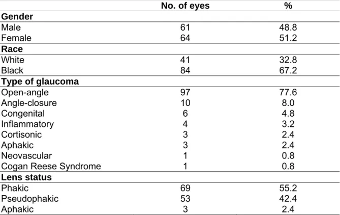
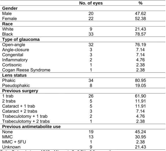
Information regarding gender, race, type of glaucoma, lens status and previous surgery is shown in Table 1.
TABLE 1. Demographics and characteristics of the study population
No. of eyes %
Gender
Male 61 48.8
Female 64 51.2
Race
White 41 32.8
Black 84 67.2
Type of glaucoma
Open-angle 97 77.6
Angle-closure 10 8.0
Congenital 6 4.8
Inflammatory 4 3.2
Cortisonic 3 2.4
Aphakic 3 2.4
Neovascular 1 0.8
Cogan Reese Syndrome 1 0.8
Lens status
Phakic 69 55.2
Pseudophakic 53 42.4
Aphakic 3 2.4
Previous surgery
1 trab 55 44
2 trabs 9 7.2
3 trabs 1 0.8
Cataract + 1 trab 47 37.6
Cataract + 2 trabs 9 7.2
Trabeculotomy + 1 trab 3 2.4
Trabeculotomy + 2 trabs 1 0.8
Trab = Trabeculectomy
Surgical Technique
surrounding skin were prepared with a 10% povidone-iodine solution, and a drop of the 5% povidone-iodine solution was applied to the conjunctiva. A lid speculum was placed, and the patient was asked to look down. The surgeon drew up 0.2 ml of 2% lidocaine with epinephrine and 0.1 ml of a 0.25 mg/ml MMC solution into a 1.0 ml syringe, for a final MMC concentration of 0.08 mg/ml. The needle was changed to a 26-gauge, and it was bent bevel up at the hub to an angle of 45º. Only 0.1 ml from the lidocaine-MMC mixture was injected into the subconjunctival space (MMC dose of 8 μg), 10 mm away from the posterior lip of the scleral flap (or from the place it was expected to be). The lid speculum was withdrawn and a gentle massage was applied to the superior eyelid for 5 minutes. The speculum was inserted again, and the needle was introduced superiorly as far as possible from the bleb; the needle was carefully advanced, using a side-to-side motion, beneath the Tenon’s space, breaking episcleral adhesions around the bleb and over the scleral flap until the bleb was reformed. We never introduced the needle under the scleral flap or into the anterior chamber. We verified reestablishment of flow by one of the following signs: softening of the eyeball, release of aqueous humor into the subconjunctival space, which created a raised conjunctival bleb, and, occasionally, a small reflux of blood into the anterior chamber. The conjunctival wound was not sutured, because of the oblique nature of the needle entry. One drop of a broad-spectrum topical antibiotic was applied after the procedure.
postoperative day and after 1 week, and 1, 3, 6, 12, 18, and 24 months, or more often as necessary, and complications were documented.
Needling was repeated using the same technique if the initial revision was not successful for controlling IOP. We permitted as many needling procedures as were necessary, but did not perform more than four needlings per eye. Some eyes received additional slit-lamp subconjunctival injections of antimetabolites during follow-up based on bleb morphology (elevation and extent) and signs of scarring (vascularization and increasing IOP). Moderately vascularized blebs received 5 mg of 5-FU (0.2 ml at a concentration of 25 mg/ml) and severely vascularized blebs received 8 μg of MMC (0.1 ml at a concentration of 0.08 mg/ ml).
Statistical Analysis
Reestablishment of aqueous flow with a resultant raised bleb was classified as an immediate success. At the last visit we defined an absolute success as an IOP ≤ 16 mmHg without medication, a qualified success as an IOP ≤ 16 mmHg with the aid of hypotensive agents, and a failure as an IOP > 16 mmHg despite medication, or if further conventional surgery was required to lower IOP.
RESULTS
We performed 186 needling procedures on 125 eyes (a mean of 1.49 needlings per eye; SD, 0.64; range, 1−4). Seventy-three eyes (58.4%) were needled once, 44 (35.2%) were needled twice, seven (5.6%) were needled three times, and one (0.8%) was needled four times. When we performed more than one needling, the mean interval between the first and second needling was 5.12 months (SD, 7.26; range, 4 days−33.57 months). The average follow-up from the first needling was 20.80 months (SD, 11.96; range, 1−57). Thirty-one eyes (24.8%) received 5-FU injections during the postoperative period, with an average of 4.10 applications per eye (SD, 2.80; range, 1−14). Four eyes (3.2%) received additional slit-lamp MMC injections during the postoperative period, with an average of 1.25 applications per eye (SD, 0.50; range, 1−2).
We reestablished aqueous flow and obtained a raised bleb in 115 eyes (92%), which were classified as immediate successes.
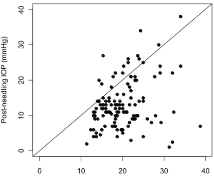
Mean IOP decreased from 20.07 mmHg (SD, 5.20; range, 11.3−38.7; median, 19 mmHg) preoperatively to 13.15 mmHg (SD, 6.77; range, 1−38; median, 12 mmHg) at the last follow-up (Wilcoxon signed rank test; P < 0.001). The change in IOP after needling is shown in Figure 1. The mean number of hypotensive agents per eye decreased from 2.35 (SD, 1.14; range, 0−5; median, 3) at baseline to 0.78 (SD, 1.30; range, 0−4; median, 0) at last follow-up (Wilcoxon signed rank test; P < 0.001).
subsequently restarted the medical therapy, 14 underwent another trabeculectomy, and three a Molteno implantation.
0 10 20 30 40
01 0 2 0 3 0 4 0
Pre-needling IOP (mmHg)
P os t-needl ing I O P ( m m H g)
FIGURE 1. Change in intraocular pressure (IOP) after all bleb needle revisions.
Points to the right of the equality line indicate pressures improved.
TABLE 2. Data regarding IOP and number of hypotensive medications
Pre-needling Post-needling P
value*
Mean (±SD) Median Mean (±SD) Median
Absolute success group (73 eyes; 58.4%)
IOP (mmHg) 19.74 (±5.14) 18.7 9.22 (±3.50) 10 < 0.001
No. of medications 2.26 (±1.09) 2 0 0 < 0.001 Qualified success group (22 eyes; 17.6%)
IOP (mmHg) 17.01 (±3.02) 16.4 12.57 (±1.50) 13 < 0.001
No. of medications 2.32 (±1.21) 3 2.36 (±1.09) 3 0.949
Failure group (30 eyes; 24.0%)
IOP (mmHg) 23.13 (±5.15) 22.5 23.13 (±4.67) 22 0.940 No. of medications 2.60 (±1.19) 3 1.50 (±1.55) 1 0.001
0 5 10 15 20 25
1 day 1 week 1 month 3 months 6 months 18 months 1 year 2 years 3 years
Follow-up
Mean
IOP
(m
m
H
g
)
Failure Qualified sucess Absolute sucess
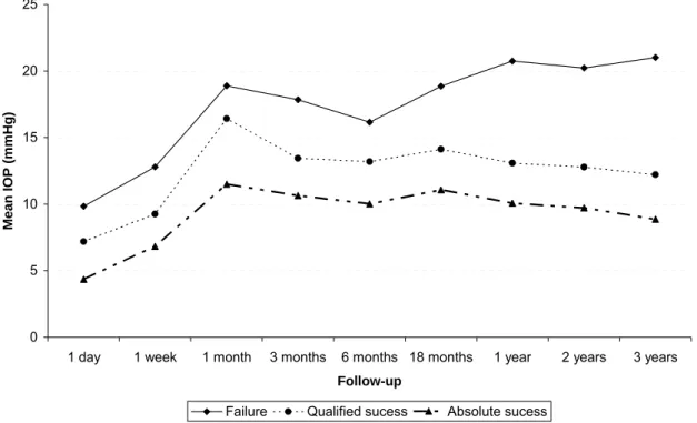
FIGURE 2. Mean intraocular pressure (IOP) during postoperative period.
needling procedure. Eleven eyes (8.8%) gradually developed cataracts some months after needling. There was no correlation between the number of needling procedures and late hypotony (Fisher’s exact test; P = 0.152) or cataract formation (Fisher’s exact test; P = 0.896). None of the eyes developed corneal decompensation, suprachoroidal hemorrhage, blebitis, or endophthalmitis.
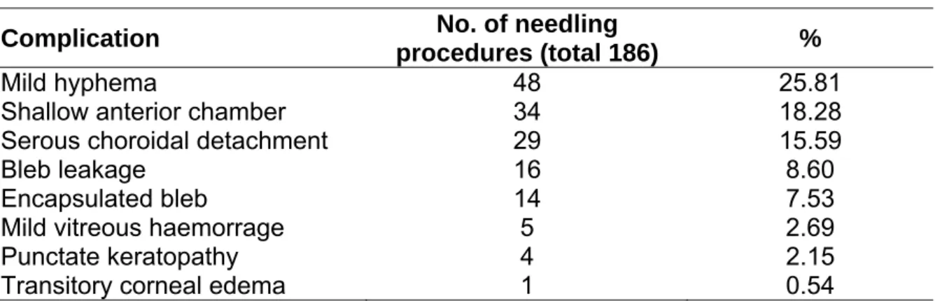
TABLE 3. Postoperative complications
Complication No. of needling
procedures (total 186) %
Mild hyphema 48 25.81
Shallow anterior chamber 34 18.28
Serous choroidal detachment 29 15.59
Bleb leakage 16 8.60
Encapsulated bleb 14 7.53
Mild vitreous haemorrage 5 2.69
Punctate keratopathy 4 2.15
Transitory corneal edema 1 0.54
0 10 20 30 40 50 60 0.0 0.2 0.4 0.6 0.8 1.0 Months Cu m u la ti v e S u rv iv a l
0 10 20 30 40 50 60
0.0 0.2 0.4 0.6 0.8 1.0 Months Cu m u la ti v e S u rv iv a l Multiple needlings One single needling
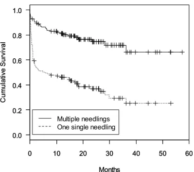
FIGURE 3. Kaplan-Meier cumulative survival curves for all needlings vs. the first
needling (125 eyes). Success criteria: IOP ≤ 16 mmHg.
DISCUSSION
Failure of filtration surgery is a very common problem. In the 5-fluorouracil filtration study,15 the early success rate was 80%, but after a follow-up period of 5 years, it dropped to 48%. The probability of success is even lower after repeat trabeculectomies, ranging from only 36 to 64%, depending on the study.16-18
31%.19-21 Subconjunctival MMC seems to be more effective than 5-FU for needle revision22 and offers the advantage of a single intraoperative application. However, 5-FU allows for a gentle modulation of the healing response during the postoperative period.23 In 1997, Apostolov and Siarov24 proposed the use of subconjunctival MMC injections during the early postoperative period in some cases of failing trabeculectomies. In our study, 31 eyes (24.8%) received postoperative 5-FU injections and four eyes (3.2%) received additional slit-lamp MMC injections based on bleb morphology and signs of scarring. We believe that these additional antifibrotic injections are a useful tool for reducing the healing response in high risk eyes.
There are many studies reporting that bleb needling can rectify a failing bleb during the early postoperative phase,5-10 but few reports confirm its effect during the late postoperative period.11-14 In the present study, the average time between the last failed trabeculectomy and needle revision was 5.31 years, ranging from 6 months to 30 years, which means that all needle revisions were performed in a late postoperative period.
Our criteria for success were more stringent than those reported by previous studies.4,6,8,9,25-30 We chose an IOP level of 16 mmHg as a cutoff point for success because most of the patients involved in this study had advanced glaucoma and required low IOP levels to prevent further damage to the optic nerve.
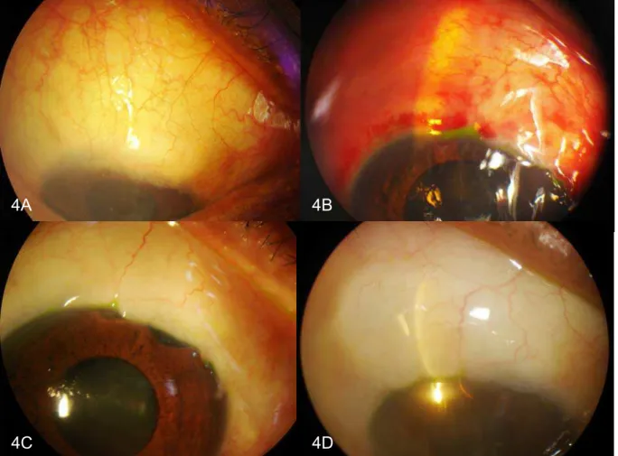
FIGURE 4. Bleb appearance before and after bleb needling with MMC. 4A:
Preoperative aspect (flat bleb, IOP 20 mmHg). 4B: First postoperative day (raised bleb, IOP 6 mmHg). 4C: Six months after needling (diffuse bleb, IOP 12 mmHg). 4D: Two years after needling (diffuse bleb, IOP 10 mmHg).
We revived 92% of all needled blebs (immediate success), which is excellent effectiveness for a relatively simple procedure. The appearance of resultant blebs after needling varied, but the most common type was large, diffuse, and discretely vascularized (Fig. 4). Our results are similar to those of Nascimento et al.,31 who obtained an elevated bleb in 90.3% of 84 eyes after needle revision with MMC. 4A 4B
Interestingly, in their study, they found that success was positively associated with the absence of an elevated bleb preoperatively. Our results also show that a completely flat bleb can provide excellent filtration, even several years after the original surgery.
It is difficult to compare studies because of differences in surgical techniques, success criteria, antimetabolite type and dose, timing of the needling procedure, and follow-up period, but our overall success rate of 76% at the latest visit agrees with previous reports4,27,28,31 that ranged between 71.6 and 76%. Needling proved to be highly effective. We achieved a significant reduction in mean IOP (from 20.07 mmHg preoperatively to 13.15 mmHg at the latest visit) and in the mean number of hypotensive agents (from 2.35 per eye preoperatively to 0.78 at last follow-up). In 58% of our patients (absolute success group), we obtained more than a 50% reduction in mean IOP. In 17.6% of our patients (qualified success group), even if the number of hypotensive medications remained the same, mean IOP dropped by 26%. In the failure group, although the mean IOP remained the same, there was a significant reduction in the mean number of medications. For most of the patients the procedure provided the chance to reduce the cost and morbidity associated with medical therapy and contributed to a better quality of life.
that most failures occurred within the first month and caused a slight elevation in mean postoperative IOP at the 1 month follow-up (Fig. 2). After that, a significant proportion of the eyes were needled again, which reduced mean IOP.
Bleb needle revision with MMC was first reported by Mardelli et al.4 who injected 4 µg of MMC at a concentration of 0.13 mg/ml and performed an average of 1.9 revisions per eye. In our study, we used a higher dose (8 µg of MMC at a concentration of 0.08 mg/ml) and performed 1.49 revisions per eye. Shetty et al.30 used a much higher MMC dose (40 µg of MMC at a concentration of 0.20 mg/ml), and required even fewer needle revisions (1.05 per eye). It is possible that fewer repeat procedures would have been required in our study if a higher MMC dose had been used.
Complications were similar to those seen after a trabeculectomy. They included small hyphemas, transient choroidal effusion, temporary conjunctival wound leaks, and shallowing of the anterior chamber. Most were minor, tolerable, and transient and required no treatment. However, the potential for more serious complications should not be underestimated. “Kissing” choroidal detachment that required surgical drainage,14,26 suprachoroidal hemorrhage,4,5,10,22,25,26,32 malignant glaucoma22,33,34 and endophthalmitis5 have been reported after bleb needling. None of the eyes in our study developed blebitis or endophthalmitis. In fact, the incidence of infectious events related to needling is very low. Pasternack et al.,26 Nascimento et al.,31 and Anand et al.22 reported a few cases of successfully treated blebitis, and Greenfield et al.5 reported the only case of endophthalmitis after bleb needling in the literature.
can be minimized by careful technique; the MMC solution must be injected slowly and some millimeters away from the bleb, to avoid a pressure gradient from the subconjunctival space to the anterior chamber, and some minutes should elapse between injection and needling. Even if the entire amount of MMC used in our study inadvertently entered the anterior chamber, it would be insufficient to cause endothelial toxicity.35 In the present study, only one eye experienced transient and mild corneal edema, which subsided within 1 week, but its preoperative endothelial cell density was very low (869 cells/mm2) and remained similar during the postoperative period (814 cells/mm2 after 6 months). MMC may also be toxic to the ciliary body epithelium and lead to persistent ocular hypotony.36-38 In our study, bleb formation was essential for reduction of IOP, which suggests that subconjunctival injection of MMC may not in itself lower IOP. Eleven eyes developed late hypotony, but all were related to large, avascular, and clearly overfiltering blebs. Only one required surgical treatment: a scleral patch graft 6 months after the needling procedure. The others were asymptomatic. There was no case of hypotonous maculopathy. Moreover, none of the eyes in our study had any evidence of scleral or conjunctival necrosis.
In conclusion, needling revision with adjunctive MMC is relatively safe and highly effective for reviving flat filtering blebs and controlling IOP, even several years after the original trabeculectomy.
REFERENCES
1. Skuta GL, Parrish RK II. Wound healing in glaucoma filtering surgery. Surv Ophthalmol 1987;32:149-70.
2. Ferrer H. Conjunctival dialysis in the treatment of glaucoma recurrent after sclerectomy. Am J Ophthalmol 1941;24:788-90.
3. Ewing RH, Stamper RL. Needle revision with and without 5-fluorouracil for the treatment of failed filtering blebs. Am J Ophthalmol 1990;110:254-9.
4. Mardelli PG, Lederer CM, Murray PL, et al. Slit-lamp needle revision of failed filtering blebs using mitomycin C. Ophthalmology 1996;103:1946-55.
5. Greenfield DS, Miller MP, Suner IJ, Palmberg PF. Needle elevation of the scleral flap for failing filtration blebs after trabeculectomy with mitomycin C. Am J Ophthalmol 1996;122:195-204.
6. Chang SH, Hou CH. Needling revision with subconjunctival 5-fluorouracil in failing filtering blebs. Chang Gung Med J 2002;25:97-103.
7. Ophir A, Wasserman D. 5-Fluorouracil-needling and paracentesis through the failing filtering bleb. Ophthalmic Surg Lasers 2002;33:109-16.
8. Fagerli M, Lofors KT, Elsas T. Needling revision of failed filtering blebs after trabeculectomy: a retrospective study. Acta Ophthalmol Scand 2003;81:577-82.
10. Rotchford AP, King AJ. Needling revision of trabeculectomies: bleb morphology and long-term survival. Ophthalmology 2008;115:1148-53 e4.
11. Ung CT, Von Lany H, Claridge KG. Late bleb needling. Br J Ophthalmol 2003;87:1430-1.
12. Paris G, Zhao M, Sponsel WE. Operative revision of non-functioning filtering blebs with 5-fluorouracil to regain intraocular pressure control. Clin Experiment Ophthalmol 2004;32:378-82.
13. Kapasi MS, Birt CM. The efficacy of 5-fluorouracil bleb needling performed 1 year or more posttrabeculectomy: a retrospective study. J Glaucoma 2009;18:144-8. 14. Passos AF, Cardozo AS, Mendes AG, Batista DMP. Late episcleral needling with adjunctive mitomycin-C for failed filtering blebs [in Portuguese]. Rev Bras Oftalmol 2002;61:622-38.
15. Five-year follow-up of the Fluorouracil Filtering Surgery Study. The Fluorouracil Filtering Surgery Study Group. Am J Ophthalmol 1996;121:349-66.
16. Inaba Z. Long-term results of trabeculectomy in the Japanese: an analysis by life-table method. Jpn J Ophthalmol 1982;26:361-73.
17. You YA, Gu YS, Fang CT, Ma XQ. Long-term effects of simultaneous subconjunctival and subscleral mitomycin C application in repeat trabeculectomy. J Glaucoma 2002;11:110-18.
18. Mietz H, Raschka B, Krieglstein GK. Risk factors for failures of trabeculectomies performed without antimetabolites. Br J Ophthalmol 1999;83:814-21.
20. Meyer JH, Guhlmann M, Funk J. How successful is the filtering bleb needling? [in German]. Klin Monatsbl Augenheilkd 1997;210:192-6.
21. Durak I, Ozbek Z, Yaman A, et al. The role of needle revision and 5-fluorouracil application over the filtration site in the management of bleb failure after trabeculectomy: a prospective study. Doc Ophthalmol 2003;106:189-93.
22. Anand N, Khan A. Long-term outcomes of needle revision of trabeculectomy blebs with mitomycin C and 5-fluorouracil: a comparative safety and efficacy report. J Glaucoma 2009;18:513-20.
23. Mastropasqua L, Carpineto P, Ciancaglini M, et al. Delayed post-operative use of 5-fluorouracil as an adjunct in medically uncontrolled open angle glaucoma. Eye 1998;12 ( Pt 4):701-6.
24. Apostolov VI, Siarov NP. Subconjunctival injection of low-dose Mitomycin-C for treatment of failing human trabeculectomies. Int Ophthalmol 1997;20:101-5.
25. Hawkins AS, Flanagan JK, Brown SV. Predictors for success of needle revision of failing filtration blebs. Ophthalmology 2002;109:781-5.
26. Pasternack JJ, Wand M, Shields MB, Abraham D. Needle revision of failed filtering blebs using 5-Fluorouracil and a combined ab-externo and ab-interno approach. J Glaucoma 2005;14:47-51.
27. Iwach AG, Delgado MF, Novack GD, et al. Transconjunctival mitomycin-C in needle revisions of failing filtering blebs. Ophthalmology 2003;110:734-42.
29. Broadway DC, Bloom PA, Bunce C, et al. Needle revision of failing and failed trabeculectomy blebs with adjunctive 5-fluorouracil: survival analysis. Ophthalmology 2004;111:665-73.
30. Shetty RK, Wartluft L, Moster MR. Slit-lamp needle revision of failed filtering blebs using high-dose mitomycin C. J Glaucoma 2005;14:52-6.
31. Nascimento GN, Passos AF, Cardozo AS, Zandonade E. Episcleral needling with adjunctive mitomycin-C: long term results [in Portuguese]. Rev Bras Oftalmol 2007;66:181-90.
32. Howe LJ, Bloom P. Delayed suprachoroidal haemorrhage following trabeculectomy bleb needling. Br J Ophthalmol 1999;83:757.
33. Mathur R, Gazzard G, Oen F. Malignant glaucoma following needling of a trabeculectomy bleb. Eye 2002;16:667-8.
34. Ramanathan US, Kumar V, O'Neill E, Shah P. Aqueous misdirection following needling of trabeculectomy bleb. Eye 2003;17:441-2.
35. McDermott ML, Wang J, Shin DH. Mitomycin and the human corneal endothelium. Arch Ophthalmol 1994;112:533-7.
36. Schraermeyer U, Diestelhorst M, Bieker A, et al. Morphologic proof of the toxicity of mitomycin C on the ciliary body in relation to different application methods. Graefes Arch Clin Exp Ophthalmol 1999;237:593-600.
37. Mietz H, Addicks K, Diestelhorst M, Krieglstein GK. Extraocular application of mitomycin C in a rabbit model: cytotoxic effects on the ciliary body and epithelium. Ophthalmic Surg 1994;25:240-4.
CONCLUSÃO DO PRIMEIRO TRABALHO, FORMULAÇÃO DE
NOVAS HIPÓTESES E OBJETIVOS DO SEGUNDO TRABALHO
O primeiro trabalho demonstrou, portanto, que o agulhamento com mitomicina C, realizado em uma fase tardia do pós-operatório, é eficaz na recuperação de fístulas antiglaucomatosas falidas, mesmo após muitos anos da realização da trabeculectomia e perante a ausência total de bolsa fistulante. Quando realizado em nível apenas episcleral, é relativamente seguro, pois suas complicações geralmente são transitórias e têm resolução espontânea. O presente trabalho demonstrou, também, a necessidade de, muitas vezes, se repetir o agulhamento para a obtenção de adequado controle da pressão intraocular. A sobrevivência de apenas um agulhamento mostrou-se bem menor quando comparada à sobrevivência de múltiplos agulhamentos.
Diante dos resultados obtidos, foram feitas então novas formulações. Gostaríamos de saber quais seriam os fatores que poderiam influenciar a taxa de sucesso do agulhamento. Já se conhecem vários fatores que interferem no resultado da trabeculectomia. Gostaríamos de saber se eles também influenciariam a taxa de sucesso do agulhamento e se haveria outras variáveis, estas já específicas do agulhamento, que também poderiam afetar seu sucesso ou seu fracasso.
O objetivo principal do segundo trabalho foi, portanto, identificar os fatores pré e pós-operatórios que pudessem estar associados ao sucesso do agulhamento com MMC na recuperação tardia de fístulas antiglaucomatosas falidas.
SEGUNDO TRABALHO
“Predictors of Success in Needle Revision of Filtering
Blebs with Mitomycin C: A Prospective Study”
“Fatores Preditivos para o Sucesso do Agulhamento com
RESUMO DO SEGUNDO TRABALHO
OBJETIVO: Identificar os fatores associados ao sucesso do agulhamento com
mitomicina C (MMC) na recuperação tardia de bolsas fistulantes falidas.
MÉTODO: Foram submetidos ao agulhamento com MMC 125 olhos de 98 pacientes
portadores de glaucoma sem controle adequado. Todos tinham pelo menos uma trabeculectomia prévia, a bolsa fistulante estava totalmente plana e o óstio interno estava pérvio à gonioscopia. O agulhamento associado à injeção subconjuntival de 8 µg de MMC foi realizado no bloco cirúrgico, pela mesma cirurgiã e repetido, se necessário.
RESULTADOS: Após um seguimento médio de 20,8 ± 11,96 meses, a pressão
intraocular (PIO) média caiu de 20,07 ± 5,20 mmHg no pré-operatório para 13,15 ± 6,77 mmHg no pós-operatório (p < 0,001) e o número médio de colírios hipotensores por olho caiu de 2,35 ± 1,14 para 0,78 ± 1,30 na última consulta (p < 0,001). Noventa e cinco olhos (76%) foram classificados como sucesso (PIO ≤ 16 mmHg) na última consulta. A principal variável associada ao sucesso foi a PIO baixa antes do agulhamento (p < 0,001). O sucesso também foi correlacionado a uma PIO baixa no primeiro dia após o agulhamento (p = 0,005), um maior intervalo de tempo entre a trabeculectomia e o agulhamento (p = 0,030) e à idade (p = 0,050; significância limítrofe), sendo que quanto mais idoso o paciente, maior sua chance de sucesso. As taxas de sucesso também foram maiores em pacientes brancos, em olhos pseudofácicos e em olhos com trabeculectomias de base fórnice, mas não em um grau que atingisse significância estatística.
CONCLUSÕES: O agulhamento com mitomicina C é um método eficaz para
Predictors of Success in Needle Revision of Filtering Blebs
with Mitomycin C: A Prospective Study
AUTHORS
1) Heloisa Andrade Maestrini1, MD 2) Sebastião Cronemberger1, MD, PhD
3) Hérika Danielle de Miranda Santos Matoso1, MD 4) Graziele Umbelina Alves Ferreira2, Statistician 5) Emília Sakurai2, PhD
1
Department of Ophthalmology, Federal University of Minas Gerais, Belo Horizonte, Minas Gerais, Brazil.
2
Department of Statistics, Federal University of Minas Gerais, Belo Horizonte, Minas Gerais, Brazil.
Address for correspondence and reprints: Heloisa Andrade Maestrini, MD.
Department of Ophthalmology, Federal University of Minas Gerais
Avenida Alfredo Balena 190, Belo Horizonte, MG, Brazil. ZIP Code: 30.130-100 Phone: 55 31 2535-5061 Fax: 55 31 3227-1911
e-mail: [email protected]
Financial Support: None.
ABSTRACT
Purpose: To identify factors associated with success of needle revision of failed
filtering blebs with adjunctive mitomycin C.
Patients and Methods: We investigated 125 eyes in 98 patients with uncontrolled
glaucoma. All had at least one failed trabeculectomy, a flat filtering bleb, and a patent internal ostium on gonioscopy. Needle revision with subconjunctival injection of 8 µg mitomycin C was performed in the operating room. Needling was repeated if necessary.
Results: After an average follow-up of 20.80 ± 11.96 months, mean intraocular
pressure (IOP) decreased from 20.07 ± 5.20 mmHg preoperatively to 13.15 ± 6.77 mmHg (P < 0.001), and the mean number of hypotensive agents per eye decreased from 2.35 ± 1.14 at baseline to 0.78 ± 1.30 (P < 0.001) at the latest visit. Overall, 95 eyes (76%) qualified as successes (IOP ≤ 16 mmHg) at the latest visit. The most important variable associated with success was a lower pre-needling IOP (P < 0.001). Successful outcomes also correlated significantly with a lower IOP on the first postoperative day (P = 0.005), a longer time between trabeculectomy and needling (P = 0.030), and older age (P = 0.050; borderline significance). The success rates tended to be greater in whites, pseudophakic eyes, and in eyes with a previous fornix-based conjunctival flap, but not to a degree that was statistically significant.
Conclusions: Needle revision with mitomycin C is effective to revive failed filtering
INTRODUCTION
Failure of trabeculectomy is a common problem and is usually the result of subconjunctival fibrosis.1 As the process of wound healing continues to occur indefinitely after initial surgery, trabeculectomy may fail at any time in the postoperative period. Needle revision is a simple and effective procedure to revive failing and failed filtering blebs,2-4 even several years after the original filtration surgery.5-7 Antimetabolites, such as 5-flurouracil (5-FU) and mitomycin C (MMC), are usually used as adjunctive therapy to inhibit fibroblast proliferation and preserve bleb function. MMC seems to be more effective than 5-FU8 and offers the advantage of a single intraoperative application, instead of a series of postoperative injections for 5-FU.
Risk factors such as young age,9 black race,10,11 high preoperative intraocular pressure (IOP),9 previous conjunctival surgery,12 uveitis,13 neovascular glaucoma13 and aphakia14 have been reported to reduce the success of any filtration procedure. The objective of this prospective study was to identify preoperative and postoperative factors associated with success or failure of needle revision of filtering blebs with adjunctive subconjunctival MMC.
PATIENTS AND METHODS
Patients
TABLE 1. Demographics and characteristics of the study population
No. of eyes %
Gender
Male 61 48.8
Female 64 51.2
Race
White 41 32.8
Black 84 67.2
Type of glaucoma
Open-angle 97 77.6
Angle-closure 10 8.0
Congenital 6 4.8
Inflammatory 4 3.2
Cortisonic 3 2.4
Aphakic 3 2.4
Neovascular 1 0.8
Cogan Reese Syndrome 1 0.8
Lens status
Phakic 69 55.2
Pseudophakic 53 42.4
Aphakic 3 2.4
TABLE 2. Previous surgery and antimetabolite exposure
No. of eyes %
Previous surgery
1 trab 55 44
2 trabs 9 7.2
3 trabs 1 0.8
Cataract + 1 trab 47 37.6
Cataract + 2 trabs 9 7.2
Trabeculotomy + 1 trab 3 2.4
Trabeculotomy + 2 trabs 1 0.8
Previous conjunctival flap
Limbus-based 59 47.2
Fornix-based 52 41.6
Unknown 14 11.2
Previous antimetabolite use
None 49 39.2
MMC 31 24.8
5-FU 2 1.6
MMC + 5-FU 9 7.2
Unknown 34 27.2
Information regarding gender, race, type of glaucoma, and lens status is shown in Table 1. Data from previous surgery are listed in Table 2. The average age of the study population was 61.64 years [standard deviation (SD), 18.81; range, 5−91; median, 66 years]. The average time interval between the original filtration surgery and the needling revision was 5.31 years (SD, 5.29 years; range, 6 months−30 years).
The study protocol was approved by the Federal University of Minas Gerais, Brazil, and all patients provided written informed consent.
Surgical Technique
given to avoiding inadvertent perforation of the overlying conjunctiva. The needle was never introduced under the scleral flap or into the anterior chamber. Reestablishment of aqueous flow was verified by one of the following signs: release of aqueous humor into the subconjuntival space, which created a raised bleb, softening of the eyeball and, occasionally, a small influx of blood into the anterior chamber. The conjunctival wound was not sutured. One drop of ciprofloxacin was applied immediately after the procedure.
All antiglaucoma medications were discontinued and the patient was placed on a topical combination of 0.1% dexamethasone and ciprofloxacin six times daily for 1 week. Thereafter, the dose was tapered according to the appearance (especially vascularization) of the bleb. Patients were examined on the first postoperative day and after 1 week, and 1, 3, 6, 12, and 24 months, or more often, as necessary, and complications were documented.
If the initial revision was not successful in controlling IOP, needling was repeated, using the same technique. Based on bleb morphology and signs of scarring, some eyes received subconjunctival injections of 5-FU (0.2 ml, at a concentration of 25 mg/ml).
Outcomes
We analyzed several variables that might affect the success rate: age, race, type of glaucoma, lens status, pre-needling number of hypotensive agents, previous conjunctival flap (limbus or fornix-based), number and type of previous operations, use of antimetabolites in previous trabeculectomy, time elapsed between trabeculectomy and needling, pre-needling IOP, number of needling procedures, occurrence of complications, IOP on the first postoperative day, and use of 5-FU in the postoperative period.
Statistical analysis
We used SPSS for Windows, version 15.0 (SPSS Inc., Chicago, IL, USA) for statistical analyses. To compare preoperative and postoperative IOP and number of hypotensive agents (non-parametric data), we used Levene’s test (to confirm variance homogeneity) and the Wilcoxon signed rank test with continuity correction. To test the association between success and variables measured in the study, we used the Pearson χ2 test with Yates’ continuity correction and the Fisher’s exact test for categorical variables, and the Mann-Whitney U-test for continuous variables; all tests were two-tailed. Finally, we performed a stepwise multiple logistic regression analysis. Because it requires a dichotomous response, absolute success and qualified success were categorized as success. P values < 0.05 were deemed to indicate statistical significance.
RESULTS
times. Thirty-one eyes (24.8%) received 5-FU injections in the postoperative period, with an average of 4.10 applications per eye (SD, 2.80; range, 1−14).
There was a statistically significant reduction in the mean IOP and the mean number of hypotensive agents after bleb needle revision. The mean IOP decreased from 20.07 mmHg (SD, 5.20; range, 11.3−38.7; median, 19 mmHg) preoperatively to 13.15 mmHg (SD, 6.77; range, 1−38; median, 12 mmHg) at last follow-up (Wilcoxon signed rank test; P < 0.001). The mean number of hypotensive agents per eye decreased from 2.35 (SD, 1.14; range, 0−5; median, 3) at baseline to 0.78 (SD, 1.30; range, 0−4; median, 0) at last follow-up (Wilcoxon signed rank test; P < 0.001).
After an average follow-up from the first needling of 20.80 months (SD, 11.96; range, 1−57 months), 95 eyes (76%) were classified as successes (IOP ≤ 16 mmHg) at the latest visit. Overall, 73 eyes (58.4%) achieved an absolute success, 22 (17.6%) a qualified success, and 30 (24%) were classified as failures.
TABLE 3. Continuous variables in success and failure groups
Variable Success group Failure group P-value*
Mean (±SD) Median Mean (±SD) Median
Age (years) 63.31 (±18.35) 68 56.41 (±19.58) 53 0.050
Pre-needling no. of medications
2.27 (±1.12) 2 2.60 (±1.19) 3 0.174 No. of previous
surgeries
1.48 (±0.68) 1 1.53 (±0.86) 1 0.895 Time
trab-needling (years)
5.70 (±5.49) 4.17 4.01 (±4.75) 2.42 0.030
Pre-needling IOP (mmHg)
19.11 (±4.86) 18.7 23.13 (±5.15) 22.5 < 0.001
No. of needling procedures
1.49 (±0.67) 1 1.47 (±0.57) 1 0.971 IOP first postop.
day (mmHg)
5.02 (±6.55) 3 9.83 (±9.43) 6 0.005 *Two-tailed Mann-Whitney U test with continuity correction. Bold P-values are statistically significant. SD = Standard Deviation. Trab = Trabeculectomy. Postop. = postoperative.
TABLE 4. Categorical variables and success rates
Variable Success rate
n (%) P-value
Race White (n=41)
Black (n=84)
35 (85.37)
60 (71.43) 0.136* Type of glaucoma Open-angle (n=97)
Other types (n=28)
75 (77.32)
20 (71.43) 0.520* Lens status Pseudophakic (n=53)
Phakic (n=69) Aphakic (n=3) 45 (84.91) 48 (69.57) 2 (66.67) 0.086** Previous trabeculectomy conjunctival flap Fornix-based (n=52) Limbus-based (n=59) 43 (82.69)
41 (69.49) 0.163* Previous use of
antimetabolites
Yes (n=42) No (n=49)
33 (78.57)
35 (71.43) 0.589* Post-needling complications Yes (n=62)
No (n=63)
48 (77.42)
47 (74.60) 0.874* Post-needling
use of 5-FU
Yes (n=31) No (n=94)
24 (77.42)
71 (75.53) 0.977*
*Two-tailed Pearson χ2 test with Yates’ continuity correction. **Two-tailed Fisher’s exact test.
Complications did not affect the success rate (Table 4), and included mild hyphema (25.8%), shallow anterior chamber (18.3%), serous choroidal detachment (15.6%), bleb leakage (8.6%), and encapsulated bleb (7.5%). Most were minor, transient, and required no treatment.
DISCUSSION
Factors associated with the success or failure of needling have been evaluated in previous studies, with variable findings.3,4,12,15-23 This variability may be explained by differences in surgical techniques, success criteria, antimetabolite type and dose, specific factors evaluated, differences in study design, and methods of data collection and statistical analyses. Some studies have been based on retrospective analyses,3,12,15,16,18-20 which is subject to various limitations, while others have evaluated risk factors prospectively,4,17,21,22 which provides stronger results. Some risk factors have been recognized for the failure of trabeculectomy, such as young age, black race,3,19 previous conjunctival incisions,12 and aphakia,18 whereas, others are more specific to needling revision, such as the interval between filtration surgery and needling,3,4,17,20 high pre-needling IOP,6,12,15,21 multiple needlings,12,22,23 and high IOP immediately after the procedure.6,8,15,21,23
In the present prospective study, the most important variable associated with success was pre-needling IOP. The lower the IOP, the better the needling result (P < 0.001). Our results are consistent with those of Kapasi and Birt,6 Broadway et al.,21 Greenfield et al.,12 and Shin et al.,15 who found that failure correlated directly with higher pre-needling IOP.
success was the immediate attainment of an IOP < 11 mmHg, a finding also reported by Shin et al.,15 who found that, if needle revision produced an IOP ≤ 10 mmHg, the procedure could be judged a preliminary success, and could predict future success. Rotchford et al.,23 Kapasi and Birt6 and Anand and Khan8 also found that immediate reduction in IOP was a good prognostic sign. We think that a low immediate IOP reflects a good aqueous flow and the reestablishment of a well-functioning filtering bleb.
and Stamper,2 Pasternack et al.,19 Kapasi and Birt,6 Greenfield et al.,12 Broadway et al.21 and Nascimento et al.22 reported that this interval did not affect the success rate.
Older age was also positively related with success in our study, although this tendency was of borderline significance (P = 0.050). Higher success rates in older patients have been attributed to a weaker healing response.1 Our results also tended to be better in pseudophakic eyes, but this perhaps reflected only the fact that these patients were older than the phakic ones. Shetty et al.20 also found a higher chance of success in pseudophakic eyes and postulated that needling was safer and better tolerated in pseudophakic eyes with a deeper chamber than in phakic eyes. On the other hand, in 1993, Shin et al.18 reported that needling tended to be more successful in phakic than aphakic and pseudophakic eyes, but, at that time, most cataract surgery was performed by extracapsular extraction, which necessitated a large conjunctival incision. In 1996, Greenfield et al.12 found unsuccessful outcomes in needle revisions performed in eyes with prior surgery that involved conjunctival incisions. In our study, the number and type of prior operations did not influence the chance of success.
Needling tended to be more successful in whites versus blacks, although this tendency was not statistically significant, a finding also reported by Broadway et al.21 Mardelli et al.3 obtained a higher success rate in whites than blacks, and Pasternack et al.19 found that African-American race was the only significant risk factor for needle revision failure. Blacks have been shown to have a lower success rate in any filtration surgery because of a more exuberant healing response.1
hand, Nascimento et al.22 found that success was positively related with the absence of an elevated bleb preoperatively. Fagerli et al.25 found no difference in success between eyes with a flattened bleb and those with a cystic bleb. Hawkins et al.16 and Broadway et al.21 also showed that pre-needling bleb characteristics had no effect on outcome. In the present study, we could not analyze this variable, because all blebs were completely flat before needle revision, but our results show that a flat bleb can provide excellent filtration, even several years after the original trabeculectomy.
In the present study, needling revision of fornix-based blebs tended to be more successful, although not to a statistically significant degree. It is easier, technically, to perform a needle revision in these blebs, and resultant post-needling blebs are also more diffuse, because there are no restrictive scarred lines in the conjunctiva, like those seen in limbus-based blebs. However, Hawkins et al.,16 in a retrospective study, found that fornix-based blebs were more likely to fail needle revision, compared with limbus-based blebs.
In previous studies, greater success has been achieved in eyes that required only one needle revision, compared with those that required multiple revisions.12,22,23 In the present study, the number of needling procedures did not affect the success rate.
and signs of scarring, especially bleb vascularization, elevation, and extent. We think that these additional antifibrotic injections are a safe and useful tool to reduce the healing response in these eyes, and perhaps some of them could have failed without this aid.
The role of antiglaucoma medication is also interesting. Kapasi and Birt6 observed that patients in their success group had used a lower number of hypotensive agents before needling, compared with the failure group. Chang and Hou26 found that the only risk factor associated with failure of needling of filtering blebs with 5-FU was the number of antiglaucoma drugs used before the filtering surgery. The use of more topical agents could result in a greater inflammatory reaction in the conjunctiva, leading to failure of the procedure.27 In the present study, however, we found no significant correlation between success and the number of pre-needling hypotensive agents.
Complications were not associated with poorer outcome and were similar to those seen after trabeculectomy. They included small hyphemas, transient choroidal effusion, temporary conjunctival wound leaks, and shallowing of the anterior chamber. Most of these were minor, tolerable, transient, and required no treatment. Finally, no significant correlation was observed between success and type of glaucoma or gender.