Revealing Host Factors
Important for Hepatocyte
Infection by Plasmodium
Cristina Dias Rodrigues
Faculdade de Ciências
Departamento de Biologia Vegetal
Doutoramento em Biologia
Biologia Celular
Cover image | An Anopheles gambiae female mosquito feeding on a mammalian host (adapted from http://www.abc.net.au/science/news/img/health/mosquito190804.jpg).
Universidade de Lisboa Faculdade de Ciências
Departamento de Biologia Vegetal
Revealing Host Factors Important for
Hepatocyte Infection by Plasmodium.
Cristina Dias Rodrigues
Dissertation submitted to obtain a PhD Degree in Biology, specialityof Cellular Biology by the Universidadede Lisboa.
Supervisor Maria Manuel Mota, MsD, PhD.
Principal Investigator of Institutode MedicinaMolecular and Auxiliary Professor at Faculdadede Medicina, Universidadede Lisboa.
Co-Supervisor Maria da Graça Alves Vieira, PhD.
Auxiliary Professor of Faculdadede Ciências, Universidadede Lisboa.
Research is to see what everybody else has seen, and to think what nobody else has thought.
Albert Szent-Györgi
All truths are easy to understand once they are discovered; the point is to discover them.
Preface
The present thesis embraces the data obtained during my Ph.D. research project developed from November 2003 to June 2007.The experimental work was supervised by Prof. Doutora Maria Manuel Mota and was carried out at Instituto de Medicina Molecular, in Lisboa and Instituto Gulbenkian de Ciência, in Oeiras, Portugal. The RNAi screens were performed at Cenix BioScience, Dresden, Germany.
This Ph.D. was supervised by Prof. Maria da Graça Alves Vieira from Departamento de Biologia Vegetal, Faculdade de Ciências, Universidade de Lisboa, Portugal.
The financial support was provided by the portuguese Fundação para a Ciência e Tecnologia, through the Ph.D. fellowship grant SFRH/BD/14232/2003.
This thesis is structured in 5 chapters, which are preceded by a summary, both in Portuguese and in English.
Chapter 1 comprises a general introduction to malaria and its current world situation, followed by an overview on the Plasmodium life cycle, the state of the art on the liver stage biology and the objectives of this thesis.
Chapters 2 to 4 consist of the results obtained throughout the research project, which are presented in a publication format. Each results chapter includes an abstract, an introduction, the results and discussion, as well as the methods, acknowledgments and references.
Chapter 5 encloses a general discussion and the future perspectives of the work develop.
In Appendix are supplied my Curriculum Vitae and the publications in which I have participated until the date of printing this thesis.
Acknowledgements
Ao longo do meu doutoramento “cresci” em termos científicos mas acima de tudo como pessoa. Estes quatro anos foram muito intensos em sentimentos totalmente diferentes: entusiasmo vs. desânimo, alegria vs. tristeza, partilha vs. inibição, convicção vs. dúvida, sorrisos vs. lâgrimas, companheirismo vs. momentos de solidão. Durante esta jornada foram várias as pessoas que me acompanharam e às quais faço questão de agradecer.
À Maria.
Um bem-haja à minha orientadora científica e amiga.
É díficil conseguir expressar em palavras tudo pelo qual gostaria de te agradecer. Obrigada pela oportunidade de poder trabalhar contigo e pela tua constante presença e apoio. Obrigada pela orientação científica e por me ensinares a fazer Ciência, pelo teu entusiasmo contagiante, pelas discussões científicas, por compreenderes os meus erros, por toda a preocupação, carinho e amizade e por respeitares as minhas ideias e o meu ritmo de trabalho e de escrita.
À Professora Graça.
Obrigada por ter aceite ser a minha orientadora interna ao nível da Faculdade de Ciências da Universidade de Lisboa. Foi um prazer partilhar e discutir o meu trabalho consigo... Muito obrigada por todo o apoio, ajuda e carinho.
Ao Miguel.
Quando começámos a trabalhar juntos foi um pouco difícil para mim. De repente ter de partilhar ideias, opiniões, horários, material, preocupações, definir prioridades e até partilhar uma vida/casa num país frio foi um desafio. Aprendi imenso contigo durante o meu doutoramento e sem ti os 6 meses em Dresden teriam sido ainda mais difíceis. Crescemos como colegas e amigos! Miguelito, muito obrigada por todo o apoio científico e pessoal.
À Sónia.
Muito obrigada pela constante boa disposição, companheirismo, ânimo, amizade no lab e principalmente durante as nossas mil viagens épicas. Estás nas minhas memórias de NY, Dresden, Berlim, Praga, Budapeste e Londres. E espero que em breve de Barcelona, mas desta vez espera-se que sem quedas!!
To Michi.
When I think about the first time that I went to Cenix BioScience, it comes to my mind the picture of you and Christian. Two friendly guys!! At that time I had never thought that we would still be working together after 4 years, which is spectacular. We were involved since the beginning and together we have been able to build an amazing story. Thank you for your
support to the development of malaria research at Cenix. Many thanks for all the help, your work and input, your enthusiasm and all the scientific discussions.
To Cécilie.
I have learned a lot working with you, especially in what regards a good laboratory methodology. Thank you for your excellent work, your availability to answer my questions, your support and friendship and for the evenings and dinners while we were in Dresden. I hope to see you and Régis soon here in Lisbon.
To Chris.
Thank you for embracing the malaria research. To be able to work directly with you is a great honor and I have been learning constantly. Thanks for all your amazing input to our work and I will never forget your support and advices at the Keystone Meeting.
À Sílvia.
Agradeço-te pela tua constante disponibilidade em ajudares e especialmente por o fazeres com um sorriso. Sei que posso contar sempre contigo. Muito obrigada pelo apoio, energia e positivismo em termos profissionais e pessoais. O teu bom humor e espírito de amiga têm-me contagiado! Si, mil “thanks”.
À Sabrina.
Muito obrigada pela ajuda no trabalho, pelo apoio na recente cruzada de se fazer hepatócitos primários, pela tua amizade repleta de energia e sabedoria de vida. Desejos de muitas felicidades e havemos de nos cruzar algures no mundo! Não desanime nunca!
À Lígia.
Muito obrigada pelos teus maravilhosos hepatócitos primários de ratinhos, por me ensinares o protocolo que tão arduamente optimizaste e por estares sempre disponível a ajudar. Obrigada por todo o apoio e carinho. Mantém esse sorriso lindo!
A todos os meus colegas que constituem a Unidade de Malária.
Vocês estiveram presentes durante o meu doutoramento em fases diferentes... Se não me falha a memória, aqui vai por ordem de chegada ao grupo.
Patrícia, muito obrigada por toda a ajuda, constante disponibilidade, amizade e contagiante
gosto de se fazer Ciência sem deixar de se viver e explorar o Mundo e os sentidos. Admiro a tua capacidade de conciliar tantas actividades. Bjinho gde Linda!
Daniel, muito obrigada pela paciência e muitos ensinamentos aquando da minha chegada ao
grupo. Foste espectacular!
Casanova, pelo apoio inicial e por me ensinares princípios laboratoriais básicos. Força para o
teu doutoramento. Pensamento positivo!
Bruno, muito obrigada por todo o apoio e força de que tudo vai correr bem. Desejos de
Margarida, marcaste-me desde o primeiro dia em que fui ao IGC. Ainda me lembro da tua
disponibilidade e simpatia a falares comigo. Foi muito bom trabalhar contigo, muito obrigada por todos os teus concelhos. Fica bem!
Ana, o teu percurso constitui para mim um exemplo de como é possível mudar. Admiro a
tua coragem, perseverança e luta. Obrigada por todo o apoio, ombro amigo e concelhos. Coragem, energia e sorriso estampado!
Marta, a tua forte personalidade em prosseguires as tuas convicções é marcante. Muito
obrigada pelas constantes perguntas e pelo apoio e compreensão em momentos difíceis. Desejos de muitas felicidades na tua nova etapa de vida em Londres and “see u”!
Nuno, aprendi muito ao trabalhar contigo e obrigada por toda a ajuda. Espero que alcances
todos os teus objectivos profissinais e pessoais. Nunca desistas!
Cristina, muito obrigada pela tua visão do que é fazer Ciência, pelo exemplo de organização
e capacidade de trabalho, pelo apoio e amizade. Ao te observar parece tão fácil!
Ataíde, obrigada pelo trabalho indispensável com os nossos amigos mosquitos, pela
constante boa disposição, companheirismo e gargalhadas. Boa sorte na Austrália e vê-se estás por lá em Fevereiro para fazeres as honras. Arrasa!
Iana, thanks for your example of courage for going after of what you really wanted. I wish
you all the best.
Carina, és um exemplo de perseverança e insistência. Não percas estas qualidades e apesar
de o tempo passar a correr lembra-te que durante um doutoramento também se deve aproveitar. Força!
Kirsten, thanks for all the scientific questions, conversations about different subjects and help
with the english. A smile!
Catarina, obrigada pelo teu empenho no trabalho de produção de mosquitos. Bjinho!
Luís, muito obrigada por toda a tua ajuda preciosa nesta fase final e por respeitares a minha
agitação. Boa sorte por NY e aproveita ao máximo esta nova fase da tua vida.
Inês e Ana, parte essencial do grupo sem vocês tudo seria muito mais difícil. Ana, um
especial obrigado por todo o teu apoio. A special thanks to all at Cenix BioScience.
Thank you for the exceptionable working environment, all the help and company outside the lab that has allowed me to enjoy Dresden.
Thank you to Geert-Jan Gemert and Robert Sauerwein for providing precious P. berghei infected mosquitoes.
Thank you to Jean-François Franetich and Dominique Mazier for the primary human hepatocytes and P. falciparum sporozoites.
Thank you to Hans-Peter Vornlocher from Alnylam for providing us the siRNAs and experimental protocol used in the RNAi in vivo experiments.
Muito obrigada a todos que constituem o IMM e o IGC e que tornam estes dois institutos locais excepcionais para se fazer investigação.
Um especial obrigada à Unidade Patogénese Viral no IMM com quem partilhámos o espaço, material e o dia-a-dia a fazer experiências e a todos os membros dos grupos de Malária no IGC por toda a ajuda.
Muito obrigada à Alina e Dolores, responsáveis pelo biotério do IMM e IGC. O vosso trabalho é essencial à realização e sucesso das nossas experiências in vivo. Obrigada pela vossa compreensão e ajuda em momentos em que os resultados são para ontem!
Muito obrigada à Margarida Vigário e Moises Mallo, membros do meu comité de tese no IGC, pelo apoio e discussão científica.
Obrigada à Fundação para a Ciência e Tecnologia pelo financiamento da minha bolsa doutoramento e por ter patrocinado a minha participação em encontros científicos internacionais.
À Isa e Maf muito obrigada pela vossa amizade e pelos bons momentos de lazer que foram indispensáveis para descomprimir. É sempre garantida muita diversão e muitas gargalhadas. À Vera, apesar de estarmos em países diferentes, a tua presença nos bons e maus momentos foi uma constante durante estes anos de doutoramento. Através de um mail, um telefonema, uma viagem senti sempre o teu apoio e amizade. Á minha grande amiga ;)
À Graça e Jorge Henriques por me terem acolhido como parte integrante da vossa família. Todo o vosso apoio e carinho tem sido imprescendíveis. Muito obrigada!
Ao meu mano David, por estar sempre presente, pela sua contagiante boa-disposição, pelos seus sorrisos e carinho, pelo seu imenso apoio à nossa família, por toda a força e apoio e pelo teu exemplo de ambição e constante luta. Ao meu pai Henrique por me ter dado o possível e impossível, por todos os “patrocínios”, pela sua visão sonhadora, a sua força de que basta querermos para sermos capazes, por me ensinar a nunca desistir e por todo o seu carinho. À minha mãe Branca por ter me ter transmitido os valores por que me guio, pela educação que me proporcionou e sempre acompanhou de perto, por valorizar o que sou e faço, por me apoiar em tudo e por todos os pequenos gestos que transmitem o seu amor por mim. Dou graças todos os dias por vos ter... Amo-vos muito!
E a ti, Ricardo... por viveres comigo todos os momentos que marcaram este projecto. Pela tua paciência, disponibilidade, compreensão, ajuda e força. Pela tua constante presença e disponibilidade para ouvir e me “guiar” durante os momentos complicados. Pelas tuas tentativas de me alegrares e por apoiares as minhas decisões. Por todo o teu amor... Que seria de mim sem ti... Amo-te! Te!!
Malaria remains the most important parasitic infection in humans and one of the most prevalent infectious diseases worldwide. It is estimated that 350–500 million people become infected and one million of children under the age of five years die every year. This disease is caused by a protozoan parasite of the genus Plasmodium and is transmitted by mosquitoes of the genus Anopheles. During a malaria infection, the parasite undergoes intracellular development within two different host cells, hepatocytes and erythrocytes. The first stage of a malaria infection, the liver stage, is asymptomatic while the second phase, the blood stage, is responsible for all the disease-associated symptoms. Although clinically silent, the liver stage is an obligatory and extremely important step in Plasmodium life cycle. Therefore, understanding the parasite’s requirements during this period is crucial for the development of any form of early intervention. Despite this awareness, the strategies developed by Plasmodium to allow its survival and development inside host liver cells remain poorly understood. We have sought to disclose some of these strategies through the identification of host factors which are important for hepatocyte infection by Plasmodium. For this purpose, RNA interference (RNAi) was extensively applied to an in vitro model of Plasmodium infection. The infection outcome of the rodent P.
berghei parasite was addressed in the context of individual gene loss of function of a
total of 830 different genes.
In chapter 2 of this thesis, it was studied the functional relevance of a selection of 50 genes differentially expressed by hepatocytes throughout Plasmodium infection, revealed by a microarray approach. Two host transcription factors, the Activating Transcription Factor 3 (Atf3) and the Myelocytomatosis oncogene (c-Myc), were recognized as important for sporozoite infection. It was observed that their individual silencing led, respectively, to an increase and to a decrease in P. berghei hepatocyte infection levels. In addition, Atf3’s functional relevance was confirmed in vivo.
In chapter 3, the role of host kinases and kinase-interacting proteins was explored. From the 727 genes investigated at least six host kinases, namely the Met proto-oncogene (MET), Protein Kinase C iota (PKCι), Protein Kinase C zeta (PKCζ), WNK lysine deficient protein kinase 1 (PRKWNK1), Serum/Glucocorticoid Regulated Kinase 2 (SGK2) and Serine/Threonine Kinase 35 (STK35) were identified as playing important roles during Plasmodium sporozoite infection. Moreover, the importance of one of these kinases, PKCζ, was further demonstrated for in vivo P. berghei infection,
and also for human primary hepatocytes infection by P. falciparum, the deadliest of the human malaria parasites.
Finally, in chapter 4, the function of 53 host lipoprotein pathway genes was investigated and the Scavenger Receptor Class B member 1 (SR-BI) was identified as being crucially required for hepatocyte infection by P. berghei. In addition, SR-BI’s importance in Plasmodium infection was also observed in vivo for P. berghei and ex vivo for P. falciparum. Further detailed analyses revealed that SR-BI is required for both sporozoite invasion and development within the hepatocyte.
Altogether, the work presented in this thesis reveals different host factors that play important roles during hepatocyte infection by Plasmodium sporozoites and, therefore, offers new insights into the processes underlying Plasmodium liver infection.
Keywords:
Resumo
A malária é uma das doenças infecciosas em humanos com maior prevalência em todo o mundo, sendo a mais importante de todas as doenças parasitárias. Esta doença afecta cerca de 40% da população mundial e estima-se que, todos os anos, entre 250 a 500 milhões de pessoas ficam infectadas e que morrem aproximadamente um milhão de crianças com menos de cinco anos. O parasita responsável por esta doença é um protozoário que pertence ao género Plasmodium e é transmitido por mosquitos do género Anopheles. O parasita humano P. falciparum provoca as formas mais severas da doença e é responsável pela maior parte das mortes por malária.
O parasita Plasmodium, no estadio de esporozoíto, após ter sido transmitido ao hospedeiro mamífero através da picada de um mosquito infectado, migra até ao fígado onde infecta hepatócitos. No interior dos hepatócitos, cada parasita replica-se e diferencia-se no estadio seguinte, em merozoítos. Estes são libertados na corrente sanguínea e infectam eritrócitos. Assim sendo, uma infecção de malária caracteriza-se pelo desenvolvimento intracelular do parasita em dois tipos de células hospedeiras, hepatócitos e eritrócitos. A primeira fase de uma infeccção de malária, a fase hepática, é assintomática, enquanto que a segunda, a fase sanguínea, é responsável pelos sintomas associados à doença. Apesar do desenvolvimento de esporozoítos de
Plasmodium em hepatócitos decorrer sem qualquer sintoma associado, a fase hepática
é um passo importante devido à sua obrigatoriedade para o estabelecimento da infecção. Como tal, a compreensão das necessidades do parasita durante este período é fundamental para o desenvolvimento de qualquer tipo de intervenção precoce.
Apesar disso, as estratégias que o parasita Plasmodium desenvolveu para conseguir sobreviver e desenvolver-se no interior dos hepatócitos são ainda desconhecidas. O presente trabalho teve como objectivo revelar algumas destas estratégias através da identificação de factores do hospedeiro importantes para a infecção de hepatócitos pelo parasita Plasmodium. Para tal, recorreu-se a utilização da técnica conhecida por RNA de interferência (RNAi). Esta técnica baseia-se no mecanismo celular em que moléculas de RNA na forma de dupla cadeia silenciam o gene alvo através da degradação específica do seu RNA mensageiro. Deste modo, através da inserção em células de duplas cadeias de RNA é possível induzir especificamente o silenciamento do gene desejado e analisar o seu papel funcional ao nível de um determinado fenótipo. No presente trabalho aplicou-se a técnica RNAi a um modelo in vitro de infecção com Plasmodium e avaliou-se qual o efeito do silenciamento de genes
específicos em termos de infecção do parasita de ratinhos, P. berghei. Ao todo foram analisados 830 genes, de diferentes categorias, nomeadamente genes expressos ao longo de uma infecção por Plasmodium, genes que codificam cinases e proteínas associadas e ainda genes associados ao metabolismo de lipoproteínas. Estes estudos são apresentados como parte integrante da presente tese em três capítulos distintosde resultados.
No capítulo 2, foi estudada a relevância funcional de 50 genes que são expressos diferencialmente por hepatócitos ao longo de uma infecção por Plasmodium, genes que foram seleccionados a partir de um estudo anterior em que se utilizaram microarrays. Através da utilização de RNAi procedeu-se ao silenciamento individual de cada um dos 50 genes seleccionados e, após diferentes passos experimentais de confirmação, os factores de transcrição, Activating Transcription Factor 3 (Atf3) e Myelocytomatosis
oncogene (c-Myc), foram reconhecidos como sendo importantes para a infecção
hepática por esporozoítos, dado que quando silenciados, a infecção com P. berghei aumenta e diminui, respectivamente. Adicionalmente, foi confirmada in vivo a relevância funcional para o factor de transcrição Atf3. Em ratinhos da estirpe BALB/c observou-se cinco horas depois da infecção com esporozoítos de P. berghei um aumento na quantidade de RNA mensageiro do gene Atf3 ao nível do fígado. E ratinhos Atf3 knock-out quando infectados com esporozoítos de P. berghei apresentam um nível de infecção mais elevado que ratinhos controlo, sem qualquer deficiência ao nível da expressão do gene Atf3.
No capítulo 3, foi explorado o papel de um total de 727 cinases do hospedeiro ou proteínas que interagem com cinases. A relevância de cada gene foi estudada através do seu silenciamento individual com três small interfering RNAs (siRNAs), tendo sido realizadas três experiências de rastreio em que os genes candidatos foram seleccionados progressivamente de acordo com os resultados obtidos. Na última experiência, confirmou-se ainda o nível de silenciamento atingido, através da quantificação do nível de RNA mensageiro específico para uma das cinases identificadas como sendo potencialmente importante(s) para a infecção da linha celular de hepatócitos humanos (Huh7) por esporozoítos de P. berghei. No final, foram seis os genes de cinases para os quais se observou uma correlação entre o nível de silenciamento e um fenótipo em termos da infecção, mais especificamente Met
proto-oncogene (MET), Protein Kinase C iota (PKCι), Protein Kinase C zeta (PKCζ), WNK lysine deficient protein kinase 1 (PRKWNK1), Serum/Glucocorticoid Regulated Kinase 2 (SGK2) e Serine/Threonine Kinase 35 (STK35). Para as cinases MET, PKCζ, SGK2, PRKWNK1 e
células infectadas com P. berghei, enquanto que para a cinase PKCι observou-se que o seu silenciamento leva a um aumento do nível de células infectadas com P. berghei. A importância de uma destas cinases, PKCζ, foi ainda explorada através da realização de experiências adicionais. O tratamento de células Huh7 in vitro com um inibidor para a cinase PKCζ, que funciona como um pseudo-substracto, teve como resultado uma redução no nível de infecção. Este resultado foi também observado ex vivo em hepatócitos primários de ratinhos da estirpe C57BL/6 e em hepatócitos primários humanos sujeitos ao tratamento com o inibidor da PKCζ, e que foram posteriormente infectados com esporozoítos do parasita P. berghei ou P. falciparum, respectivamente. Através da realização de RNAi in vivo para a cinase PKCζ, em que se induziu uma redução da expressão desta cinase ao nível do fígado, foi demonstrado que esta cinase também desempenha um papel na infecção de malária in vivo, já que se observou uma significativa diminuição no nível de parasita no fígado. Todos estes resultados adicionais para a cinase PKCζ apoiam de certo modo a validade dos genes candidatos identificados através da utilização de RNAi, e demonstram também que esta cinase específica é importante para a infecção de Plasmodium.
Finalmente, no capítulo 4, foi investigada a função de 53 genes associados a vias metabólicas que envolvem lipoproteínas, e o receptor Scavenger Receptor Class B
member 1 (SR-BI) foi identificado como sendo fundamental para a infecção de
hepatócitos por P. berghei. A relevância deste receptor para a infecção in vitro de P.
berghei foi demonstrada através da realização de diferentes experiências em que se
recorreu ao tratamento da linha celular Huh7 com diversos reagentes que permitiram silenciar o gene SR-BI ou bloquear a actividade do receptor SR-BI. O silenciamento do gene SR-BI foi realizado através da utilização de três siRNAs diferentes, e o nível de silenciamento obtido foi comprovado ao nível de RNA mensageiro e também ao nível da quantidade de proteína. Para bloquear a actividade de SR-BI recorreu-se a um anticorpo contra este receptor e a compostos que bloqueiam a actividade de transporte de lípidos (BLTs de blocker lipid transport) pelo receptor SR-BI. Em todas as experiências realizadas observou-se que o silenciamento ou bloqueio de SR-BI conduz a uma redução ao nível da infecção por P. berghei. O papel deste receptor na infecção de P. berghei foi confirmado em hepatócitos primários de ratinhos C57BL/6 sujeitos ao silenciamento de SR-BI ou ao bloqueio da sua actividade através do tratamento com o composto BLT-1 e ainda em experiências in vivo, em que ratinhos C57Bl/6 foram tratados por RNAi para induzir o silenciamento do gene SR-BI. Em todas estas experiências observou-se uma redução na infecção de P. berghei. A importância de SR-BI foi também observada em experiências ex vivo para o parasita P. falciparum. Uma análise extensiva revelou que o receptor SR-BI é necessário não só para a invasão dos
esporozoítos, mas também para o seu desenvolvimento no interior dos hepatócitos. O papel exacto que cada um dos factores do hospedeiro identificados desempenha na infecção de hepatócitos por Plasmodium tem ainda de ser determinado.
O trabalho apresentado nesta tese identifica já diferentes factores do hospedeiro que desempenham papéis importantes durante a infecção de hepatócitos por esporozoítos de Plasmodium, oferecendo assim uma nova perspectiva acerca dos processos que decorrem durante a fase hepática de uma infecção de malária.
Palavras Chave:
A13009K04RIK RIKEN cDNA A13009K04 gene acLDL acetylated low density lipoprotein AMA-1 apical membrane antigen 1 protein aPKCs atypical protein kinase C
ApoA1 apolipoprotein A-I
ApoA1BP apolipoprotein A-I binding protein ApoE apolipoprotein E
Atf2 activating transcription factor 2 Atf3 activating transcription factor 3 BLTs blockers of lipid transport
Bst1 bone marrow stromal cell antigen 1 cAMP cyclic adenosyl monophosphate CD36 CD36 molecule
cDNA complementary deoxyribonucleic acid
Cebpb CCAAT/enhancer binding protein (C/EBP), beta CelTOS cell traversal protein for ookinete and sporozoite c-Jun Jun oncogene
c-Myc myelocytomatosis oncogene CO2 carbondioxide
CREB cAMP-response-element-binding protein cRNA complementary ribonucleic acid
CSP circumsporozoite protein CTL cytotoxic T-cell DAPI 4'6-diamidino-2-phenylindole DCs dendritic cells DDT Dichloro-Diphenyl-Trichloroethane DEPC diethylpyrocarbonate
DMEM Dulbecco’s MEM medium DMSO dimethyl sulfoxide
dsRNA double stranded RNA molecules e.g. exempli gratia (for example)
EBA 175 erythrocyte-binding antigen 175 EDTA ethylenediamine tetraacetic acid
EEF exoerythrocytic form
EGF epidermal growth factor
EURYI European Science Foundation Young Investigator Award FACS fluorescence activated cell sorting
FC fold change
FCT Fundação para a Ciência e Tecnologia Fos FBJ osteosarcome oncogene
gadd153/CHOP10 DNA-damage inducible transcript 3 GAS genetically attenuated sporozoites GFP green fluorescent protein GFP+ GFP-positive population
HCV Hepatitis C virus
HDL high density lipoprotein HGF hepatocyte growth factor
HPRT hypoxanthine guanine phosphoribosyltransferase Hsp70 heat shock protein 70
HSPGs heparan sulphate proteoglycans
i.v. intra-venous
IC50 half maximal inhibitory concentration
ICAM-1 intercellular adhesion molecule 1 ICAM-2 intercellular adhesion molecule 2 IPA ingenuity pathway software ITNs insecticide-treated bednets
JunB Jun-B oncogene
Kif5c kinesin family member 5C
KO knock-out
LDL low density lipoprotein
LDLR low density lipoprotein receptor L-FABP liver-fatty acid binding protein LPDS lipoprotein-deficient serum
LRP low density lipoprotein receptor-related protein
MET met proto-oncogene
modLDL modified forms of low density lipoprotein
mRNA messenger RNA
MSR1 macrophage scavenger receptor 1
NCBI national center for biotechnology information NF-κB nuclear factor kappa-B
op osteopetrosis
oxLDL oxidized low density lipoprotein
P. Plasmodium
p.i. post infection
PBS phosphate buffered saline solution pen/strep penicillin/streptomycin
PFA paraformaldehyde
PfEMP3 Plasmodium falciparum erythrocyte membrane protein 3
PK protein kinase
PKC protein kinase C
PKCι protein kinase C iota
PKCζInh PKCζ pseudo-substrate inhibitor
PL phospholipase
PPLP1 Plasmodium perforin-like protein 1
PRKWNK1 WNK lysine deficient protein kinase 1
PV parasitophorous vacuole
qRT-PCR quantitative real-time polimerase chain reaction RAS radiation attenuated sporozoites
RNA ribonucleic acid
RNAi RNA interference
rRNA ribosomal RNA
RT room temperature
s.d. standard deviation
SGK2 serum/glucocorticoid regulated kinase 2 siRNA small interfering RNA
Slc7a11 solute carrier family 7, member 11
SPATR secreted protein with altered thrombospondin repeat SPECT sporozoite microneme protein essential for cell traversal
spp. species
SR-AI scavenger receptor class A member 1 SR-AII scavenger receptor class A member 2 SR-BI scavenger receptor class B member 1 SR-BII scavenger receptor class B member 2
Src Rous sarcoma oncogene
STARP sporozoite-threonine-asparagine-rich protein STK35 serine/threonine kinase 35
Syn syndecan
TNF tumor necrosis factor
TRAP thrombospondin related anonymous protein TRSP thrombospondin related sporozoite protein
TSR thrombospondin motif
UIS upregulated in infective sporozoites UV ultraviolet light
Preface i Agradecimentos / Aknowledgements iii Abstract vii Resumo ix Abbreviations xiii Table of Contents xvii
Chapter 1 General Introduction 1 Chapter 2 Results 49
Identification of host molecules involved in the liver stage of malaria infection using transcriptional profiling followed by RNAi analysis
Chapter 3 Results 135
Kinome-wide RNAi screen identifies host PKCζ as a critical kinase for Plasmodium sporozoite infection
Chapter 4 Results 195
SR-BI is a crucially required host factor with a dual role in the establishment of malaria liver infection
Chapter 5 General Discussion 257 Appendix 279
Curriculum Vitae
Publications
Dissecting in vitro host cell infection by Plasmodium sporozoites using flow cytometry
Heme oxygenase-1 and carbon monoxide suppress the pathogenesis of experimental cerebral malaria
The relevance of host genes in malaria
Chapter 2 | Results
Chapter 3 | Results
Chapter 4 | Results
Chapter 5 | General Discussion
1.1. Malaria: from ancient times to modern scientific discoveries
Malaria is one of the most important infectious diseases in the world, and its history dates back to ancient times. The characteristic periodic fevers of malaria are recorded from every civilized society from China in 2700 BC through the writings of Greek, Roman, Assyrian, Indian, Arabic, and European physicians up to the 19th century (Cox, 2002). The earliest detailed accounts are those of Hippocrates in his Book of Epidemics in the 5th century BC, and thereafter there are increasing numbers of references to the disease in Greece and Italy and throughout the Roman Empire (Sherman, 1998). Over this period, it became clear that malaria was associated with marshes, and there were many ingenious explanations to enlighten the disease in terms of the miasmas rising from the swamps (Cox, 2002). In fact the disease was initially called ague or marsh fever due to its association with swamps and only later, in 1740, was the term malaria coined from the italian “mala aria” meaning "bad air" (Bruce-Chuvatt, 1981).
Malaria scientific studies made their advance in the 19th century. The German Heinrich Meckel, in 1847, noted the presence of a brown pigment in the organs of people who died of fever and proposed that the pigment was the malaria cause (Bruce-Chuvatt, 1981). However, in 1880, it was the French Charles Louis Alphonse Laveran who associated malaria to a living organism when he observed the malaria parasite in a fresh blood specimen from patients with fever episodes. Laveran named these parasites Oscillaria malariae (Bruce-Chuvatt, 1981; Sherman, 1998). In 1907 Laveran was awarded the Nobel Prize for Physiology or Medicine for these discoveries [see http://nobelprize.org/nobel_prizes/medicine/laureates/ and (Nobel, 1967)]. In 1884, the Italians Ettore Marchiafava and Angelo Celli, with the help of microscopes with oil immersion lenses, were also able to observe the parasite development within the red blood cell, confirming Laveran's theory of a parasite. However, since the parasite they observed had no resemblance to the one described by Laveran, they named it as Plasmodium malariae (Sherman, 1998). The genus name
Plasmodium of Marchiafava and Celli became the one chosen for the malaria parasite.
The Italian Camillo Golgi, in 1886, established that there were at least two forms of the disease, one with tertian periodicity (fever every other day) and one with quartan periodicity (fever every third day). Golgi also noticed that the fever coincided with the rupture and release of new parasites into the blood stream (Sherman, 1998).
The malaria transmission mystery was solved independently by the British Ronald Ross and the Italians Giovanni Battista Grassi, Amico Bignami, and Giuseppe Bastianelli. In 1897, when studying malaria in birds, Ronald Ross described oocysts of
the malarial parasite in the walls of the stomach of an unclassified mosquito. Giovanni Battista Grassi and colleagues confirmed experimentally that the human malaria parasites go through the same stages as the bird parasites and are transmitted by mosquito bite. However, by the time the Italians began working in human malaria transmission, Ross’s proof was complete and parts had already been published (Sherman, 1998; Capanna, 2006). Ross received the 1902 Nobel Prize in Medicine for his achievements [see http://nobelprize.org/nobel_prizes/medicine/laureates/ and (Nobel, 1967)]. For nearly 50 years, the life cycle in humans remained poorly understood as it was not known where the parasites, which could not be seen in the blood in the first days after infection, developed during this period. Finally, in 1947, Henry Shortt and Cyril Garnham showed that a phase of division in the liver preceded the development of parasites in the blood (Shortt and Garnham, 1948).
1.2. The malaria burden reality
The malaria burden is not evenly distributed since the global pattern of malarial transmission suggests a disease centered in the tropics, but with a reach into subtropical regions in five continents. As of 2004, 107 countries and territories were reported as exposed to malaria transmission (Figure 1.1) with 40% of the world total population at risk (WHO and UNICEF, 2005). Current estimates are that around 350– 500 million clinical disease episodes occur annually, with about 90% of these occurring in sub-Saharan Africa. This inequality is due to the fact that the majority of infections in Africa are caused by Plasmodium falciparum, the most deadly of the human malaria parasites, and also because the Anopheles gambiae mosquito, the most effective malaria vector and difficult to control, is largely widespread in this continent. Malaria is thought to kill between 1.1 and 2.7 million people worldwide each year, about 1 million of which are children under the age of 5 years (WHO and UNICEF, 2000). These estimates render malaria the most pre-eminent tropical parasitic disease and one of the top three killers among communicable diseases. Moreover, malaria has also a major economic and social burden in endemic areas. Malaria endemic countries are not only poorer than non-malarious countries, but they also have lower rates of economic growth (Sachs and Malaney, 2002). The malaria burden falls disproportionately on poor and vulnerable individuals (Barat et al., 2004).
Figure 1.1 | Global distribution of malaria incidence in 2003.
Malaria is found in tropical regions throughout sub-Saharan Africa, Southeast Asia, the Pacific Islands, India, and Central and South America [adapted from (Todryk and Hill, 2007), with original data taken from World Malaria Report, WHO and UNICEF, 2005].
During the 20th century, the Global Malaria Eradication Programme attempted to reduce the malaria spread through vector control combined with improved access to treatment. The Programme’s effort was able to reduce or eliminate malaria transmission but only in areas where transmission occurred at low intensity, such as the Americas, Asia, Europe and Transcaucasia. Recent evidence shows that the burden of malaria increased in several areas in terms of proportions of population at risk, of the severity of infections and of the number of deaths. Factors contributing to malaria increase consist of parasite resistance to the common used antimalarial drugs, breakdown of control programmes, complex emergencies, collapse of local primary health services and resistance of mosquito vectors to insecticides (WHO and UNICEF, 2005).
As consequence of the present malaria reality, a wide-ranging coalition of interests has been gathering to fight this disease, namely: the World Health Organization’s “Roll Back Malaria” campaign, which aims at halving the burden of disease by 2010 in the participating countries through interventions that are adapted to local needs and by reinforcement of the health sector; pharmaceutical industry support; research coordination, with the main funding agencies coming together in the Multilateral Initiative on Malaria; and philanthropy, most notably the Malaria Vaccine Initiative
supported by the Bill and Melinda Gates Foundation (Nabarro and Tayler, 1998; WHO and UNICEF, 2000; Remme et al., 2001; Carucci, 2004).
1.3. Overview of the Plasmodium life cycle
Plasmodium, the causative agent of malaria belongs to the Apicomplexa phylum. The Apicomplexa are named after their distinctive organelle, the apical complex, which
plays a central role in cell invasion. Several other intracellular animal parasites belong to this phylum, for example Cryptosporidium, Eimeria, Leishmania, Theileria, and
Toxoplasma.
The Plasmodium genus includes more than 100 species that infect a wide range of vertebrate hosts such as reptiles, birds, rodents, non-human primates and humans. Under natural conditions only four Plasmodium (P.) species can infect humans: P.
falciparum, P. vivax, P. ovale and P. malariae. The first two species are responsible for
most infections worldwide.
P. falciparum, most prevalent in Africa south of the Sahara and in certain areas of
South-East Asia and the Western Pacific, is the agent of severe, potentially fatal malaria, causing around 1.1 - 2.7 million deaths annually, most of them in young children in Africa. The second most common species, P. vivax, found in Asia, Latin America and in some parts of Africa, can cause symptoms that are incapacitating and only exceptionally leads to death (often due to rupture of an enlarged spleen). P. vivax contributes substantially to malaria social and economic burden. P. ovale is found in West Africa and the islands of the western Pacific. P. vivax and P. ovale form resting stages in the liver, called "hypnozoites", which can reactivate leading to a clinical relapse several months or years after the infecting mosquito bite. P. malariae, found worldwide, produces long-lasting infections and if untreated can persist asymptomatically in the human host for lifetime [reviewed in (Despommier et al., 2000)].
The Plasmodium life cycle comprises two different hosts, a mammalian and a female anopheline mosquito. The life cycle is rather complex and begins when a Plasmodium infected mosquito probes for a blood source under the skin of the mammalian host. During this process sporozoites, the parasite’s designation at this stage, are deposited in the skin together with anticoagulant saliva that ensures an even flowing meal (Ponnudurai et al., 1991; Matsuoka et al., 2002). These sporozoites reach the circulatory system and are transported to the liver (Sidjanski and Vanderberg, 1997; Vanderberg and Frevert, 2004; Amino et al., 2006). Once in the liver, sporozoites migrate through several hepatocytes by breaching their plasma membranes until they infect a final one
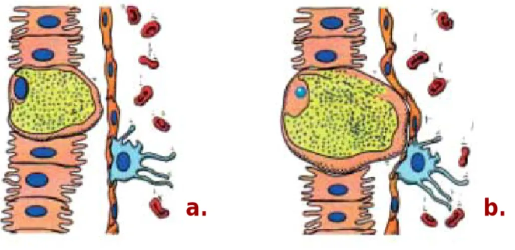
with the formation of a parasitophorous vacuole (PV) (Mota et al., 2001a; Frevert et al., 2005). After several days of development inside a hepatocyte, between 2 and 16 days depending on the Plasmodium species (spp.), 10000 to 30000 merozoites per invading sporozoite are released via budding of parasite-filled vesicles (merosomes) into the blood stream [reviewed in (Prudêncio et al., 2006)]. Each released merozoite invades an erythrocyte, again with the formation of a PV, and undergoes a replication cycle that ends with the release of new 16 to 32 merozoites from the mature infected erythrocyte (schizont), which go on to infect other erythrocytes [reviewed in (Sturm and Heussler, 2007)]. This cyclic blood stage infection occurs with a periodicity of 48 to 72 hours, depending on the Plasmodium spp.. Malaria associated symptoms only occur during the blood stage of infection. Furthermore, some merozoites develop into sexual parasite stages, the male and female gametocytes (micro- and macro-gametocytes, respectively), which can be taken up when another female mosquito feeds off an infected mammalian host. The Plasmodium life cycle continues in the mosquito midgut, where the exflagellation of microgametocytes occurs and the macrogametocytes are fertilized. The resulting ookinete migrates through the mosquito midgut into the hemocele and develops into an oocyst, where sporozoites are formed. When the oocyst is fully matured it bursts and the released sporozoites migrate into the mosquito’s salivary glands, where they become more infective and, therefore, ready for the next transmission step [reviewed in (Barillas-Mury and Kumar, 2005; Matuschewski, 2006; Vlachou et al., 2006)] (Figure 1.2).
a.
b.
c.
d.
e.
f.
g.
a.
b.
c.
d.
e.
f.
g.
Figure 1.2 | Plasmodium life cycle in the human and mosquito hosts.
(a.) The parasite's life cycle in the human host begins when sporozoites are inoculated into
the host's skin through the bite of an infected female Anopheles mosquito. The sporozoites are rapidly transported to the liver as soon as they reach the blood stream. (b.) In the liver sporozoites migrate through several hepatocytes before infection of a final host cell with the formation of a parasitophorous vacuole. (c.) The liver schizonts, named exoerythrocytic forms (EEFs), proliferate and differentiate into thousands of merozoites that will ultimately be released through merosomes into the blood stream. (d.) Each merozoite will recognize, bind to, and invade an erythrocyte, initiating a replication cycle that ends with the release of new merozoites. (e.) Some merozoites develop sexual parasite morphologies (male and female gametocytes), which can be ingested by a mosquito during a blood meal. (f.) Once in the mosquito midgut, the parasite undergoes a series of transformations, culminating in the development of new sporozoites that invade the salivary glands and (g.) consequently can be inoculated into another host .
1.4. A glimpse at the malaria clinical features and pathogenesis
Malaria infection manifestations include a wide variety of symptoms, ranging from absent or very mild symptoms to severe disease and even death. Symptoms and signs of uncomplicated malaria comprise sensation of cold, shivering, fever, headaches, vomiting, sweats and tiredness. Severe malaria manifestations include metabolic acidosis which leads to respiratory distress, severe anemia, thrombocytopenia (blood platelets decrease), organ failure, cerebral malaria (impairment of consciousness, seizures, coma, or other neurological abnormalities) and placental malaria in pregnant
women. Overall patterns of disease depend markedly on the age and the previous immunological experience of the host [reviewed in (Greenwood et al., 2005; Schofield and Grau, 2005)]. The multiplicity of severe malaria syndromes has confounded the identification of unifying mechanisms of the disease. Research studies support the idea that these several syndromes arise from the intersection of a few basic processes: rapid expansion of infected erythrocytic mass; destruction of both infected and uninfected erythrocytes; adhesion and sequestration of infected erythrocytes in the vasculature; release of bioactive parasite products in host tissues molecules; local and systemic production of cytokines and chemokines by the innate and adaptive immune systems in response; and activation, recruitment and infiltration of inflammatory cells (Table 1.1) [reviewed in (Malaguarnera and Musumeci, 2002; Clark and Cowden, 2003; Rasti et al., 2004; Hisaeda et al., 2005; Schofield and Grau, 2005; Boutlis et al., 2006)]. Over the last few years, significant progress has been made towards the identification of both parasite and host molecules that actively participate in these processes. This is particularly important because understanding the biology associated with the malaria disease is an essential key for the development of successful tools for intervention.
Table 1.1 | Severe malaria clinical features and underlying mechanisms.
Severe malaria comprises a variety of diverse syndromes that present singular and distinguishing clinical features. The comprehension of the mechanisms that lead to the development of each syndrome constitutes a leading area of malaria research. A remarkable knowledge has been achieved, however there is still a great deal to be understood [adapted from (Schofield and Grau, 2005)].
1.5. The fight against malaria: available tools
1.5.1. Anti-malarial tools: the presentCurrently, the fight against malaria is focused on mosquito eradication, reduction of human–vector contact and disease prevention and treatment using antimalarial drugs. A viable vaccine is not yet available, despite the significant efforts that have been made to develop one.
Mosquito eradication is mainly achieved through indoor residual spraying and
environmental management to eliminate breeding sites. Global control efforts from the 1950s to the 1970s, mainly through the use of the insecticide Dichloro-Diphenyl-Trichloroethane (DDT), virtually eliminated malaria transmission in the subtropics (Greenwood et al., 2005). However, the programme was abandoned due to the negative impact of the use of high concentrations of DDT, namely resistence developed by mosquitoes and toxic effects to humans (Turusov et al., 2002). A deeper reason for abandoning the campaign might have been geopolitical (Sachs, 2002). Despite attempts to ban DDT completely, the use of small amounts of DDT (allowed under the Stockholm Convention on persistent organic pollutants) still plays a major role in malaria control (Schapira, 2004; Greenwood et al., 2005). A new and alternative approach, the deployment of mosquito-killing fungi, has been recently shown as possible (Blanford et al., 2005; Scholte et al., 2005) and has been extensively discussed [see (Michalakis and Renaud, 2005; Kanzok and Jacobs-Lorena, 2006; Thomas and Read, 2007)]. Although vector control strategy is very effective in reducing malaria in some regions of Africa, it is also expensive, logistically demanding and has been undermined by problems of insecticide resistance, environmental contamination and risks to human health.
Reduction of human–vector contact is achieved through insecticide-treated bed nets
(ITNs) and is more appropriate for malaria control in Africa (Klausner and Alonso, 2004). Randomized trials of the ITNs in diverse settings have established their effectiveness at cutting malaria-related morbidity and mortality (Lengeler, 2000). Nonetheless, though ITNs are inexpensive and effective, fewer than 2% of Africans sleep under them, which means that considerable campaigns to increase their use are urgently required (Monasch et al., 2004). Furthermore, the regular ITN re-treatment with insecticide has proved difficult to sustain on a large scale. However, this might be overcome by the development of long lasting insecticidal ITNs and in fact different prototypes are being produced and two have already been approved by the WHO and are undergoing large-scale production (WHO and UNICEF, 2004).
Antimalarial drugs have been quite essential in the combat against malaria, but
parasite resistance problems are arising. The used antimalarials are quinolines (amodiaquine, piperaquine, primaquine, quinine, mefloquine and chloroquine), antifolate drugs (pyrimethamine, chloroguanide,sulfadoxine, sulfalene and dapsone), artemisinins and derivatives (artemether, arteether, artesunate and dihydroartemisinin), atoquavone, and antibiotics (such as, tetracycline, doxycycline, and clindamycin) [reviewed in (Cunha-Rodrigues et al., 2006b; Schlitzer, 2007; Vangapandu et al., 2007)]. The choice of the drug to use is usually driven by what drugs the parasites in the area are resistant to, as well as their side-effects. To counteract the rapid development of resistance some drugs are used in fixed combinations [reviewed in (Fidock et al., 2004)].
The increasingly serious problem of malaria parasite resistance to the currently used antimalarials discloses the urgent need to develop new and effective antimalarial molecules. This goal can be achieved in two ways: either by focusing on validated targets in order to generate new drug candidates or by identifying new potential targets for malaria chemotherapy [see (Jana and Paliwal, 2007)].
1.5.2. Anti-malarial tools: the future
Sadly, the available tools against malaria mentioned above provide no strategy to sustainably reduce or eliminate the burden of malarial disease. A vaccine against malaria could lead the way given that this has been the most cost-effective health intervention for a range of other infectious diseases. However, despite the extensive research that is developed in this area, there is still no vaccine available. Malaria vaccine research has focused on different approaches: an anti-infection vaccine aimed at protecting malaria-naive travelers or residents of low endemic areas from becoming infected; an anti-disease/anti-mortality vaccine aimed at children, pregnant women and migrants living in endemic areas; and an anti-mosquito-stage vaccine aimed at preventing the transmission of malaria from one person to another. Different pre-erythrocytic, blood stage, and transmission-blocking vaccines are the focus of ongoing research [reviewed in (Richie and Saul, 2002; Moorthy et al., 2004; Todryk and Hill, 2007)]. The currently most advanced malaria vaccine candidate in development is the pre-erythrocytic RTS,S/AS02A [state of the art addressed in (Alonso, 2006; Hill, 2006)]. RTS,S/AS02A comprises a hybrid molecule in which the circumsporozoite protein of P. falciparum is expressed with hepatitis B surface antigen in yeast (Stoute et
al., 1997). This is the only vaccine candidate shown in field trials to prevent malaria
and, in one instance, to limit disease severity. RTS,S/AS02A has provided substantial, short-lived protection in volunteers, exposed experimentally to bites by infected
mosquitoes (Kester et al., 2001), and substantial (71%) but only short-term protection in naturally exposed, semi-immune adults from The Gambia (Bojang et al., 2001). In a subsequent trial in Mozambican children, RTS,S/AS02A gave 30% protection against the first clinical episode of malaria and 58% protection against severe malaria (Alonso
et al., 2004).
Another approach for malaria control focuses on the development of transgenic or genetically modified genetic mosquitoes in order to convert them into inefficient parasite vectors (Moreira et al., 2002; Marrelli et al., 2006; Marrelli et al., 2007).
In addition, the availability of genome sequences, such as the ones from the three most relevant organisms to malaria, Homo sapiens (Nature, 2001; Science, 2001),
Anopheles gambiae (Holt et al., 2002) and Plasmodium falciparum (Gardner et al., 2002),
together with bioinformatics tools and high-throughput technologies (microarrays, RNA interference, proteomics) will ultimately provide an integrated picture of the parasite biology and malaria pathogenesis and hopefully facilitate the development of the existing and new approaches [see (Hoffman et al., 2002; Ghosh et al., 2003; Carucci, 2004; Johnson et al., 2004)].
1.6. A close look at the pre-erythrocytic stage
Although the clinical symptoms only appear during the erythrocytic stage of
Plasmodium’s life cycle it should not be disregarded that the asymptomatically
pre-erythrocytic stage (also referred to as liver stage) is essential for the malaria infection outcome. During this stage Plasmodium develops inside hepatocytes and there is an amazing parasite multiplication. Still, relatively little knowledge exists on
Plasmodium-hepatocytes interactions, which is due to the fact that the blood stage, being the pathogenic stage of the parasite’s life cycle, has soon attracted much more attention than the asymptomatic liver stage. Also, large and detailed studies of liver stage development are difficult because of the prerequisite of freshly extracted infectious sporozoites (breeding of infectious mosquitoes is a necessity) and to the low infection rates obtained in vitro (Prudêncio et al., 2007) and in vivo (Heussler et al.,
2006). Despite these restrictions, malaria researchers have been working towards an
understanding of the biology behind the malaria liver stage and, during this journey, huge steps have been made to disclose the processes involved in this stage.
1.6.1. Plasmodium models for liver stage research
Liver stage research of human malaria is not feasible in vivo, therefore, several studies have successfully accessed the complete development of hepatic stages of human
Plasmodium sps. in human primary hepatocytes (Mazier et al., 1984; Smith et al., 1984;
Mazier et al., 1985; Mazier et al., 1987). Still, these are technically challenging because they do not grow continuously in culture and, consequently, their availability is dependent on often scarce and unpredictable material. Thus, several studies have focused on the development of model systems using human hepatocyte cell lines (Uni
et al., 1985; Calvo-Calle et al., 1994; Karnasuta et al., 1995; Sattabongkot et al., 2006).
Initially, P. vivax and P. falciparum development in vitro was shown to be possible,
although in different cell lines. Complete liver stage development for P. vivax was shown in HepG2-A16 cells (Uni et al., 1985) while P. falciparum liver stage growth was achieved in Huh1 cells (Calvo-Calle et al., 1994) as well as HHS-102 cells (Karnasuta et
al., 1995). Only recently has a human hepatocyte cell line, HC-04, been shown to support the complete liver stage development of both P. vivax and P. falciparum (Sattabongkot et al., 2006). The main advantage of this cell line over the ones previously mentioned is the fact that it allows the development of the two most prevalent human malaria parasites with greatly improved infection rates. In addition, mouse models with humanized livers have been shown to represent a promising new tool for P. falciparum in vivo studies (Morosan et al., 2006; Sacci et al., 2006). These recent developments are extremely important because they might finally bring new avenues to human malaria liver stage research. Nevertheless, it should not be disregarded that human malaria research also requires the production and handling of human malaria parasite infectious mosquitoes, which involves very controlled conditions.
As a result of the difficulties mentioned above, liver stage malaria research progress has been achieved through the use of rodent Plasmodium spp., in particular P. berghei and P. yoelii. These parasites are established as well-suited models for Plasmodium pre-erythrocytic stage biology, immunology and vaccine development (Hafalla et al., 2006; Prudêncio et al., 2006). P. berghei and P. yoelii share more than 90% genome identity (Kooij et al., 2005). Although the P. yoelii model is generally thought to reflect human malaria better than the P. berghei model (Calvo-Calle et al., 1994; Doolan and Hoffman, 2000; Mota et al., 2001b), the latter is the most widely used because the technologies to enable its transfection were developed earlier (van Dijk et al., 1996) than for P. yoelii (Mota et al., 2001b; Jongco et al., 2006). Moreover, green or red fluorescent protein-tagged parasites have been developed first for P. berghei (Natarajan et al., 2001; Franke-Fayard et al., 2004; Frevert et al., 2005) and these have already allowed extensive in vivo studies focused in sporozoite transmission by mosquito bite (Frischknecht et al., 2004), its subsequent journey from the skin to the liver (Vanderberg and Frevert, 2004; Frevert et al., 2005; Amino et al., 2006) and liver stage
development (Sturm et al., 2006). Only recently were GFP-tagged P. yoelii parasites developed (Tarun et al., 2006; Ono et al., 2007) and used for in vivo liver stage studies (Tarun et al., 2006).
Both P. berghei and P. yoelii are able to infect primary mouse hepatocytes (Meis et al., 1984; Millet et al., 1985; Davies et al., 1989; Long et al., 1989) and these cells have often been used in liver stage research. However, since primary cells have to be freshly prepared and can only be maintained in culture for a short period of time, established cell lines have been more widely used. Cell lines, easily maintained in culture through several passages, constitute an extremely important tool that provides useful information to be subsequently tested ex vivo and/or in vivo.
The hepatoma cell lines that present a relatively high level of infectivity for P. berghei and P. yoelii and, therefore, are widely used in in vitro studies are the human hepatoma cell lines, HepG2 and Huh7 (Aikawa et al., 1984; Calvo-Calle et al., 1994) and the murine hepatoma cell line, Hepa1-6 (Mota and Rodriguez, 2000). P. berghei is known to be more promiscuous than P. yoelii, since the former is able to efficiently infect both the human and the murine hepatoma cell lines (Aikawa et al., 1984; Calvo-Calle et al., 1994; Mota and Rodriguez, 2000) whereasthe latter only infects the murine Hepa1-6 cells efficiently (Mota and Rodriguez, 2000). P. berghei sporozoite invasion
and exoerythrocytic forms (EEFs) development has also been observed in some non-hepatic cell lines, namely the human lung cell line WI38 (Hollingdale et al., 1981; Hollingdale et al., 1983) and the human epithelial cells HeLa (Calvo-Calle et al., 1994). Additionally, it has been shown that P. berghei and P. yoelii sporozoites incubated, without any host cells, at 37º C in the presence of serum develop into early EEFs (Kaiser et al., 2003; Wang et al., 2004). Although at the morphological level axenically cultured EEFs are indistinguishable from those that develop within hepatocytes, intracellular residence is essential for parasite’s further growth.
P. berghei and P. yoelii display different infection efficiencies in vitro and in vivo. There
is an inverse relationship between the two species in their in vitro and in vivo infection rates. P. yoelii infection rate is high in vivo but low in vitro, whereas P. berghei stands in the opposite situation with the highest infection rate of malaria models in vitro, both in primary hepatocytes and hepatoma cell lines, and comparatively low in vivo success in mice (Khan and Vanderberg, 1991; Briones et al., 1996; Druilhe et al., 1998). Moreover, P. berghei and P. yoelii infectivity differences in vivo not only depend on the parasite but also on the clone and the genetic background of the rodent host (Jaffe et
al., 1990; Scheller et al., 1994; Belmonte et al., 2003). All these aspects should be
considered while extrapolating results obtained with the different experimental models.
1.6.2. Introducing the Plasmodium sporozoite
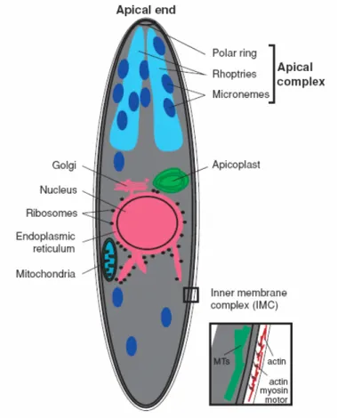
The invasive Plasmodium stage that is transmitted by mosquitoes and is responsible for initiating the infection in the vertebrate host is called sporozoite and it is distinguished by specific features. Sporozoites are small unicellular organisms, about 10 μm long and 1 μm wide. At the anterior cell pole, they possess the apical polar ring, which serves as a microtubule-organizing center, and a unique set of secretory organelles, termed micronemes and rhoptries, that belong to the apical complex. Micronemes are small vesicles of varying electron density while rhoptries are large, usually paired, pear-shaped organelles. Both these organelles discharge at the anterior pole and their contents are involved in the three basic types of tissue interaction in the mammalian host cell environment: gliding motility (a substrate-dependent form of locomotion), migration through cells by membrane rupture wounding and invasion of the host cell with the formation of a PV. The micronemes, rhoptries and also dense granules (microspheres of approximately 200 nm in diameter) are characteristic organelles of other invasive forms of apicomplexa parasites. Interestingly, dense granules have not yet been observed for the sporozoite invasive form. Another unique organelle of the sporozoite is the inner membrane complex, a flattened vesicle underneath the cell membrane that is associated with a set of subpellicular microtubules. An actin-myosin motor essential for sporozoite motility and invasion is located in the narrow space between the plasma membrane and the outer membrane of the inner membrane complex [reviewed in (Kappe et al., 2004)] (Figure 1.3). The
Plasmodium sporozoite’s features outlined above are essential for sporozoite
Figure 1.3 | The Plasmodium sporozoite.
Schematic representation of a Plasmodium sporozoite, showing some of its organelles and subcellular structures. At the anterior cell pole is positioned the apical complex which is formed by the apical polar ring and the secretory organelles, micronemes and rhoptries. Underneath the sporozoite’s cell membrane is the inner membrane complex with an actin-myosin motor and associated to subpellicular microtubules [adapted from (Kappe et al., 2004)].
1.6.3. The sporozoite journey from the skin to the liver
Malaria transmission takes place when an infected mosquito bites a mammalian host while probing for a blood source under the skin (Matsuoka et al., 2002). During the mosquito bite saliva containing sporozoites, vasodilators and anticoagulants is released (Griffiths and Gordon, 1952). Although mosquitoes can harbor thousands of sporozoites in their salivary glands, the number of sporozoites delivered in each bite rarely exceeds 200 and most estimates from experimental infections record numbers around 20 (Vanderberg, 1977; Rosenberg et al., 1990; Ponnudurai et al., 1991; Rosenberg, 1992). Recently, 2 different studies (Frischknecht et al., 2004; Medica and Sinnis, 2005) report higher numbers (sporozoite means 114 and 123, respectively). Nevertheless, considering the total number of sporozoites present in the salivary glands the number of sporozoites injected is quite low.
Sporozoites are deposited into the skin (Sidjanski and Vanderberg, 1997; Amino et al., 2006), where they migrate for at least 30 min (Vanderberg and Frevert, 2004; Amino et
al., 2006). Recently, a study using quantitative Real-Time Polimerase Chain Reaction
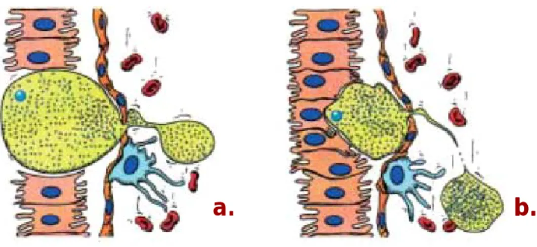
(qRT-PCR) has determined the kinetics with which P. yoelii sporozoites leave the injection site and arrive in the liver and has shown that the majority of infective sporozoites can remain in the skin for hours (Yamauchi et al., 2007). A sporozoite surface phospholipase (PL) was shown to be required for host cell membrane breaching during migration in the skin (Bhanot et al., 2005). Sporozoites migrate extensively through the avascular dermis until they reach a vessel and enter the circulatory system, which transports them into the liver. It has been proposed that sporozoites may also travel using the lymphatic system (Vaughan et al., 1999; Krettli and Dantas, 2000). Intravital microscopy observations have showed that within 1 hour after P. berghei injection a proportion of sporozoites invades blood vessels and gets carried away by the blood flow, whereas others actively enter lymph vessels or remain in the skin after exhaustion of their motility (Amino et al., 2006) (Figure 1.4).
Figure 1.4 | Plasmodium sporozoites are deposited in the skin and enter the circulatory and lymphatic systems.
Sporozoites (in green) are placed under the skin of the mammalian host while an infected female Anopheles mosquito probes for a blood meal. Sporozoites migrate in the skin until they come into contact with a blood vessel (in red) and enter the circulatory system, being then transported into the liver. A small proportion of sporozoites can enter the lymphatic system (in yellow). Adapted from the Plasmodium‘s life cycle presented on Figure 1.2.
Most of the sporozoites that enter the lymphatic vessels do not reach the circulatory system; instead, they are trapped in the lymph nodes. Some of these sporozoites
partially develop into small-sized EEFs before eventually being degraded (Amino et
al., 2006). The relevance of presence of parasites in such an important organ of the
immune system has been under investigation by Chakravaraty and Zavala. It has been observed a sporozoite-specific cytotoxic T-cell (CTL) response in the draining lymph node just 2 days after intradermal immunization with irradiated sporozoites. When the draining lymph node was surgically ablated to prevent early local priming, the number of sporozoite-specific CTLs in the liver was significantly reduced, highlighting the importance of skin-draining lymph nodes in the initiation of the immune response to sporozoites during natural infection [Chakravaraty and Zavala, unpublished data referenced in (Sinnis and Coppi, 2007)].
1.6.4. Sporozoite arrest in the liver
Once in the circulatory system, sporozoites are rapidly arrested in the liver. Sporozoites are found in hepatocytes within 2 minutes after intravenous injection into rats (Shin et al., 1982). The speed and selectivity of this process suggests specific interactions between parasite surface protein(s) and host molecule(s).
The major surface protein of Plasmodium sporozoites, the circumsporozoite protein (CSP), appears to have an essential role by interacting with the heparan sulphate proteoglycans (HSPGs) of liver cells. It has been reported that recombinant CSP binds specifically to HSPGs from the basolateral cell surface of hepatocytes in the Disse space (region that separates the sinusoidal endothelium from hepatocytes) and that this interaction occurs between the CSP’s conserved I and II-plus regions and heparin-like oligosaccharides and/or heparan sulfate (Cerami et al., 1992; Pancake et al., 1992; Frevert et al., 1993; Cerami et al., 1994; Sinnis et al., 1994; Rathore et al., 2002; Ancsin and Kisilevsky, 2004). Although HSPGs are present in most tissues, liver HSPGs are known to be more highly sulphated than those in other tissues (Lyon et al., 1994). This feature has been proposed to be responsible for the selective recognition of recombinant CSP and Plasmodium sporozoites in the liver (Ying et al., 1997; Pinzon-Ortiz et al., 2001).
Between the blood and hepatocytes there is a layer of liver endothelial cells that have open fenestrations which allow direct contact between the circulation and the space of Disse (Wisse et al., 1985). Lipoproteins, because of their small size (90 nm of diameter), are able to freely diffuse through the endothelial fenestrations and access directly the space of Disse while sporozoites cannot due to their larger size (1µm). It has been proposed that hepatocyte HSPGs may extend through endothelial fenestrations to the lumen of blood vessels where they would sequester sporozoites (Sinnis et al., 1996). More recently, it has been shown that CSP and another parasite protein,