1
© Global Society of Scientific Research and Researchers
http://asrjetsjournal.org/
Benefits of Sports for Arterial Distensibility in Youths
Filipe Fernandes
a*, Cátia Correia
b, Emanuel Nabais
c, Virginia Fonseca
d, João
Lobato
e, Gilda Cunha
f, João O’Neill
g, Valentina Vassilenko
h a,b,c,d,eScientific Area of Cardiopneumology, Department of Sciences and Technologies of Radiation and Health Biossignals, Lisbon School of Health Technology - Polytechnic Institute of Lisbon (ESTeSL-IPL), Lisbon,
Portugal
f
Scientific Area of Mathematics, Department of Natural and Exact Sciences, ESTeSL-IPL, Lisbon, Portugal
g
Department of Anatomy, NOVA Medical School, NOVA University of Lisbon, Portugal
h
Department of Physics, LIBPhys –FCT NOVA, Faculdade de Ciências e Tecnologia, NOVA University of Lisbon, Caparica, Portugal
a
Email: filipefernandes2@gmail.com
Abstract
Arterial distensibility (AD) measures the ability of relaxation for arteries in response to pressure changes. Its reduction is closely associated with an increased risk of cardiovascular disease and higher pulse wave velocity (PWV), the gold standard method for AD evaluation. Several factors can decrease AD and therefore increase arterial stiffness, such as atherosclerosis which begins in childhood. Exercise has been related to beneficial effects in blood pressure and heart rate which are associated to AD. The main objective of this study was to compare carotid-femoral PWV (PWVcf) between two populations of youths, sportsmen and non-sportsmen, in order to identify potential benefits of sports practice in AD. Additionally, systolic (SBP), diastolic (DBP) blood pressure and heart rate (HR) were also compared. 78 individuals with ages between 15 and 20 years old were divided into two samples: Sportsmen (n= 43, 79% male and 21% female) and Non-Sportsmen (n= 35, 71% male and 29% female). Sociodemographic data, cardiovascular risk factors and weekly physical load were assessed using an individual survey. PWVcf, SBP, DBP and HR were measured with validated automatic devices. Samples were homogeneous as to gender, race and body mass index. Mean PWVcf was lower in the Sportsmen
although without statistical significance (Sportsmen: 6,21 ±0,95m/s; Non-Sportsmen: 6,33 ±0,84m/s). The samples were heterogeneous as to HR (Sportsmen: 62,44 ±9,77bpm; Non-Sportsmen: 88,03 ±15,43bpm) and DBP (Sportsmen: 67,70 ±6,59mmHg; Non-Sportsmen: 78,29 ±9,35mmHg).
--- * Corresponding author.
2
The results are consistent with physiological adaptations to exercise which may be characterized by increased vagal tone and production of nitric oxide. Lower PWVcf in Sportsmen was observed and is consistent with improved AD, although non-significant between our samples, which may vary depending on each individual type and period of sports practice. A beneficial effect of sport in the Sportsmen sample was obverse, with significantly lower mean values of HR, SBP and DBP and a trend towards lower values of PWVcf, in keeping with an overall better preservation of arterial distensibility in youths.
Keywords: Arterial distensibility; Pulse wave velocity; Sportsmen; Blood pressure; Youths.
1. Introduction
Cardiovascular diseases (CVD) are the leading cause of death in contemporary society, which determines the need to understand the pathophysiological mechanisms in order to develop preventive strategies. These mechanisms which precede the development of CVD occur in the arterial wall and affect its distensibility [1–4]. Arterial distensibility (AD), or compliance, is a measure of the distension capability of arteries in response to pressure changes. Its decline demonstrates stiffening of the arteries and is closely associated with increased risk of cardiovascular disease [1,5].
Pulse wave velocity (PWV) is a measure of regional arterial stiffness of the arterial territory between the two measurement sites [6]. It is a parameter dependent on structural changes that modify the elasticity/stiffness of the arterial wall [7–12]. This parameter is related not only to the elastic modulus (E) of the arterial wall (which represents the intrinsic stiffness of the wall), but also to the arterial geometry (thickness: h and radius: r) and blood density (ρ) [6].
At the end of the 19th century, Moens and Korteweg formulated this relationship as: PWV2=E.h/2rρ. Later on, Bramwelland Hill applied the Moens- Korteweg equation to arterial physiology and described the relationship in terms of relative change in volume (ΔV/V) and pressure (ΔP) during ex vivo experiments: PWV2=ΔPV/ΔVρ.
Thus, PWV is a direct measurement of arterial stiffness since it is the square value of 1/distensibility. In this aspect, it differentiates itself from indirect methods based on the models of circulation. The assessment of PWV involves measurement of two parameters: transit time of the arterial pulse along the analyzed arterial segment and estimated distance on the skin between both recording sites [5,6].
Thus, the determination of PWV is the gold standard method and the simplest non-invasive way to assess arterial stiffness, making PWV an independent early marker of CVD and mortality [10,13]. Studies carried out by Laurent and his colleagues[5] and Mancia and his colleagues [7] have demonstrated the usefulness and effectiveness of this method in assessing arterial distensibility, associating increased values of PWV to greater arterial wall stiffness which is related to the development of cardiovascular events. The determination PWV in large elastic arteries is representative of the arterial system. The thoracic and abdominal segments of the aorta contribute mainly to the arterial damping function and by the fact that there are few branches in this region, these segments are affected by an early atherosclerotic process. Research studies on this matter conclude that at carotid-femoral territory PWV (PWVcf), values above 10m/s are at increased risk for developing CVD [5,7,14].
3
Sport is a known factor likely to delay or even prevent the changes in the arterial wall, acting as a protective factor for CVD. High levels of cardiorespiratory fitness and physical activity (PA) from moderate and vigorous intensity (brisk walking, jogging, aerobics, and other sports) activities have been shown to attenuate arterial stiffening [3,8,12,15].
Knowing that the first changes of these diseases begin in childhood, it becomes important to measure the impact of physical activity in these populations and whether this presents benefits in the prevention of CVD, since pulse transit time and blood pressure relationship is known to be altered by active changes to vascular tone, via for example sympathetic activity or a particular duration of the cardiac cycle [3,16,17].
It is therefore important to understand whether populations of youth sports groups have greater arterial distensibility then individuals, with similar socio-demographic characteristics, that do not practice sports. Thus, the main objective of this study was to compare the values of PWVcf between two populations of school aged adolescent students, being sportsmen and non-sportsmen. The secondary objectives were to compare blood pressure parameters as SBP and DBP as well as the heart rate (HR) between these population samples.
2. Methodology
A quantitative approach method was used with descriptive-comparative study typology. Participants were selected using convenience sampling method from a population of about 900 students of secondary school in Lisbon area.
43 individuals were selected from Sports class (33 males and 10 females), whose sport load was ≥420 minutes weekly for the sportsmen sample for at least the last 6 months. Similarly, 35 individuals were selected within the remaining classes for the sample of non-sportsmen, including 25 males and 10 females, whose sport load did not exceed 180 minutes weekly. All subjects were aged between 15 and 20 years old with the Sportsmen sample consisting of 95.3% white individuals and 4.7% black. In the Non-sportsmen sample, 85.7% of individuals were white, 8.6% blacks and 2.9% of other races. Smokers and subjects that had ingested any stimulants (e.g.: power drinks) two hours prior to data collection were excluded.
Prior to performing any procedure, a written informed consent was obtained from parents for underage individuals, and by the participants themselves, when aged 18+ years old. Privacy was guaranteed to the participants during the procedures as well as confidentiality of personal data by codification methods. Through a previously established authors’ protocol, all subjects were examined for height and weight and calculated the body mass index (BMI) using the equation (1):
BMI = Weight / Height2 (1)
A survey was conducted to obtain individual data on risk factors and weekly physical load. After a rest period of 5 minutes, three consecutive blood pressure (BP) and heart rate (HR) measurements were made using calibrated and validated OMRON digital automatic M6 Comfort® device, considering the arithmetic average of these measurements as the resting BP and HR values [18].
4
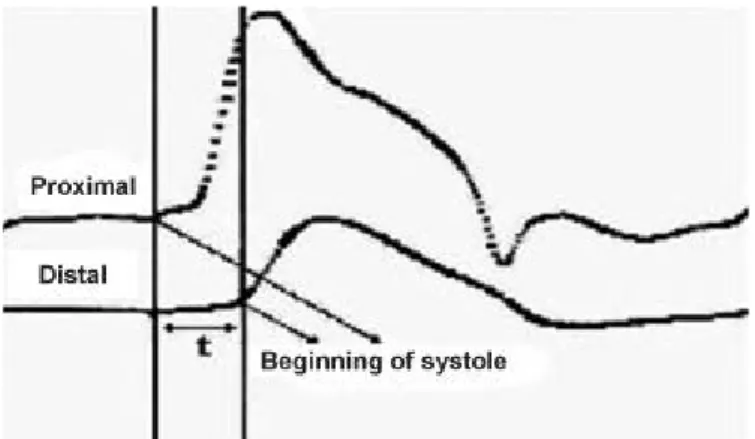
PWVcf measurements were conducted by two previously trained researchers supervised by an experienced operator. After 10 minutes of rest in supine position, PWVcf was obtained with the automatic device Complior® SP, (Artech Medical) [19,20]. In this equipment, the pressure waves of the right carotid and femoral arteries are simultaneously recorded using pressure sensitive transducers. The PWVcf is calculated by means of the foot-to-foot method (figure 1), i.e., the original deflection is determined as corresponding to systole in both simultaneous pressure waves and the transit time (t) between the two sites is calculated, having the distance been estimated by direct tape measurement [21].
Figure 1: Foot-to-foot method – calculates the transit time (t) between the beginning of the deflection corresponding to systole pressure wave in the carotid artery and the pressure wave of the femoral artery in the
same cycle.
Three PWV measurements were recorded and arithmetic mean value was calculated in post-processing. The PWV calculation is then carried out by dividing 80% of the measured distance between the two acquisition sites [14] and the transit time between the two waves at the same cardiac cycle according to the equation derived from the adapted equation (2) for velocity calculation [21]:
PWV= CF Distance / CF Transit Time (2)
The data were processed and analyzed using statistical analysis software SPSS® 15 (Statistical Package for the Social Sciences).
The characterization of the variables was carried out through descriptive analysis, using statistics of location and dispersion. t-test was performed to compare the mean values for a significance level of 5% (p ≤0.05).
3. Results
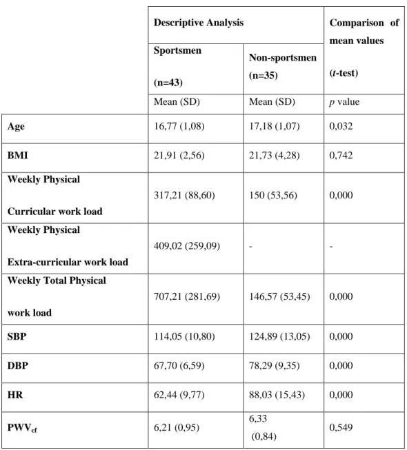
Table 1 summarizes the results of the characterization of the samples, presenting the mean values and standard deviation (SD) of the variables Age, BMI, Weekly Physical Curricular work load, Weekly physical extra-curricular work load, Weekly total physical work load, systolic and diastolic blood pressure, HR and PWVcf, as well as the results of t-tests to compare the mean values between the two samples (p value).
5
Table 1: Characterization of quantitative variables as to the mean and SD and comparison of mean values between the two samples.
Descriptive Analysis Comparison of
mean values (t-test) Sportsmen (n=43) Non-sportsmen (n=35)
Mean (SD) Mean (SD) p value
Age 16,77 (1,08) 17,18 (1,07) 0,032
BMI 21,91 (2,56) 21,73 (4,28) 0,742
Weekly Physical
Curricular work load
317,21 (88,60) 150 (53,56) 0,000
Weekly Physical
Extra-curricular work load
409,02 (259,09) - -
Weekly Total Physical
work load 707,21 (281,69) 146,57 (53,45) 0,000 SBP 114,05 (10,80) 124,89 (13,05) 0,000 DBP 67,70 (6,59) 78,29 (9,35) 0,000 HR 62,44 (9,77) 88,03 (15,43) 0,000 PWVcf 6,21 (0,95) 6,33 (0,84) 0,549
Legend: SD – Standard Deviation; BMI – Body Mass Index; SBP – Systolic Blood Pressure; DBP – Diastolic Blood Pressure; HR – Heart Rate; PWVcf – Carotid to Femoral Pulse Wave Velocity.
The samples are homogeneous as to gender, race, and BMI. Although there are significant differences in the average age, the median is equal in both samples. The differences are explained by the fact that the average values for the sample Non-Sportsmen include data from two 20 years old individuals, causing a greater average value for age in this sample. BMI values are more scattered in the sample of non-sportsmen, which does not determine significant differences between samples. According to the inclusion criteria listed, there are also significant differences in the Weekly Total Physical work load.
6
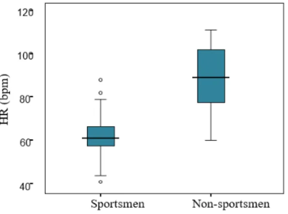
There are significant differences for the HR, systolic and diastolic BP. Regarding the HR, 25% of the sample exhibited bradycardia (HR <60 bpm) since the 25th percentile being 57 bpm (figure 2). The HR values vary between 41 and 88 bpm in the sample of Sportsmen and between 60 and 111 bpm in the sample of Non-sportsmen.
Figure 2: Comparison of percentiles of HR between the two samples.
Regarding PWVcf, there are no statistically significant differences between the two samples (p= 0.549). The maximum and minimum values ranged between 4.3 and 8.6 m/s for the Sportsmen sample and between 5.1 and 8.20 m/s for the Non-sportsmen sample.
Figure 3: Comparison of PWVcf percentiles of the two samples.
4. Discussion
The pulse wave is originated from ventricular ejection, and propagates through the arterial tree, acquiring a certain speed, which is greater at lower arterial distensibility [1,5,6]. This property of the arterial wall is dependent on factors that influence its compliance such as aging of the arteries, high blood pressure, atherosclerosis, heart rate, smoking and physical exercise [11,21,22].
The regular practice of physical exercise prevents the development of atherosclerotic processes, that begin in childhood, which are responsible for the deterioration of endothelial function and therefore the AD [8,12,15]. By
7
increasing cardiac output and vascular changes in arterial wall shear stress that occurs during exercise, there is stimulation of endothelial production of molecules with vasodilator characteristics such as nitric oxide (NO). Hence, with continuous training there is a stimulation of anti-atherosclerotic physical properties of NO, increased chronic NO synthase and plasma levels of its oxidation products (nitrate and nitrite), which are likely to contribute as beneficial effects of physical exercise in relation to CVD [2,15,23–25]. Numerous direct mechanisms have been suggested as promotors of the BP lowering effects of exercise, including neurohumeral, vascular and structural adaptations [3].
Several authors have studied the implications of the practice of sport in PWVcf, verifying that individuals who perform aerobic/resistance exercises have lower PWVcf than sedentary and those who exercise strength [2,17,25]. In the present study, there were differences in the average value of PWVcf between the two samples, but these were not statistically significant. However, there was greater dispersion of the sample values of this parameter for Sportsmen, having lower mean and minimum values, unlike what is observed in the sample of Non-sportsmen, in which values occur more homogeneous and with higher mean value. These results can be explained by the fact that individuals belonging to this young Sportsmen sample practice several kinds of sports, and the nature of these arrangements may be responsible for different adaptations of the cardiovascular system, verifying this way, differences in PWVcf values [2,8,12,15]. The differences in the time each individual spends weekly practicing sport and the years of practice of each sport can also be the basis of the results obtained. A number of cases were identified where young sportsmen began practicing sport since childhood and others only recently, considering a minimum of the last 6 months of practice.
Despite the atherosclerotic process being started in childhood, the expression of endothelial changes occurs mostly in adult life. Individuals from this study were young and all carry a minimum of weekly physical load (Non-sportsmen hold physical education classes). Thus, according to our results, all participants in the study had normal PWVcf values, only verifying a tendency towards lower values in the Sportsmen sample. Leary and his colleagues [3] referred that teen individuals have low perception of the physical exercise that they practice. In order to circumvent this limitation, the author monitored the physical load of 5505 youth with a portable accelerometer for seven days. Use of this equipment allows obtaining reliable and accurate volume and intensity of performed exercise.In our study, the physical burden was assessed weekly in minutes, from an individual inquiry, constituting a possible limitation to the study [3].
Due to continuous, frequent training, sports practitioners are submitted to physiological adaptations of the cardiac autonomic nervous system as reflected by the increased vagal tone or decreased expression of sympathetic activity. The adaptations are manifested at different levels, namely the decrease in resting HR (sinus bradycardia) [26]. In this study, 25% of the sample of Sportsmen demonstrated bradycardia, which reflects the adjustments described.
Several studies verify the existence of an inverse relationship between BP and physical activity in youth and therefore lower BP values in physically active individuals [16,27].The results obtained in our study regarding the BP are similar to those described by Leary and his colleagues observing lower mean SBP (114.05 mmHg) and DBP (67.70 mmHg) in the Sportsmen sample, compared to non-sportsmen (SBP and DBP= 153/93 mmHg).
8
Limiting factors should be consider in this study, such as the fact that the measurements were carried out by two different investigators, using the same automatic measuring device for PWV. This may constitute a limitation due to inter-observer reproducibility as not all measurements were taken by the same operator. However, several studies using the same methodology and device have shown a high intra and inter-observer reproducibility and accuracy for PWVcf measurements [5,19,20].
During data collection, the methodology and laboratory conditions were replicated to all the participants in the study. However, data over the two samples were collected at different locations, as in a cardiovascular assessment laboratory (Sportsmen) and in their school’s adapted laboratory (Non-Sportsmen), which may constitute a constraint, as per possible higher levels of anxiety for the Sportsmen sample that needed to travel outside their usual school facilities. This would possibly increase the mean values of BP and PWVcf in this group, attenuating the differences between the two samples. Additionally, the data from the Sportsmen sample were obtained from October to February and the other sample taking place between March and May. Despite the best efforts to preserve similar environmental room conditions, the exposure to colder weather temperatures experienced by the Sportsmen individuals, could possibly induces vasoconstriction mechanisms through the release of norepinephrine [28], which would result in increased BP and consequently higher values of PWVcf. The reverse applies when there is exposure to heat, with the release of nitric oxide that causes vasodilation [24]. The measured values of PWVcf in the two samples could have been influenced by these environmental and seasonal factors, which are often difficult to control in prospective studies.
We recommend the use of an accelerometer as a complementary method to assess exercise load in order to determine more reliable and accurate measurement of this variable for further similar studies [3]. Additionally, other investigations in youths are suggested using parameters in association with the BMI, such as the abdominal perimeter measurement and determining the percentage of body fat mass, intimately associated with increased risk of cardiovascular diseases and raised PWVcf related to adipocyte volume and obesity [7,29].
5. Conclusion
Although the difference between the mean values of PWVcf was not statistically significant, a trend towards lower values in the Sportsmen sample was identified, demonstrating the beneficial effects of sport since adolescence in this population, by preservation of optimal arterial distensibility. There were also significant differences between the Sportsmen and Non-Sportsmen sample concerning the variables HR, SBP and DBP. The Sportsmen had lower average values for all these variables, reflecting physiological adaptations inherent to higher physical activity. Regarding our results in this particular population, we determined that exercise shows beneficial effects in physically active young individuals, appearing to perform as a protective factor against cardiovascular diseases, in keeping with an overall better preservation of arterial distensibility in youths.
References
[1] T. Pereira, “Princípios e utilidade da velocidade da onda de pulso na avaliação da árvore arterial,” Cardiopulmonar, vol. 1, pp. 41–47, 2007.
9
[2] J. Maldonado, T. Pereira, J. Polónia, and L. Martins, “Modulation of arterial stiffness with intensive competitive training,” Rev. Port. Cardiol., vol. 25, no. 7–8, pp. 709–714, 2006.
[3] S. D. Leary, A. R. Ness, G. D. Smith, C. Mattocks, K. Deere, S. N. Blair, and C. Riddoch, “Physical activity and blood pressure in childhood: Findings from a population-based study,” Hypertension, vol. 51, no. 1, pp. 92–98, 2008.
[4] M. da Saúde, Plano Nacional de Saúde 2004-2010: Vol. II - Orientações estratégicas. Portugal: Direcção Geral da Saúde, 2004.
[5] S. Laurent, J. Cockcroft, L. Van Bortel, P. Boutouyrie, C. Giannattasio, D. Hayoz, et al., “Expert consensus document on arterial stiffness: methodological issues and clinical applications.,” Eur. Heart J., vol. 27, no. 21, pp. 2588–605, Nov. 2006.
[6] P. Boutouyrie, M. Briet, C. Collin, S. Vermeersch, and B. Pannier, “Assessment of pulse wave velocity,” Artery Res., vol. 3, no. 1, pp. 3–8, Feb. 2009.
[7] G. Mancia, R. Fagard, K. Narkiewicz, J. Redon, A. Zanchetti, M. Böhm, et al., “2013 ESH/ESC guidelines for the management of arterial hypertension: the Task Force for the Management of Arterial Hypertension of the European Society of Hypertension (ESH) and of the European Society of Cardiology (ESC).,” Eur. Heart J., vol. 34, no. 28, pp. 2159–219, Jul. 2013.
[8] K. D. Currie, S. G. Thomas, and J. M. Goodman, “Effects of short-term endurance exercise training on vascular function in young males,” Eur. J. Appl. Physiol., vol. 107, no. 2, pp. 211–218, 2009.
[9] P. Boutouyrie and S. J. Vermeersch, “Determinants of pulse wave velocity in healthy people and in the presence of cardiovascular risk factors: Establishing normal and reference values,” Eur. Heart J., vol. 31, no. 19, pp. 2338–2350, 2010.
[10] M. W. Rajzer, W. Wojciechowska, M. Klocek, I. Palka, M. Brzozowska-Kiszka, and K. Kawecka-Jaszcz, “Comparison of aortic pulse wave velocity measured by three techniques: Complior, SphygmoCor and Arteriograph.,” J. Hypertens., vol. 26, no. 10, pp. 2001–7, Oct. 2008.
[11] P. Lantelme, C. Mestre, M. Lievre, A. Gressard, and H. Milon, “Heart rate: An important confounder of pulse wave velocity assessment,” Hypertension, vol. 39, no. 6, pp. 1083–1087, 2002.
[12] T. Otsuki, S. Maeda, M. Iemitsu, Y. Saito, Y. Tanimura, R. Ajisaka, and T. Miyauchi, “Relationship Between Arterial Stiffness and Athletic Training Programs in Young Adult Men,” Am. J. Hypertens., vol. 20, no. 9, pp. 967–973, 2007.
[13] P. Boutouyrie, A. Achouba, P. Trunet, and S. Laurent, “Amlodipine-valsartan combination decreases central systolic blood pressure more effectively than the amlodipine-atenolol combination: the
10
EXPLOR study.,” Hypertension, vol. 55, no. 6, pp. 1314–22, Jun. 2010.
[14] L. M. Van Bortel, S. Laurent, P. Boutouyrie, P. Chowienczyk, J. K. Cruickshank, T. De Backer, J., et al., “Expert consensus document on the measurement of aortic stiffness in daily practice using carotid-femoral pulse wave velocity.,” J. Hypertens., vol. 30, no. 3, pp. 445–8, Mar. 2012.
[15] T. Okamoto, M. Masuhara, and K. Ikuta, “Effect of low-intensity resistance training on arterial function,” Eur. J. Appl. Physiol., vol. 111, no. 5, pp. 743–748, May 2011.
[16] T. F. HK So, RYT Sung, AM Li, KC Choi, EAS Nelson, J Yin, PC Ng, “Higher exercise frequency associated with lower blood pressure in Hong Kong adolescents: a population-based study.,” J. Hum. Hypertens., vol. 24, no. 10, pp. 646–651, 2010.
[17] A. E. Mark and I. Janssen, “Dose-response relation between physical activity and Blood pressure in youth,” Med. Sci. Sports Exerc., vol. 40, no. 6, pp. 1007–1012, 2008.
[18] J. a Topouchian, M. a El Assaad, L. V Orobinskaia, R. N. El Feghali, and R. G. Asmar, “Validation of two automatic devices for self-measurement of blood pressure according to the International Protocol of the European Society of Hypertension: the Omron M6 (HEM-7001-E) and the Omron R7 (HEM 637-IT).,” Blood Press. Monit., vol. 11, no. 3, pp. 165–171, 2006.
[19] I. B. Wilkinson, S. a Fuchs, I. M. Jansen, J. C. Spratt, G. D. Murray, J. R. Cockcroft, and D. J. Webb, “Reproducibility of pulse wave velocity and augmentation index measured by pulse wave analysis.,” J. Hypertens., vol. 16, no. 12 Pt 2, pp. 2079–84, 1998.
[20] C. J. Huck, U. G. Bronas, E. B. Williamson, C. C. Draheim, D. A. Duprez, and D. R. Dengel, “Noninvasive measurements of arterial stiffness: Repeatability and interrelationships with endothelial function and arterial morphology measures,” Vasc. Health Risk Manag., vol. 3, no. 3, pp. 343–349, 2007.
[21] E. D. Lehmann, K. D. Hopkins, and R. G. Gosling, “Assessment of arterial distensibility by automatic pulse wave velocity measurement.,” Hypertension, vol. 27, no. 5. pp. 1188–1191, 1996.
[22] L. M. Van Bortel, D. Duprez, M. J. Starmans-kool, M. E. Safar, C. Giannattasio, J. Cockcroft, et al., “Clinical Applications of Arterial Stiffness, Task Force III: Recommendations for User Procedures Luc,” Am J Hypertens, vol. 7061, no. 1, pp. 445–452, 2002.
[23] J. F. Arnal, a T. Dinh-Xuan, M. Pueyo, B. Darblade, and J. Rami, “Endothelium-derived nitric oxide and vascular physiology and pathology.,” Cell. Mol. Life Sci., vol. 55, no. 8–9, pp. 1078–87, 1999.
[24] D. L. Kellogg, J. L. Zhao, and Y. Wu, “Roles of nitric oxide synthase isoforms in cutaneous vasodilation induced by local warming of the skin and whole body heat stress in humans.,” J. Appl.
11 Physiol., vol. 107, no. 5, pp. 1438–1444, 2009.
[25] M. J. Joyner, “Effect of exercise on arterial compliance,” Circulation, vol. 102, no. 11, pp. 1214–1215, 2000.
[26] D. Corrado, A. Pelliccia, H. Heidbuchel, S. Sharma, M. Link, C. Basso, et al., “Recommendations for interpretation of 12-lead electrocardiogram in the athlete,” Rev. Port. Cardiol., vol. 28, no. 12, pp. 1505–1506, 2009.
[27] L. B. Andersen, M. Harro, L. Sardinha, K. Froberg, U. Ekelund, S. Brage, and S. Anderssen, “Physical activity and clustered cardiovascular risk in children: a cross-sectional study (The European Youth Heart Study),” Lancet, vol. 368, no. 9532, pp. 299–304, 2006.
[28] C. S. Thompson-Torgerson, L. A. Holowatz, N. A. Flavahan, and W. Larry Kenney, “Rho kinase-mediated local cold-induced cutaneous vasoconstriction is augmented in aged human skin,” AJP Hear. Circ. Physiol., vol. 293, no. 1, pp. 30–36, 2007.
[29] P. Arner, J. Bäckdahl, P. Hemmingsson, P. Stenvinkel, D. Eriksson-Hogling, E. Näslund, et al., “Regional variations in the relationship between arterial stiffness and adipocyte volume or number in obese subjects,” Int. J. Obes., vol. 39, no. 2, pp. 222–227, Feb. 2015.