Cilia at the node of mouse embryos sense fluid flow for left-right
determination via Pkd2
Satoko Yoshiba1, Hidetaka Shiratori1, Ivana Y. Kuo2, Aiko Kawasumi1,*, Kyosuke
Shinohara1, Shigenori Nonaka3, Yasuko Asai1, Genta Sasaki1, Jose Antonio Belo4, Hiroshi Sasaki5, Junichi Nakai6, Bernd Dworniczak7, Barbara E. Ehrlich2, Petra Pennekamp7,8, and Hiroshi Hamada1
1Developmental Genetics Group, Graduate School of Frontier Biosciences, Osaka University, and
CREST, Japan Science and Technology Corporation (JST), 1–3 Yamada-oka, Suita, 565-0871, Osaka, Japan
2Department of Pharmacology, Yale University School of Medicine, New Haven, CT 06510, USA 3Laboratory for Spatiotemporal Regulations, National Institute for Basic Biology, Nishigonaka 38,
Myodaiji, Okazaki, 444-8585, Aichi, Japan
4Regenerative Medicine Program, Departamento de Ciências Biomédicas e Medicina, and
IBB-Institute for Biotechnology and Bioengineering, Universidade do Algarve, Campus de Gambelas, 8005-135 Faro, Portugal
5Laboratory for Embryonic Induction, RIKEN Center for Developmental Biology, Kobe, 650-0047,
Japan
6Saitama University Brain Science Institute, 255 Shimo-Okubo, Sakura-ku, Saitama 338-8570,
Japan
7Universitätsklinikum Münster, Institut für Humangenetik, Vesaliusweg 12–14, 48149, Münster,
Germany
8Universitaetsklinikum Muenster, Klinik für Kinder- und Jugendmedizin, Allgemeine Pädiatrie,
Albert-Schweitzer-Campus 1, 48149 Muenster, Germany
Abstract
Unidirectional fluid flow plays an essential role in the breaking of left-right (L-R) symmetry in mouse embryos, but it has remained unclear how the flow is sensed by the embryo. We report that
the Ca2+ channel Pkd2 is required specifically in the peri-nodal crown cells for sensing the nodal
flow. Examination of mutant forms of Pkd2 shows that the ciliary localization of Pkd2 is essential for correct L-R patterning. Whereas Kif3a mutant embryos, which lack all cilia, failed to respond to an artificial flow, restoration of primary cilia in crown cells rescued the response to the flow. Our results thus suggest that nodal flow is sensed in a manner dependent on Pkd2 by the cilia of crown cells located at the edge of the node.
(Please start the introduction with a more general sentence about LR asymmetry to draw in the broad reader —which organs are asymmetric, etc.— and define the ventral node for the nonspecialist.) Most of the visceral organs in vertebrates exhibit left-right (L-R) asymmetry
Correspondence and requests for materials should be addressed to P.P. ([email protected]) or H.H. ([email protected]).
NIH Public Access
Author Manuscript
Science
. Author manuscript; available in PMC 2013 July 15.Published in final edited form as:
Science. 2012 October 12; 338(6104): 226–231. doi:10.1126/science.1222538.
NIH-PA Author Manuscript
NIH-PA Author Manuscript
in their shape or position. The breaking of L-R symmetry in the embryos of many vertebrates is mediated by a unidirectional fluid flow in the ventral node (an embryonic cavity at the midline filled with extra-embryonic fluid) or its equivalent structure (1, 2). The flow may transport a determinant molecule or provide mechanical force. However, it has remained unclear how the flow is sensed. We have now addressed this issue with the use of
mice lacking Polycystin-2 (Pkd2, also known as TRPP2), a gene encoding a Ca2+-permeable
cation channel implicated in polycystic kidney disease in humans(3, 4).
Pkd2 is required only in peri-nodal crown cells
The Pkd2−/− mouse was previously shown to lose Nodal expression in the lateral plate
mesoderm (LPM) and to exhibit typical L-R patterning defects(5). Pkd2 is expressed ubiquitously at the early somite stage of mouse embryos(5) (fig. S1); however, it has been unclear whether the function of Pkd2 in L-R patterning is executed in the node, LPM, or
other region of the embryo. We first examined if the LPM of the Pkd2−/− embryo is
competent for Nodal signaling. When an expression vector for Nodal was introduced to a
small region of the right LPM of the Pkd2−/− embryo, expression of endogenous Nodal gene
was induced in the entire region of the right LPM (fig. S2), suggesting that the LPM of the
Pkd2−/− embryo remains competent to express Nodal. To further localize the site of Pkd2
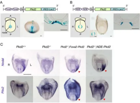
action, we established three types of transgenic mice that express Pkd2 specifically in the node and examined whether such transgenes were able to prevent the L-R defects of the
Pkd2−/− mouse (Fig. 1; fig. S3). In one transgene, Foxa2NNE-hsp-Pkd2-IRES-LacZ (Fig.
1A), Pkd2 expression is driven by a node/notochord-specific enhancer of the mouse Foxa2 gene. This transgene is expressed in the midline including the node (Fig. 1A), and it restored
left-sided expression of Nodal and Pitx2 in the LPM of Pkd2−/− embryos (rescued/tested
embryos: 6/6 for Nodal and 6/6 for Pitx2) (Fig. 1C).
The ventral node contains two different types of ciliated cells: pit cells located in the central region of the node and crown cells located at the edge (fig. S3A)(1). The second Pkd2 transgene studied, NDE-hsp-Pkd2-IRES-LacZ (Fig. 1B), is driven by the node-specific enhancer (NDE) of Nodal, which is active specifically in the crown cells. This transgene
was able to rescue left-sided expression of Nodal and Pitx2 in the LPM of Pkd2−/− embryos
(6/6 and 3/3, respectively) (Fig. 1C). Finally, expression of Pkd2 specifically in the pit cells with dgFoxa2-hsp-Pkd2-IRES-LacZ (fig. S3B) was unable to rescue the left-sided
expression of Nodal and Pitx2 (0/1 and 0/3 embryos, respectively) (fig. S3C). These results thus suggested that Pkd2 is required exclusively in crown cells of the node for correct L-R determination.
Cerl2 is the major target of Pkd2-mediated signaling
Initial breaking of L-R symmetry by leftward nodal flow is followed by asymmetric (R>L) expression of Cerl2 in crown cells(6, 7). Given that Pkd2 is required specifically in crown
cells, we examined crown cell–specific markers in Pkd2−/− embryos. Gdf1, which is
bilaterally coexpressed in crown cells and is required for subsequent Nodal expression in left
LPM(8, 9), was normally expressed in Pkd2−/− embryos (fig. S4A). However, L-R
asymmetric gene expression in crown cells was impaired in the mutant embryos. Expression of Cerl2 in crown cells is greater on the right side in wild-type embryos (fig. S4A)(6), but it
was expressed equally on the two sides in Pkd2−/− embryos (fig. S4A). Nodal expression,
which shows a subtle asymmetry in the wild-type embryo (right expression lower than left)
(10), was bilaterally equal in Pkd2−/− embryos (fig. S4A). These results suggested that
crown cells are correctly specified but lose asymmetric gene expression in the absence of Pkd2.
NIH-PA Author Manuscript
NIH-PA Author Manuscript
The higher expression of Cerl2 on the right in crown cells is required for subsequent asymmetric expression of Nodal in LPM(6). To clarify the relation between Pkd2 and Cerl2,
we examined possible interaction between the two genes by analyzing Pkd2−/−;Cerl2−/−
double-mutant embryos (fig. S4B). Nodal expression in LPM was always absent in Pkd2−/−
embryos (5/5), whereas it was either left-sided (2/11), right-sided (3/11), bilateral (2/11), or
absent (4/11) in Pkd2−/−;Cerl2−/− embryos (fig. S4B, C). The Nodal expression pattern in
the double mutant was thus similar to that in Cerl2−/− embryos (fig. S4B, C), indicating that
Cerl2 is the major target of Pkd2-mediated signaling for L-R asymmetric patterning (fig. S4D).
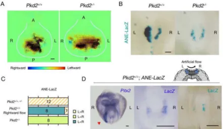
Pkd2 in crown cells is required for sensing nodal flow
Particle image velocimetry (PIV) revealed that nodal flow is maintained in the Pkd2−/−
mutant (Fig. 2A), suggesting that Pkd2 might serve to sense a flow-derived signal(s) in crown cells. We tested this possibility with the use of a transcriptional enhancer that responds to nodal flow. This enhancer (ANE) is derived from the human Lefty1 gene and exhibits asymmetric (L>R) activity in crown cells (Fig. 2B)(11). Furthermore, ANE was found to respond to artificial flow. The relative L-R activity of ANE was thus reversed when
a rightward flow was imposed on the wild-type embryo (Fig. 2C, D). In Pkd2−/− embryos,
ANE activity was R = L (Fig. 2B), despite the fact that the mutant possesses
morphologically normal cilia (fig. S5) and functional flow (Fig. 2A), suggesting that crown
cells of Pkd2−/− embryos fail to sense the flow.
Given that Pkd2 encodes a Ca2+ permeable ion channel, we examined Ca2+ signaling in
crown cells by generating a transgenic mouse that expresses Ca2+ indicator GCaMP2, which
exhibits highly cooperative Ca2+ binding over the physiological range of cellular free Ca2+
(KD= 146 nM)(12), specifically in crown cells. Indeed Ca2+ signaling was detected in
crown cells, but it was present bilaterally and was retained in Pkd2−/− embryos (fig. S6). It
should be noted that Ca2+ asymmetry previously observed with exogenous dyes (13, 14)
occurs in the endoderm, a different tissue around the node. To clarify the role of Ca2+
signaling, transgenic embryos harboring ANE-lacZ were incubated with various Ca2+
signaling blockers. GdCl3(an inhibitor of stretch-sensitive TRP channels), 2-ABP (an
inhibitor of IP3 receptor) and Thapsigargin (an inhibitor of Ca2+-ATPase in ER) disrupted
L>R asymmetry of ANE activity, whereas Ruthenium Red (a potent inhibitor of intracellular
Ca2+release by ryanodine receptors) did not (Fig. 3). Treatment with Thapsigargin did not
significantly reduce the level of Ca2+ signaling within crown cells (fig. S7), likely because a
single inhibitor would only affect a portion of Ca2+ signaling within a cell. Rotational
movement of pit cell cilia and ciliary localization of Pkd2 were maintained by treatment
with GdCl3and Thapsigargin (fig. S8). These results suggest that Ca2+ signaling mediated by
Pkd2 and IP3 receptor is essential for generating L-R asymmetry at the node (Fig. 3B).
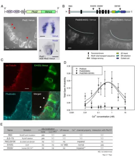
Ciliary localization of Pkd2 is essential for correct L-R decision
Pkd2 protein resides in the primary cilia of renal epithelial cells(15). In crown cells as well as pit cells of the node, endogenous Pkd2 was also localized in the cilia (fig. S9A; (13)). We also examined the subcellular localization of Pkd2 with the use of a transgenic mouse that expresses a Pkd2::Venus fusion protein in crown cells (Fig. 4A). This fusion protein was shown to be functional by the observation that its expression in crown cells corrected the
L-R defects of Pkd2−/− embryos, and was preferentially localized to the cilia of crown cells
(Fig. 4A). Expression of Pkd2::Venus protein did not influence motility of cilia (movie S1). Notably, live imaging of Pkd2::Venus-labeled cilia at the presomite stage showed that most of the cilia were immotile (fig. S10; movie S2). The frequency of motile cilia gradually increased between the presomite stage and the three-somite stage (fig. S10). However, most
NIH-PA Author Manuscript
NIH-PA Author Manuscript
(90%~) crown cell cilia were immotile while ~60% of pit cell cilia are already motile at the presomite stage (fig. S10), the stage at which Cerl2 expression in crown cells begins to exhibit L-R asymmetry (11).
Ciliary localization of Pkd2 may depend on its interaction with Pkd1l1, another ciliary protein whose deficiency in mice and zebrafish results in L-R defects similar to those of the
Pkd2−/− mutant(16, 17). To test if Pkd2 may function in cilia of crown cells, various mutant
forms of Pkd2 were examined for their abilities to localize to crown cell cilia and to correct
the L-R defects of Pkd2−/− embryos (Fig. 4B-E; fig. S9; fig. S11). Although a missense
mutation of Pkd2 (E442G) induces L-R defects identical to those of Pkd1l1 mutant mouse(18), a Pkd2(E442G)::Venus fusion protein was unable to localize to the cilia of crown cells (Fig. 4B, C) and cilia of cultured LLC-PK1 cells (fig. S12). Pkd2(E442G) can interact with Pkd1l1(fig. S13), suggesting that ciliary localization of Pkd2 requires Pkd1l1-independent mechanisms. Pkd2(R6G), a missense mutant unable to localize to primary cilia in cultured LLC-PK1 cells(19), was found to localize to the cilia of crown cells (fig. S9B)
and rescued L-R patterning in the Pkd2−/− mutant (fig. S11). Pkd2( 5–73), which lacks the
NH2-terminal region (residues 5 to 73) including the R6VxP motif(19), was also able to
localize to the cilium when expressed in crown cells (fig. S9C). Pkd2(R6G-G819X), which harbors the R6G missense mutation and lacks the COOH-terminal region, was unable to
localize to the crown cell cilium (fig. S9D) or to correct the L-R defects of the Pkd2−/−
mutant (fig. S11). Similarly, Pkd2(D509V), a missense mutant associated with polycystic kidney disease in humans(20), failed both to localize to cilia of crown cells (Fig. 4B) and to
normalize the Pkd2−/− phenotype (fig. S11). Two mutants that were unable to localize to
crown cell cilia (E442G and R6G-G819X) were examined for their channel activity (Fig. 4D). Although Pkd2(R6G-G819X) lost channel activity, Pkd2(E442G) retained it (Fig. 4D). Our findings that Pkd2(E442G) retains channel activity yet is unable to localize to crown cell cilia (Fig. 4B, C) indicate that ciliary localization of Pkd2 in crown cell is essential for correct L-R decision.
Crown cell cilia function as sensors of nodal flow
Finally, we examined directly whether cilia of the crown cells function as sensors of nodal flow with the use of an IFT (intraflagellar transport) mouse mutant. Kif3a and Kif3b are expressed ubiquitously in mouse embryo and encode motor proteins that are required for
formation of cilia(21–23). Kif3a−/− mutant embryos thus lack all node cilia, including those
of both pit and crown cells and exhibit L-R defects(22, 23). A transgene that confers Kif3a expression specifically in crown cells (NDE-hsp-Kif3a-IRES-LacZ) restored cilia formation exclusively in these cells (Fig. 5A). Although Nodal expression in LPM was bilateral (L=R)
in Kif3a−/− embryos (8/8), restoration of cilia formation in crown cells by the NDE-Kif3a
transgene rescued left-sided Nodal expression in LPM in Kif3a−/− embryos (3/3) (Fig. 5B,
C). PIV analysis revealed a weak leftward flow as well as vortical flow in the node of such embryos (fig. S14), likely because the number of motile cilia among crown cells is much smaller than that of immotile cilia (fig. S10). This result is consistent with our recent finding that as few as two rotating cilia are sufficient for symmetry breaking(24).
We then examined whether the embryos with cilia only in the crown cells are able to
respond to artificial fluid flow. The Kif3a−/−;NDE-Kif3a embryos, as well as control
Kif3a−/− embryos, were thus subjected to rightward artificial flow (Fig. 5B), and L-R
patterning as revealed by Pitx2 expression in LPM was examined. Whereas Kif3a−/−
embryos (5/5) failed to respond to the rightward flow, the Kif3a−/−;NDE-Kif3a embryos
(3/3) did respond by showing right-sided Pitx2 expression in LPM (Fig. 5B, C). These results thus demonstrated that nodal flow is indeed sensed by crown cell cilia located at the edge of the node.
NIH-PA Author Manuscript
NIH-PA Author Manuscript
Our results indicate that the fluid flow in the node is sensed by cilia of peri-nodal crown cells via ciliary-localized Pkd2. Many proteins are known to be localized to primary cilia, and they are implicated in cilium-mediated signaling. However, the role of their ciliary localization has not been rigorously established. Identification of Pkd2(E442G), a L-R
defect-causing mutant form of Pkd2 that retains Ca2+ channel activity yet is unable to
localize to the cilia, provides the direct evidence showing that ciliary localization of a protein is indeed essential for cilium-mediated signaling.
Several questions remain unanswered. It is still not clear what the cilia sense in the breaking of L-R symmetry: Do they sense flow-transported chemicals or flow-generated mechanical forces? Pkd2 is localized to the primary cilium of kidney cells and vascular endothelial cells and mediates mechanosensation(25, 26). Circumstantial evidence, including our recent observation that as few as two rotating cilia are sufficient for L-R symmetry breaking(24), favors the possibility that crown cell cilia sense mechanical force. Also unknown is how cilia signal to Cerl2, the major target of the signal transmitted by nodal flow and Pkd2. Finally, our results support the previous proposal (13, 27) that there are two populations of cilia in the node, motile and immotile cilia, and that the former generate nodal flow whereas the latter sense the flow. Given that most cilia of crown cells are immotile at the time when L-R symmetry is broken, it is likely that the flow is sensed by immotile cilia, although we are not able to rigorously exclude the possibility that motile cilia of crown cells also sense the flow. Further studies with diverse approaches are needed to address these issues.
Supplementary Material
Refer to Web version on PubMed Central for supplementary material.
Acknowledgments
We thank Y. Cai for YCB9 antibodies to Pkd2; S. Somlo for very helpful comments on Pkd2 function; D.P. Norris for Pkd1l1-CC-GFP plasmid and A. Fukumoto, Y. Ikawa, K. Miyama, K. Mochida, H. Nishimura, and S. Ohishi for technical assistance. This work was supported by a grant from CREST of the Japan Science and Technology Corporation to H.H., an NIH grant P30 DK090744 to B.E.E. and HFSP grant ST00246/2003C and the Deutsche Forschungsgemeinschaft grant PE 853/2 to P.P. S.Y. was supported by a fellowship from the Japan Society for the Promotion of Science for Japanese Junior Scientists. I.Y.K. was supported by the American Heart Association Postdoctoral Fellowship R10682.
References and Notes
1. Shiratori H, Hamada H. Development. 2006; 133:2095. [PubMed: 16672339]
2. Hirokawa N, Tanaka Y, Okada Y. Cold Spring Harb Perspect Biol. 2009; 1:a000802. [PubMed: 20066075]
3. Gonzalez-Perrett S, et al. Proc Natl Acad Sci U S A. 2001; 98:1182. [PubMed: 11252306] 4. Koulen P, et al. Nat Cell Biol. 2002; 4:191. [PubMed: 11854751]
5. Pennekamp P, et al. Curr Biol. 2002; 12:938. [PubMed: 12062060] 6. Marques S, et al. Genes Dev. 2004; 18:2342. [PubMed: 15466485] 7. Schweickert A, et al. Curr Biol. Apr 27.20:738. [PubMed: 20381352]
8. Tanaka C, Sakuma R, Nakamura T, Hamada H, Saijoh Y. Genes Dev. 2007; 21:3272. [PubMed: 18079174]
9. Rankin CT, Bunton T, Lawler AM, Lee SJ. Nat Genet. 2000; 24:262. [PubMed: 10700179] 10. Collignon J, Varlet I, Robertson EJ. Nature. 1996; 381:155. [PubMed: 8610012]
11. Kawasumi A, et al. Dev Biol. 2012; 353:321. [PubMed: 21419113]
12. Tallini YN, et al. Proc Natl Acad Sci U S A. 2006; 103:4753. [PubMed: 16537386]
13. McGrath J, Somlo S, Makova S, Tian X, Brueckner M. Cell. 2003; 114:61. [PubMed: 12859898] 14. Tanaka Y, Okada Y, Hirokawa N. Nature. 2005; 435:172. [PubMed: 15889083]
NIH-PA Author Manuscript
NIH-PA Author Manuscript
15. Pazour GJ, San Agustin JT, Follit JA, Rosenbaum JL, Witman GB. Curr Biol. 2002; 12:R378. [PubMed: 12062067]
16. Field S, et al. Development. 2011; 138:1131. [PubMed: 21307093] 17. Kamura K, et al. Development. 2011; 138:1121. [PubMed: 21307098] 18. Ermakov A, et al. Dev Dyn. 2009; 238:581. [PubMed: 19235720] 19. Geng L, et al. J Cell Sci. 2006; 119:1383. [PubMed: 16537653]
20. Reynolds DM, et al. J Am Soc Nephrol. 1999; 10:2342. [PubMed: 10541293] 21. Nonaka S, et al. Cell. 1998; 95:829. [PubMed: 9865700]
22. Takeda S, et al. J Cell Biol. 1999; 145:825. [PubMed: 10330409]
23. Marszalek JR, Ruiz-Lozano P, Roberts E, Chien KR, Goldstein LS. Proc Natl Acad Sci U S A. 1999; 96:5043. [PubMed: 10220415]
24. Shinohara K, et al. Nat Commun. 2012; 3:622. [PubMed: 22233632] 25. Nauli SM, et al. Nat Genet. 2003; 33:129. [PubMed: 12514735] 26. AbouAlaiwi WA, et al. Circ Res. 2009; 104:860. [PubMed: 19265036] 27. Tabin CJ, Vogan KJ. Genes Dev. 2003; 17:1. [PubMed: 12514094]
NIH-PA Author Manuscript
NIH-PA Author Manuscript
Figure 1. Specific requirement for Pkd2 in crown cells of the node for correct L-R patterning
(A, B) Schematic representation of two types of node-specific Pkd2 transgeneare shown on the top. Their expression patterns were examined by staining of transgenic embryos at embryonic day (E) 8.0 with X-gal. Schematic (left panels) and actual (middle panels) views of whole embryos, and transverse sections of the node (right panels) are shown. Inset of the middle panel in B shows node from ventral view at higher magnification. Note that
expression of the NDE-driven transgene is highly specific to crown cells of the node (B). A,
anterior; P, posterior; L, left; R, right. Scale bars, 50μm. (C) Whole-mount in situ
hybridization analysis of the expression of Nodal and Pitx2 in E8.0 embryos of the indicated
genotypes. Left-sided expression of Nodal and Pitx2 in LPM is lost in Pkd2−/− embryos but
is restored (red arrowheads) in Pkd2−/−;Foxa2-Pkd2 and Pkd2−/−;NDE-Pkd2 embryos. Scale
bar, 500μm.
NIH-PA Author Manuscript
NIH-PA Author Manuscript
Figure 2. Pkd2 is necessary for sensing of nodal flow
(A) Nodal flows in wild-type and Pkd2−/− embryos were examined by PIV analysis at E8.0.
Flow is maintained in the Pkd2−/− embryo. Each arrowhead denotes the direction and speed
of the flow at the indicated position. The color scale indicates the direction and magnitude of the flow velocity (leftward in yellow and red, rightward in blue). White lines indicate the
outlines of the node. Scale bar, 10μm. (B) E8.0 Pkd2+/+ and Pkd2−/− embryos harboring the
ANE-LacZ transgene were stained with X-gal. Scale bar, 50μm. (C) The L-R patterns of
ANE activities in embryos of the indicated genotypes either in situ or under conditions of artificial rightward flow in vitro are summarized. The numbers of embryos showing each pattern are indicated. (Adjust the color/pattern for C. The small box definitions of colors will not show the pattern of top line after figure reduction. ---response: color/patterns have been
changed and it is easy to see the difference in color/pattern) (D) Pkd2+/+ embryos with
ANE-LacZ were cultured under the influence of a rightward artificial flow from early headfold to six-somite stages. Artificial rightward flow reversed the pattern of Pitx2 expression in LPM (left panel, red arrowhead) and that of ANE activity in crown cells, as detected by in situ hybridization of LacZ mRNA (middle) and X-gal staining (right). Scale
bars, 100μm. Why does the “L” and “R” designation switch from left and right between the
figure panels? Figure 1 shows “L” on the right side of figure; Figure 2 shows “L” and “R” in different panel sides in the figure. ---Response: embryos showing gene expression in the lateral plate such as ones in Fig. 1C are frontal views, so that “L ” is shown on the right side. Embryos showing gene expression in the node such as ones in Fig. 2B were back views, so that “L ” was shown on the left side. To avoid confusion, however, all embryo photos are shown in such a way that the right side of an embryo is always shown on the left side.) (Define N and C.---response: N & C are now deleted)
NIH-PA Author Manuscript
NIH-PA Author Manuscript
Figure 3. Calcium signal is essential for L-R symmetry breaking in the node crown cells
(A) Activity of ANE is disrupted when Ca2+ signal blockers are treated. Two pathways that
lead to Ca2+ release from endoplasmic reticulum were examined (B). L>R asymmetric ANE
activity is maintained in control and Ruthenium Red- treated embryos, whereas it is
disrupted with Gd3+, 2-aminoethoxydiphenyl borate (2-APB) or Thapsigargin (TG). Scale
bar, 50μm. (C) The effects of each reagent on L-R asymmetric ANE activity are
summarized. The numbers of embryos showing each pattern are indicated.
NIH-PA Author Manuscript
NIH-PA Author Manuscript
Figure 4. Ciliary localization of Pkd2 is required for correct L-R determination
(A) Schematic representation of NDE-driven Pkd2::Venus transgene is shown on the top. Crown cell-specific expression of the transgene in an E8.0 embryo was confirmed by
whole-mount in situ hybridization with a Venus probe (upper right panel). Scale bar, 50μm.
Left-sided expression of Nodal was restored in Pkd2−/−; NDE-Pkd2::Venus embryo (lower right
panel), suggesting that Pkd2::Venus is functional. Scale bar, 500μm. Note that Pkd2::Venus
protein is preferentially localized to cilia (left panel, red arrowheads). Scale bar, 10 μm. (B)
Transgenic embryos expressing Pkd2(E442G)::Venus or Pkd2(D509)::Venus in crown cells.
Note that both proteins are unable to localize to cilia. Scale bar, 10 μm. (Define N and C.
---response: N & C are removed) (C) Localization of Pkd2(E442G)::Venus was confirmed by immunofluorescence staining with antibodies to acetylated tubulin (red—difficult to see/ read tubulin---response: we have changed the color brighter so that it is easier to see), to
Venus; GFP (green) and phalloidin (cyan). Scale bar, 10μm. (D) Open probability of
Pkd2(wt), Pkd2(E442G) and Pkd2(R6G-G819X) channels in the presence of increasing
cytosolic Ca2+ concentration. Note that Pkd2(E442G) retains normal channel activity
whereas Pkd2(R6G-G819X) loses it. Error bars represent ± SEM. (E) The relationship
between ciliary localization, Ca2+ channel activity, ability of L-R rescue and interaction with
Pkd1l1 is summarized for various Pkd2 mutants. (Please remove E from the figures section and make it a separate Table.---response: we have made E a separate Table).
NIH-PA Author Manuscript
NIH-PA Author Manuscript
Figure 5. Nodal flow is sensed by the cilia of crown cells
(A) An E8.0 transgenic embryo harboring NDE-Kif3a-IRES-LacZ was stained with X-gal,
revealing crown cell specific expression of the transgene (left). Scale bar, 50μm. In scanning
electron microscopy of the node of an E8.0 Kif3a−/− embryo with NDE-Kif3a, cilia are
apparent at the edge (boxed by the red line), but not at the center (boxed by the blue line) of the node. Pale blue lines indicate the border between the endoderm and crown cells, with the dotted circle enclosing pit cells. The boxed regions on the left are shown at a higher
magnification on the right. Scale bars, 5 μm. (B) Expression of L-R marker genes in
embryos of the indicated genotypes that were examined at E8.0 (in vivo) or cultured under the influence of a rightward artificial flow before analysis. Note that Pitx2 expression
pattern of the Kif3a−/−;NDE-Kif3a embryo responded to the flow and is right-sided (red
arrowhead). Scale bar, 500 μm. (C) The numbers of embryos showing each pattern of gene
expression are summarized.
NIH-PA Author Manuscript
NIH-PA Author Manuscript
NIH-PA Author Manuscript
NIH-PA Author Manuscript
NIH-PA Author Manuscript
Table 1
Properties of various Pkd2 mutants
Name Mutation cilia localization LR rescue Ca 2+ channel property Interaction with Pkd1l1 node LLC-PK1 R6G R6 VxP point mutation yes ND yes ND ND Δ (5–73) R6 VxP deletion yes ND ND ND ND R6G,G819X Δ coiled-coil domain no ND no impaired ND E442G Irm4 * no no no * (ND) normal yes D509V human D511V ** no no no impaired ** ND
ND, not determined * Ref.18 **
Ref4