Joint use of cervical mediastinoscopy and video-assisted
thoracoscopy for the evaluation of mediastinal lymph nodes
in patients with non-small cell lung cancer*
Utilização conjunta de mediastinoscopia cervical e videotoracoscopia para a avaliação linfática mediastinal em pacientes com carcinoma de pulmão não-pequenas células Darcy Ribeiro Pinto Filho, Alexandre José Gonçalves Avino, Suzan Lucia Brancher Brandão, Wilson Paloschi SpiandorelloAbstract
Objective: To evaluate the efficacy of the joint use of cervical mediastinoscopy and video-assisted thoracoscopy for the sampling of mediastinal lymph nodes in patients with non-small cell lung cancer (NSCLC) and candidates for pulmonary resection. Methods: Sixty-two patients diagnosed with NSCLC were submitted to cervical mediastinoscopy and video-assisted thoracoscopy. The samples obtained (from paratracheal chains, anterior and posterior subcarinal chains, paraesophageal chains and pulmonary ligament) were submitted to frozen section analysis. The following variables were also evaluated: age; gender; weight loss; diagnostic method; tomographic findings; histological type; staging; and location and size of the primary tumor. Results: In 11 patients, mediastinoscopy showed no involvement of the subcarinal chain, whereas such involvement was identified when video-assisted thoracoscopy was used: positive predictive value = 88.89% (95% CI: 51.75-99.72); negative predictive value = 94.34% (95% CI: 84.34-98.82); prevalence = 17.74% (95% CI: 9.2-29.53); sensitivity = 72.73% (95% CI: 39.03-93.98); and specificity = 98.77% (95% CI: 93.31-99.97). In 60% of the patients with involvement of the posterior subcarinal chain, the primary tumor was in the right inferior lobe. (p = 0.029) Conclusions: The joint use of cervical mediastinoscopy and video-assisted thoracoscopy for the evaluation of posterior mediastinal lymph nodes proved to be an efficacious method. When there is no access to posterior chains by means of ultrasound with transbronchial or transesophageal biopsy, which dispenses with general anesthesia, this should be the method of choice for the correct evaluation of mediastinal lymph nodes in patients with NSCLC.
Keywords: Neoplasm staging; Mediastinoscopy; Biopsy; Lymphatic metastasis.
Resumo
Objetivo: Avaliar a eficácia da utilização conjunta de mediastinoscopia cervical e videotoracoscopia para a amos-tragem linfonodal mediastinal em pacientes com câncer de pulmão não-pequenas células (CPNPC) candidatos à ressecção pulmonar. Métodos: Uma amostra de 62 pacientes com diagnóstico de CPNPC foi submetida à mediastinoscopia cervical e à videotoracoscopia. As amostras obtidas (das cadeias paratraqueais, cadeia subcarinal anterior e posterior, cadeias paraesofágicas e ligamento pulmonar) foram submetidas a exame de congelação. Foram também avaliadas as seguintes variáveis: idade, sexo, perda ponderal, método diagnóstico, achados tomo-gráficos, tipo histológico, estadiamento, localização e tamanho do tumor primário. Resultados: Em 11 pacientes, a mediastinoscopia não apresentou comprometimento da cadeia subcarinal, enquanto esse envolvimento foi detec-tado na videotoracoscopia: valor preditivo positivo = 88,89% (IC95%: 51,75-99,72); valor preditivo negativo = 94,34% (IC95%: 84,34-98,82); prevalência = 17,74% (IC95%: 9,2-29,53); sensibilidade = 72,73% (IC95%: 39,03-93,98); e especificidade = 98,77% (IC95%: 93,31-99,97). Em 60% dos pacientes com comprometimento da porção posterior da cadeia subcarinal, o tumor primário estava no lobo inferior direito (p = 0,029).Conclusões: A utilização conjunta da mediastinoscopia cervical e videotoracoscopia para avaliação linfática mediastinal posterior se mostrou um método eficaz. Quando o acesso às cadeias posteriores não for possível através de ultrassom com biópsia transbrônquica ou transesofágica, que prescinde de anestesia geral, esse deve ser o método de escolha para a correta avaliação linfática mediastinal em pacientes com CPNPC.
Descritores: Estadiamento de neoplasias; Mediastinoscopia; Biópsia; Metástase linfática.
* Study carried out in the Department of Thoracic Surgery, University of Caxias do Sul Foundation General Hospital, Caxias do Sul, Brazil.
Correspondence to: Darcy Ribeiro Pinto Filho. Rua Arcy da Rocha Nóbrega, 401/201B, Madureira, CEP 95044-000, Caxias do Sul, RS, Brasil.
Tel 55 54 3228-4882. E-mail: [email protected] Financial support: None.
lymph nodes in patients who were candidates for surgical treatment of non-small cell lung cancer (NSCLC).
Methods
In the period between June of 2006 and June of 2008, 62 patients with NSCLC, were prospectively selected for the present study, after preoperative evaluation compatible with the procedure proposed and absence of systemic disease. Patients were selected at the Department of Thoracic Surgery of the Hospital Geral da Fundação Universidade de Caxias do Sul (HG-FUCS, University of Caxias do Sul Foundation General Hospital), in the city of Caxias do Sul, Brazil. The study protocol and the study design were approved by the Research Ethics Committee of the HG-FUCS. The following patients were excluded from the present study: patients with lesions located in the upper lobe of the left lung, since lymphatic drainage of these lesions is not preferentially into subcarinal nodes; patients with pleural adhesions that precluded sampling by video-assisted thoracoscopy; and patients submitted to neoadjuvant radiotherapy with chemotherapy. The noninvasive method used for mediastinal lymph node evaluation was contrast-enhanced chest CT. The results were considered altered when the minimum trans-verse diameter of mediastinal lymph nodes was larger than 10 mm. The 62 patients underwent surgical evaluation of mediastinal lymph nodes by cervical mediastinoscopy and video-assisted thoracoscopy, regardless of the tomographic findings related to the mediastinal lymph nodes. The designation of the lymph nodes accessed was based on the Mountain-Dresler (MD) map, which was approved by the American Thoracic Society, and the definition of staging was in accordance with the tumor-node-metastasis system.(14) The following variables were also evaluated: gender; age; weight loss (>10%); histological type; staging of the disease; size of the primary tumor; location of the primary tumor (central or peripheral, by visualization through bronchoscopy); and lobe of origin of the primary tumor.
Cervical mediastinoscopy allowed the biopsy of the following lymph node stations: right upper paratracheal (MD: 2D); right lower parat-racheal (MD: 4D); and anterior and superior subcarinal (MD: 7A). The samples from these
Introduction
In patients who are candidates for surgical treatment of lung cancer, once metastatic spread of the disease has been ruled out, the determi-nation of mediastinal lymph node involvement is crucial for the selection of the best thera-peutic strategy.(1) The imaging methods defined as noninvasive and currently available for that purpose—CT, positron emission tomography (PET) and nuclear magnetic resonance—might not suffice for this evaluation, since they do not rule out the need for histological confirmation of lymphatic metastasis.(2-5) The methods known as invasive—such as mediastinoscopy, video-assisted thoracoscopy, transbronchial biopsy and, more recently, endobronchial ultrasound and transesophageal endoscopic ultrasound—can be used in combination with imaging methods for the histological or cytopathologic study of lymph nodes. Cervical mediastinoscopy, which is considered the gold standard among the inva-sive methods, is used to investigate pretracheal, paratracheal (right and left) and subcarinal (ante-rior and supe(ante-rior) lymph nodes.(6,7) This method, however, does not allow access to posterior and inferior segments of the subcarinal chain, of the paraesophageal chain and of the pulmonary ligament chain, which is a limitation of cervical mediastinoscopy.(8) Recently, the introduction of endobronchial ultrasound and transesophageal endoscopic ultrasound has allowed, through a nonsurgical method and without general anesthesia, the histological sampling of practi-cally all mediastinal lymph nodes, as well as of hilar and interlobar lymph nodes. This reinforces the idea that mediastinoscopy as an isolated method does not evaluate the entire extension of the lymph node disease.(9,10) The joint use of mediastinoscopy and video-assisted thoracos-copy was first described in the beginning of the 1990s(11) and subsequently validated in other studies(12,13); since then, it has been considered a strategy to investigate the lymph nodes that are inaccessible by cervical mediastinoscopy. However, no study with a significant number of patients has investigated the joint use of medi-astinoscopy and video-assisted thoracoscopy, regardless of the tomographic findings.
tumor was predominantly located in the right upper lobe in 27 patients, in the right lower lobe in 20 patients, in the middle lobe in 5 patients and in the left lower lobe in 10 patients. In the present study, mean tumor size was 4.1 cm. With regard to location of the tumors, 31 had a central location and 31 had a peripheral location. The joint use of mediastinoscopy and video-assisted thoracoscopy ruled out the presence of lymphatic metastasis in 51 patients (82.3%). In 11 cases (17.7%), the evaluation of anterior lymph node stations were sent for frozen section
analysis, which, when positive, was considered final. If there was no neoplasia, surgical staging was performed by video-assisted thoracoscopy during the same anesthesia. Patients were in lateral decubitus and samples of the posterior lymph node stations (subcarinal, pulmonary liga-ment and paraesophageal nodes) were obtained through three accesses. The subcarinal station was exposed by anterior traction of the upper segment of the lower lobe and opening of the mediastinal pleura, which allowed biopsy of the posterior subcarinal nodes (MD: 7P). After the pulmonary ligament was freed, samples were obtained from the pulmonary ligament nodes (MD: 9) and paraesophageal nodes (MD: 8). These samples were also sent for frozen section analysis. If there was absence of neoplasia, pulmonary resection was performed. On the other hand, positive anatomopathological results at that time determined that the patient should be excluded from isolated surgical treat-ment and referred for neoadjuvant therapy. In patients surgically treated, pulmonary resec-tion was followed by systematic mediastinal lymphadenectomy, and all lymph nodes were sent for anatomopathological examination using the paraffin method (H&E). Only for the present study, for the analysis of the results, the samples sent for frozen section analysis were also analyzed using the paraffin method.
Predictive values, sensitivity, specificity and prevalence were calculated using the StatsDirect statistical software (StatsDirect Ltd., Altrincham, UK). A 95% CI was adopted. The chi-square test was used to compare proportions, with a p value < 0.05.(15,16)
Results
The present study investigated 62 patients. Most of the patients were male (n = 40). Mean age was 64 years (variation: 45-81 years). Contrast-enhanced CT scans were considered altered in only 8 patients (13%), and weight loss > 10% was observed in 18 patients (29%). Adenocarcinoma was the most frequent histological type (52%), followed by epidermoid carcinoma (42%), undif-ferentiated large cell carcinoma (3%) and large cell neuroendocrine carcinoma (3%). Early stages (IA, IB, IIA and IIB) were present in 46 patients (74%) and more advanced stages (IIIA, IIIB and IV) were present in 16 patients. The primary
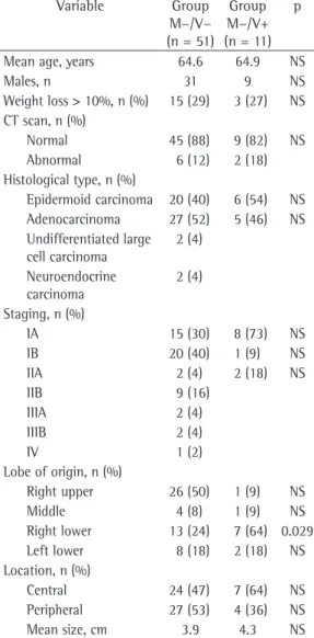
Table 1 - Variables studied, according to the results of cervical mediastinoscopy and video-assisted thoracoscopy.
Variable Group
M−/V−
(n = 51)
Group
M−/V+
(n = 11) p
Mean age, years 64.6 64.9 NS
Males, n 31 9 NS
Weight loss > 10%, n (%) 15 (29) 3 (27) NS CT scan, n (%)
Normal 45 (88) 9 (82) NS Abnormal 6 (12) 2 (18) Histological type, n (%)
Epidermoid carcinoma 20 (40) 6 (54) NS Adenocarcinoma 27 (52) 5 (46) NS Undifferentiated large
cell carcinoma
2 (4)
Neuroendocrine carcinoma
2 (4)
Staging, n (%)
IA 15 (30) 8 (73) NS IB 20 (40) 1 (9) NS IIA 2 (4) 2 (18) NS
IIB 9 (16)
IIIA 2 (4)
IIIB 2 (4)
IV 1 (2)
Lobe of origin, n (%)
Right upper 26 (50) 1 (9) NS Middle 4 (8) 1 (9) NS Right lower 13 (24) 7 (64) 0.029 Left lower 8 (18) 2 (18) NS Location, n (%)
Central 24 (47) 7 (64) NS Peripheral 27 (53) 4 (36) NS Mean size, cm 3.9 4.3 NS
The use of CT for the evaluation of medi-astinal lymph nodes in patients with NSCLC is controversial due to the high rates of false-positive and false-negative results (> 20%). Central tumors, tumors with a diameter greater than 4 cm and obstructive pneumonia are associated with a greater risk of incorrect inter-pretation.(19) In the present study, the 2 patients who presented an altered CT scan, negative mediastinoscopy results and positive video-assisted thoracoscopy results had tumors that were larger than 4 cm and centrally located in the lower lobe of the right lung. Even in the early stages of the disease, defined as T1N0M0, in which the rates of false-negative CT results can be as high as 15%, tomographic evaluation does not suffice and tissue sampling is required. (20) In a series of 291 patients with clinical stage I NSCLC submitted to routine mediastinoscopy, N2 disease and N3 disease were not detected in CT scans from 20 patients (7%). The rate of false-negative mediastinoscopy results was 9.2% (25/291). More importantly, this rate was 52% (13/25) for subcarinal nodes and 8% (2/25) for pulmonary ligament nodes.(21) There is clear evidence that posterior lymph node stations, principally the posterior subcarinal nodes, are not entirely accessed by cervical mediasti-noscopy.(8,22) In a series of 383 patients with NSCLC prospectively submitted to chest CT and PET, 199 patients were classified as N0 stage. Of those 199 patients, 28 (14%) were identi-fied as having unsuspected N2 disease after cervical mediastinoscopy and endoscopic ultra-sound-guided fine-needle aspiration.(22) A very significant finding of that study was that the posterior nodes, which are inaccessible by medi-astinoscopy, were the principal location of N2 disease that had not been detected by nonin-vasive methods (38% in posterior subcarinal nodes). The authors concluded that it was neces-sary to combine mediastinoscopy with another invasive method in order to thoroughly evaluate the mediastinum. The only methods available for histological or cytopathologic evaluation of posterior mediastinal lymph nodes are endobron-chial ultrasound-guided fine-needle aspiration, transesophageal endoscopic ultrasound-guided fine-needle aspiration and video-assisted thora-coscopy.(10) The choice of the method depends on the training of the examiner and on the availability of the method. Adenocarcinoma is subcarinal nodes by mediastinoscopy did not
detect neoplasia; however, sampling of poste-rior subcarinal nodes by mediastinoscopy and video-assisted thoracoscopy revealed neoplasia. Only 1 patient had involvement of both the pulmonary ligament nodes (MD: 9) and the posterior subcarinal nodes. Intraoperative frozen section analysis yielded 3 false-negative results (4.8%)—1 in the right paratracheal nodes and 2 in the subcarinal nodes—and 1 false-positive result (1.6%)—in the posterior subcarinal nodes. Video-assisted thoracoscopy successfully iden-tified 1 patient (1.6%) with a pleural implant, which was not visualized in the preoperative CT scan. Mean time to perform the cervical mediastinoscopy and video-assisted thoracos-copy with frozen section analysis was 45 min. Mortality and morbidity rates were zero.
The statistical analysis of the results of the present study included 95% CIs and revealed a positive predictive value of 88.89% (95% CI: 51.75-99.72), a negative predictive value of 94.34% (95% CI: 84.34-98.82), prevalence of 17.74% (95% CI: 9.20-29.53), sensitivity of 72.73% (95% CI: 39.03-93.98) and specificity of 98.77% (95% CI: 93.31-99.97).
The results that show the comparison between the two populations (negative
mediasti-noscopy + negative video-assisted thoracoscopy and negative mediastinoscopy + positive
video-assisted thoracoscopy) according to the variables analyzed are shown in Table 1. In the compara-tive analysis between these two populations, location in the right lower lobe was the only statistically significant variable (p = 0.029; 95% CI: 0.35-0.84).
Discussion
The rates of false-negative results in the frozen section analysis were 4.8%, which is in accordance with the means reported in the literature.(6,21,28) However, due to the therapeutic impact of selecting the best candidates for curative surgical treatment, we should consider performing the procedure known as surgical
staging (mediastinoscopy + video-assisted
thoracoscopy) separately from the pulmonary resection, considering the risks and costs of this choice.
Video-assisted thoracoscopy has become a routine procedure in thoracic surgery and is therefore available at all general thoracic surgery centers. The probability of performing endo-scopic ultrasound for the analysis of the posterior lymph nodes or even preferring this method over video-assisted thoracoscopy is a reality in many countries(8,9,29,30) and a near-future perspective in Brazil.
We conclude that the joint use of cervical mediastinoscopy and video-assisted thoracos-copy for the evaluation of posterior mediastinal lymph nodes was an effective method. In NSCLC patients who are candidates for pulmonary resec-tion, in which there is no access to the posterior lymph nodes by transbronchial or transesopha-geal ultrasound-guided biopsy (which dispenses with general anesthesia), the combined use of cervical mediastinoscopy and video-assisted thoracoscopy should be the method of choice to adequately evaluate mediastinal lymph nodes.
References
1. Detterbeck FC, Jantz MA, Wallace M, Vansteenkiste J, Silvestri GA; American College of Chest Physicians. Invasive mediastinal staging of lung cancer: ACCP evidence-based clinical practice guidelines (2nd edition). Chest. 2007;132(3 Suppl):202S-220S.
2. Takamochi K, Nagai K, Yoshida J, Suzuki K, Ohde Y, Nishimura M, et al. The role of computed tomographic scanning in diagnosing mediastinal node involvement in non-small cell lung cancer. J Thorac Cardiovasc Surg. 2000;119(6):1135-40.
3. Reed CE, Harpole DH, Posther KE, Woolson SL, Downey RJ, Meyers BF, et al. Results of the American College of Surgeons Oncology Group Z0050 trial: the utility of positron emission tomography in staging potentially operable non-small cell lung cancer. J Thorac Cardiovasc Surg. 2003;126(6):1943-51. Erratum in: J Thorac Cardiovasc Surg. 2007;133(4):864.
4. Lardinois D, Weder W, Hany TF, Kamel EM, Korom S, Seifert B, et al. Staging of non-small-cell lung cancer with integrated positron-emission tomography and computed tomography. N Engl J Med. 2003;348(25):2500-7.
more often associated with mediastinal involve-ment(21,22) in patients with NSCLC; however, this was not observed in the present study, since we found the same incidence for epidermoid carci-noma and adenocarcicarci-noma.
The probability of lymphatic metastasis, as well as its correlation with the lobe in which the lesion originated, is of utmost importance. Various studies (23-25) suggest the same route of spread: right upper lobe to ipsilateral paratra-cheal nodes; middle lobe to paratraparatra-cheal and subcarinal nodes; right lower lobe to paratra-cheal and subcarinal nodes; left upper lobe to subaortic and anterior mediastinal lymph nodes; and left lower lobe to subcarinal nodes. A recent retrospective analysis(26) involving 954 patients investigated the incidence and the location of N2 disease, as well as its correlation with the primary tumor location, and reported the same results as those previously reported in the literature. Based on this analysis, the authors recom-mended the use of video-assisted thoracoscopy for the evaluation of lymph node tumors in the left upper lobe (MD: 5 and 6), mediastinoscopy for the evaluation of tumors in the right upper lobe (MD: 2 and 4D) and endoscopic ultrasound for the evaluation of tumors in the middle lobe and in the lower lobe (MD: 7). The present study identified 11 cases (17.7%) in which the evalu-ation of the subcarinal nodes yielded negative results in the cervical mediastinoscopy and posi-tive results in the video-assisted thoracoscopy. Of these, 81% (9/11) had primary tumors in the lower lobes, predominantly in the right lower lobe (p = 0.029).
Mediastinal evaluation in patients with NSCLC by cervical mediastinoscopy alone requires a more thorough critical analysis. When we choose against the histological evaluation of the posterior lymph nodes, especially the posterior and lower subcarinal nodes, there is a reason-able chance (safely greater than 15%) that we are not offering the patient the best therapeutic strategy. Even presenting data that have not yet been confirmed by phase III randomized studies, recent guidelines(27) have shown that there is no scientific evidence for the use of combined
treatment (chemotherapy and radiotherapy +
19. McLoud TC, Bourgouin PM, Greenberg RW, Kosiuk JP, Templeton PA, Shepard JA, et al. Bronchogenic carcinoma: analysis of staging in the mediastinum with CT by correlative lymph node mapping and sampling. Radiology. 1992;182(2):319-23.
20. Pretreatment evaluation of non-small-cell lung cancer. The American Thoracic Society and The European Respiratory Society. Am J Respir Crit Care Med. 1997;156(1):320-32.
21. Choi YS, Shim YM, Kim J, Kim K. Mediastinoscopy in patients with clinical stage I non-small cell lung cancer. Ann Thorac Surg. 2003;75(2):364-6.
22. Cerfolio RJ, Bryant AS, Ojha B, Eloubeidi M. Improving the inaccuracies of clinical staging of patients with NSCLC: a prospective trial. Ann Thorac Surg. 2005;80(4):1207-13; discussion 1213-4.
23. Kotoulas CS, Foroulis CN, Kostikas K, Konstantinou M, Kalkandi P, Dimadi M, et al. Involvement of lymphatic metastatic spread in non-small cell lung cancer accordingly to the primary cancer location. Lung Cancer. 2004;44(2):183-91.
24. Naruke T, Tsuchiya R, Kondo H, Nakayama H, Asamura H. Lymph node sampling in lung cancer: how should it be done? Eur J Cardiothorac Surg. 1999;16 Suppl 1:S17-24.
25. Inoue M, Sawabata N, Takeda S, Ohta M, Ohno Y, Maeda H. Results of surgical intervention for p-stage IIIA (N2) non-small cell lung cancer: acceptable prognosis predicted by complete resection in patients with single N2 disease with primary tumor in the upper lobe. J Thorac Cardiovasc Surg. 2004;127(4):1100-6.
26. Cerfolio RJ, Bryant AS. Distribution and likelihood of lymph node metastasis based on the lobar location of nonsmall-cell lung cancer. Ann Thorac Surg. 2006;81(6):1969-73; discussion 1973.
27. Robinson LA, Ruckdeschel JC, Wagner H Jr, Stevens CW; American College of Chest Physicians. Treatment of non-small cell lung cancer-stage IIIA: ACCP evidence-based clinical practice guidelines (2nd edition). Chest. 2007;132(3 Suppl):243S-265S.
28. Meyers BF, Haddad F, Siegel BA, Zoole JB, Battafarano RJ, Veeramachaneni N, et al. Cost-effectiveness of routine mediastinoscopy in computed tomography- and positron emission tomography-screened patients with stage I lung cancer. J Thorac Cardiovasc Surg. 2006;131(4):822-9; discussion 822-9.
29. Yasufuku K, Chiyo M, Sekine Y, Chhajed PN, Shibuya K, Iizasa T, et al. Real-time endobronchial ultrasound-guided transbronchial needle aspiration of mediastinal and hilar lymph nodes. Chest. 2004;126(1):122-8. 30. Caddy G, Conron M, Wright G, Desmond P, Hart D, Chen
RY. The accuracy of EUS-FNA in assessing mediastinal lymphadenopathy and staging patients with NSCLC. Eur Respir J. 2005;25(3):410-5.
5. Silvestri GA, Gould MK, Margolis ML, Tanoue LT, McCrory D, Toloza E, et al. Noninvasive staging of non-small cell lung cancer: ACCP evidenced-based clinical practice guidelines (2nd edition). Chest. 2007;132(3 Suppl):178S-201S.
6. Semik M, Netz B, Schmidt C, Scheld HH. Surgical exploration of the mediastinum: mediastinoscopy and intraoperative staging. Lung Cancer. 2004;45 Suppl 2:S55-61
7. De Leyn P, Lardinois D, Van Schil PE, Rami-Porta R, Passlick B, Zielinski M, et al. ESTS guidelines for preoperative lymph node staging for non-small cell lung cancer. Eur J Cardiothorac Surg. 2007;32(1):1-8. 8. Eloubeidi MA, Tamhane A, Chen VK, Cerfolio RJ.
Endoscopic ultrasound-guided fine-needle aspiration in patients with non-small cell lung cancer and prior negative mediastinoscopy. Ann Thorac Surg. 2005;80(4):1231-9.
9. Wallace MB, Ravenel J, Block MI, Fraig M, Silvestri G, Wildi S, et al. Endoscopic ultrasound in lung cancer patients with a normal mediastinum on computed tomography. Ann Thorac Surg. 2004;77(5):1763-8. 10. Schipper P, Schoolfield M. Minimally invasive staging of
N2 disease: endobronchial ultrasound/transesophageal endoscopic ultrasound, mediastinoscopy, and thoracoscopy. Thorac Surg Clin. 2008;18(4):363-79. 11. Landreneau RJ, Hazelrigg SR, Mack MJ, Fitzgibbon LD,
Dowling RD, Acuff TE, et al. Thoracoscopic mediastinal lymph node sampling: useful for mediastinal lymph node stations inaccessible by cervical mediastinoscopy. J Thorac Cardiovasc Surg. 1993;106(3):554-8.
12. Mouroux J, Venissac N, Alifano M. Combined video-assisted mediastinoscopy and video-assisted thoracoscopy in the management of lung cancer. Ann Thorac Surg. 2001;72(5):1698-704.
13. Sebastián-Quetglás F, Molins L, Baldó X, Buitrago J, Vidal G; Spanish Video-assisted Thoracic Surgery Study Group. Clinical value of video-assisted thoracoscopy for preoperative staging of non-small cell lung cancer. A prospective study of 105 patients. Lung Cancer. 2003;42(3):297-301.
14. Mountain CF. Revisions in the International System for Staging Lung Cancer. Chest. 1997;111(6):1710-7. 15. Ingelfinger JA, Mosteller F, Thibodeau LA, Ware J.
Diagnostic testing: likelihood and odds. In: Ingelfinger JA, Mosteller F, Thibodeau LA, Ware J, editors. Biostatistics in clinical medicine. New York: McGraw-Hill; 1994. p. 26-50.
16. Fletcher RH, Fletcher S, Wagner EH, editors. Epidemiologia clínica: elementos essenciais. 3rd ed. Porto Alegre: Artes Médicas; 1996.
17. Ruckdeschel JC. Combined modality therapy of non-small cell lung cancer. Semin Oncol. 1997;24(4):429-39. 18. Kassis ES, Vaporciyan AA. Defining N2 disease
About the authors
Darcy Ribeiro Pinto Filho
Head of the Department of Thoracic Surgery, University of Caxias do Sul Foundation General Hospital, Caxias do Sul, Brazil.
Alexandre José Gonçalves Avino
Associate Surgeon. Department of Thoracic Surgery, University of Caxias do Sul Foundation General Hospital, Caxias do Sul, Brazil.
Suzan Lucia Brancher Brandão
Associate Surgeon. Department of Thoracic Surgery, University of Caxias do Sul Foundation General Hospital, Caxias do Sul, Brazil.
Wilson Paloschi Spiandorello