UNIVERSIDADE DE LISBOA
Faculdade de Medicina Veterinária
EVALUATION OF DIFFERENT EXTENDERS FOR COLD STORAGE OF MEAGRE (ARGYROSOMUS REGIUS) SEMEN
MARIANA COSTA CRUZ SANTOS
CONSTITUIÇÃO DO JÚRI
Doutora Luísa Maria Freire Leal Mateus Doutor Mário Alexandre Gonçalves Quaresma Doutor José Beirão dos Santos
ORIENTADOR Doutor José Beirão dos Santos
CO-ORIENTADOR
Doutor Fernando Ribeiro Alves Afonso
2017 LISBOA
UNIVERSIDADE DE LISBOA
Faculdade de Medicina Veterinária
EVALUATION OF DIFFERENT EXTENDERS FOR COLD STORAGE OF MEAGRE (ARGYROSOMUS REGIUS) SEMEN
MARIANA COSTA CRUZ SANTOS
DISSERTAÇÃO DE MESTRADO INTEGRADO EM MEDICINA VETERINÁRIA
CONSTITUIÇÃO DO JÚRI
Doutora Luísa Maria Freire Leal Mateus Doutor Mário Alexandre Gonçalves Quaresma Doutor José Beirão dos Santos
ORIENTADOR Doutor José Beirão dos Santos
CO-ORIENTADOR
Doutor Fernando Ribeiro Alves Afonso
2017 LISBOA
ACKNOWLEDGEMENTS
This project was financed by the projects BONAQUA (0433_BONAQUA_5_E), from the Operational Programme for Cross-border Cooperation: Spain-Portugal 2007-2013 (POCTEP 2007-2013), and AQUACOR (PROMAR 31-03-05FEP-003), co-financed by the European Regional Development Fund (FEDER, EU).
ABSTRACT
“EVALUATION OF DIFFERENT EXTENDERS FOR COLD STORAGE OF MEAGRE (ARGYROSOMUS REGIUS) SEMEN”
Semen refrigeration is usually recommended as a cheap and simple procedure that facilitates artificial reproduction techniques. The main objective of this experiment was to develop a semen refrigeration protocol for meagre that is considered a potential candidate for aquaculture diversification in Southern Europe. This thesis also contributes for the understanding of the causes of the fish sperm quality degradation during refrigeration.
Three extenders (non-activating medium (Fauvel et al., 1998); NaCl 0.9% and; NaCl 0.9% with glycine and glucose) and three different dilutions (1:4, 1:9 and 1:19, sperm:extender) were tested in a full factorial design. The following quality parameters were assessed along the storage time: sperm motility parameters; percentage of viable sperm; adenosine triphosphate (ATP); lipid peroxidation in the form of malondialdehyde (MDA); and bacteriology.
The 0.9% NaCl and the 0.9% NaCl with glycine and glucose extenders and the dilutions 1:4 and 1:9 kept a higher percentage of motile cells for longer, as well as higher sperm velocity. Sperm viability and ATP had better results with 0.9% NaCl and the 0.9% NaCl with glycine and glucose extenders. The MDA values were lower in treatments with dilution 1:4 when compared to those with dilution 1:9. In the CFU/ml values, no differences were found between extenders and dilutions. Motility parameters were strongly correlated with viability, whereas no or weak correlations existed with the remaining parameters. Thus, motility and viability seem to have the most impact in the loss of semen quality. According to the results, meagre semen could be kept refrigerated using 0.9% NaCl, in dilution 1:4, for up to 10 days.
RESUMO
“AVALIAÇÃO DE DIFERENTES DILUIDORES PARA A REFRIGERAÇÃO DE SÉMEN DE CORVINA (ARGYROSOMUS REGIUS)”
A refrigeração de sémen é tipicamente recomendada como um procedimento barato e simples que facilita as técnicas de reprodução artificial. O principal objectivo desta experiência foi desenvolver um protocolo de refrigeração de sémen para a corvina, que é considerada uma potencial candidata para a diversificação de aquacultura no sul da Europa. Esta tese contribui também para a compreensão das causas da degradação da qualidade de sémen de peixe durante a refrigeração.
Três diluidores (non-activating medium (Fauvel et al., 1998); NaCl 0,9%; e NaCl 0,9% com glicina e glucose) em três diferentes diluições (1:4, 1:9 e 1:19, sémen:diluidor) foram testados num plano factorial completo. Os seguintes parâmetros de qualidade espermática foram avaliados ao longo do tempo de armazenamento: parâmetros de mobilidade do sémen; percentagem de espermatozoides viáveis; adenosina trifosfato (ATP); peroxidação lipídica na forma de malondialdeído (MDA); e bacteriologia.
Os diluidores NaCl 0,9% e NaCl 0,9% com glicina e glucose e as diluições 1:4 e 1:9 mantiveram uma percentagem de células móveis mais elevada por mais tempo, bem como maior velocidade dos espermatozóides. A viabilidade e o ATP tiveram melhores resultados com NaCl 0,9% e NaCl 0,9% com glicina e glucose. Os valores de MDA foram mais baixos em tratamentos com as diluições 1:4, quando comparados com aqueles com as diluições 1:9. Nos valores de bacteriologia não foram encontradas diferenças entre diluidores e diluições testadas. Os parâmetros de mobilidade correlacionaram-se fortemente com a viabilidade, enquanto inexistentes ou fracas correlações foram encontradas entre os restantes parâmetros. Por conseguinte, mobilidade e viabilidade parecem ter o maior impacto na perda de qualidade do sémen. De acordo com os resultados, o sémen de corvina pode ser mantido refrigerado usando o diluidor NaCl 0,9% na diluição 1:4 até 10 dias.
TABLE OF CONTENTS
ACKNOWLEDGEMENTS ... i
ABSTRACT ... iii
RESUMO ... iv
TABLE OF CONTENTS ... v
LIST OF FIGURES ... vii
LIST OF GRAPHS ... vii
LIST OF TABLES ... viii
LIST OF ABBREVIATIONS, INITIALS AND ACRONYMS ... x
ACTIVITIES DEVELOPED IN THE SCOPE OF THE MASTER’S DEGREE ... 1
1. Internship ... 1
Faculty of Veterinary Medicine – University of Lisbon (FMV-UL) ... 1
Oceanário de Lisboa ... 1
Aquaculture Research Station of the Portuguese Institute for the Ocean and Atmosphere (EPPO – IPMA) ... 1
2. Formation and Publications ... 2
BIBLIOGRAPHIC REVISION ... 3
1. Aquaculture in the world, and in Portugal ... 3
2. Biology of the meagre ... 4
3. Meagre as an advantageous species for aquaculture ... 5
4. Overview of meagre reproduction ... 6
5. Artificial reproduction ... 7
6. Gamete storage ... 8
7. General considerations on semen and spermatozoa ... 11
8. Sperm quality and its evaluation ... 12
9. Sperm quality parameters ... 13
9.1. Concentration ... 13 9.2. Motility ... 14 9.3. Viability ... 16 9.4. ATP ... 17 9.5. Lipid Peroxidation ... 18 9.6. Bacteriology ... 18 9.7. Fertilization success ... 19 EXPERIMENTAL STUDY ... 21 1. Objectives ... 21
2. Material and methods ... 21
2.1. Experimental animals ... 21
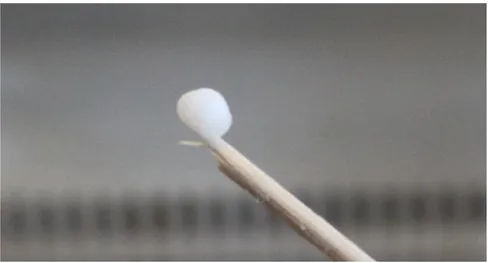
2.2. Semen collection ... 21
2.3. Refrigeration and samples’ processing ... 23
2.4. Concentration and motility ... 25
2.6. Viability ... 26
2.7. ATP ... 27
2.8. MDA... 28
2.9. Bacteriology ... 29
3. Results ... 33 3.1. Concentration ... 34 3.2. Motility ... 34 3.3. Viability ... 40 3.4. ATP ... 42 3.5. MDA ... 43 3.6. Bacteriology ... 45 3.7. Correlations ... 46 4. Discussion ... 47 4.1. Concentration ... 47 4.2. Motility ... 48 4.3. Viability ... 49 4.4. ATP ... 50 4.5. MDA ... 51 4.6. Bacteriology ... 52 5. Conclusions ... 55 BIBLIOGRAPHY ... 56 ANNEX ... 78
Annex A: Temperature of refrigeration. ... 78
Annex B: Composition of solutions used in the ATP protocol. ... 79
Annex C: Intermediate tests of the data analysis. ... 80
Annex D: Concentration values and data treatment. ... 87
Annex E: Motility values and data treatment. ... 88
Annex G: ATP values and data treatment. ... 99
Annex H: MDA values and data treatment. ... 100
LIST OF FIGURES
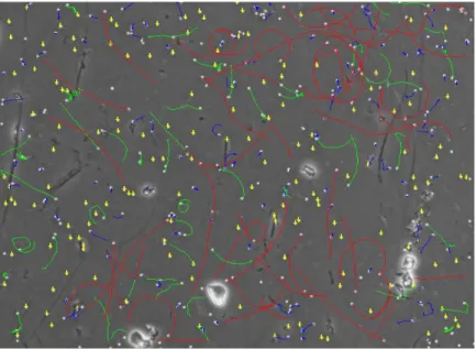
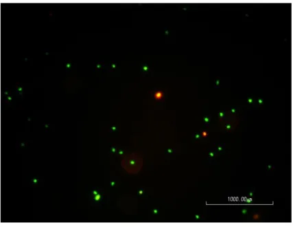
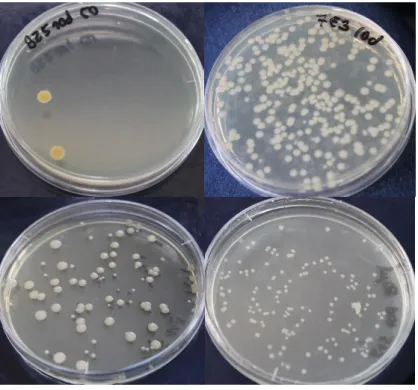
Figure 1: The process of stripping eggs from a female meagre (DIVERSIFY, n.d.). ... 8 Figure 2: Schematic representation of some motility patterns measured by a CASA system. VCL, curvilinear velocity, in the entire distance travelled; VSL, velocity in a straight line; VAP, velocity of an average path. Black circles represent the position of the cell in each frame. Adapted from Rurangwa et al. (2004). ... 16 Figure 3: Image of one tank during semen collection, showing the lowered water level and the net separating fish already sampled. ... 22 Figure 4: Two meagre on the 200 liters tank with anesthesia, showing loss of equilibrium, by being upside down. ... 22 Figure 5: Moment of semen collection, with the meagre upside down and enveloped in a wet towel, in the acrylic cylinder half. Abdominal pressure was being applied and semen was collected with a syringe. ... 23 Figure 6: The refrigerator shelf. In the front, the falcons with the various samples from three separated pools. On the left, bottles with the extenders and reagents for the ATP protocol. On the right, a falcon filled with water and with the temperature sensor. ... 24 Figure 7: Image obtained with the CASA software. Different colours represent spermatozoa with different motility speeds. Yellow dots represent immotile spermatozoa, whereas, blue, green and red represent slow, medium and fast spermatozoa respectively, and their paths. ... 26 Figure 8: Image taken in the fluorescence microscope of a sample observed after incubation, showing the difference between cells coloured green (viable cells) and red (dead cells). ... 27 Figure 9: Plates with apparent bacterial growth after 7 days of incubation. ... 30 Figure 10: Positive result of a catalase test, with the formation of gas bubbles. ... 31
LIST OF GRAPHS
Graph 1: Mean percentage of motile spermatozoa (MOT) of each treatment for each day of assessment. ... 35 Graph 2: Survival analysis until the percentage of motility reaches zero, comparing the values obtained in five pools for: A, the different combinations of extenders (NAM, NC, NCG, C); B, the different combinations of dilutions (1:4, 1:9, 1:19, Control). ... 36 Graph 3: Mean curvilinear velocity (VCL) of each treatment for each day of assessment and treatment. ... 37 Graph 4: Mean percentage of linearity (LIN) of each treatment for each day of assessment and treatment. ... 39 Graph 5: Mean values obtained for each treatment, along the days of evaluation. ... 40 Graph 6: Survival analysis until the viability reaches 50%, comparing the values obtained in five pools for: A, the different combinations of extenders (NAM, NC, NCG, C); B, the
different combinations of dilutions (1:4, 1:9, 1:19, Control). ... 41 Graph 7: Mean ATP values obtained for each treatment, from five pools, along the days of viability evaluation. ... 42 Graph 8: Mean and standard error of the mean of the values of MDA at days 0, 3, 6 and 9, in which it was quantified. ... 44
Graph 9: Relative frequency of the genera of the bacteria that were isolated at the seventh day
of incubation in the TSA and PS mediums from the three pools. N = 43. ... 46
LIST OF TABLES Table 1: Sperm motility parameters, their abbreviation, unit, and description (Rurangwa et al., 2004). ... 15
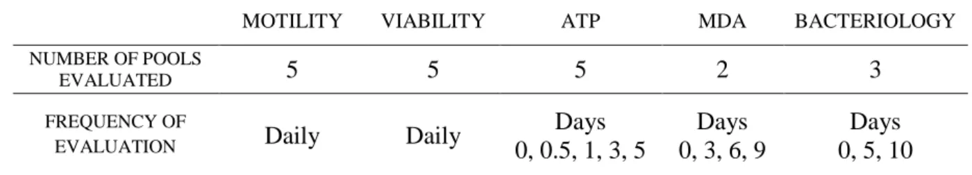
Table 2: Number of pools and frequency of evaluation of each sperm quality parameter. ... 25
Table 3: Curve points used in the ATP protocol. ... 28
Table 4: Curve points used in the MDA protocol. ... 29
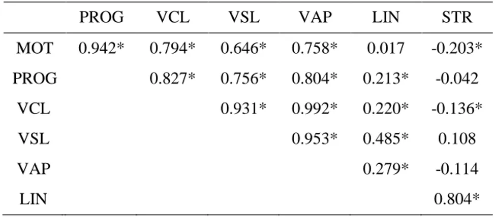
Table 5: Spearman’s rho test of correlation between the values obtained for all the sperm motility parameters measured with CASA. ... 34
Table 6: Mann-Whitney U test for the values of MOT, VCL and LIN in pairs of extenders and pairs of dilutions... 36
Table 7: Mann-Whitney U test done to the pairs of extenders. ... 40
Table 8: Mann-Whitney U test done to the pairs of extenders. ... 43
Table 9: Mann-Whitney U test done to the MDA values in pairs of different dilutions. ... 44
Table 10: Mean ± standard error of the mean of the CFU/ml obtained for each treatment (C, NAM, NC and NCG) and day, in TSA medium. Values from three pools. N = 3. ... 45
Table 11: Mean ± standard error of the mean of the CFU/ml obtained for each treatment (C, NAM, NC and NCG) and day, in PS medium. Values from three pools. N = 3. ... 45
Table 12: Spearman's rank correlation coefficient between the different parameters tested. .. 46
Table 13: Mean maximum and minimum values of temperature of semen storage, as registered daily in the temperature sensor, during the days in which the seven pools were stored.. ... 78
Table 14: Composition of the solutions that were used during the procedure of extraction and quantification of ATP from meagre semen. ... 79
Table 15: Shapiro-Wilk normality test for each motility parameter. ... 80
Table 16: Values for the Statistic of the Shapiro-Wilk normality test, done for each combination of treatment. ... 80
Table 17: Shapiro-Wilk normality test done to the percentage of viability values of each treatment... 81
Table 18: Linear regression equations, and r2 value, for the viability values along the time of each treatment from each pool. ... 82
Table 19: Shapiro-Wilk normality test done to the ATP values of each treatment. ... 83
Table 20: Shapiro-Wilk normality test done to the MDA values obtained in each treatment. 83 Table 21: Shapiro-Wilk normality test done to the CFU/ml values obtained in each treatment, medium and day. ... 84
Table 22: Shapiro-Wilk normality test done to the values obtained in all the pools for VIAB, and the respective values of MOT, VCL and LIN. ... 84
Table 23: Shapiro-Wilk normality test done between ATP and the respective values of MOT, VCL and LIN, as well as ATP and VIAB. ... 85 Table 24: Shapiro-Wilk normality test done between MDA and the respective values of MOT, VCL and LIN, as well as MDA and VIAB. ... 85 Table 25: Statistic values of the Shapiro-Wilk normality test for the different assessments of correlation, as indicated in the first row. ... 86 Table 26: Concentration values obtained for the control sample, in each and in all of the pools tested. ... 87 Table 27: Mean ± standard error of the mean of the percentage of motile cells for each
treatment and day... 88 Table 28: Results of the Mann-Whitney U of all the pairs tested of the various combinations of extenders, and of dilutions, for percentage of motility (MOT) values. ... 90 Table 29: Mean ± standard error of the mean of the curvilinear velocity for each treatment and day. ... 91 Table 30: Results of the Mann-Whitney U of all the pairs tested of the various combinations of extenders, and of dilutions, for curvilinear velocity (VCL) values. ... 92 Table 31: Mean ± standard error of the mean of the percentage of linearity for each treatment and day. ... 94 Table 32: Results of the Mann-Whitney U of all the pairs tested of the various combinations of extenders, and of dilutions, for the percentage of linearity (LIN) values. ... 95 Table 33: Mean ± standard error of the mean of the percentage of viability for each treatment and day. ... 96 Table 34: Results of the Mann-Whitney U of all the pairs tested of the various combinations of extenders, for percentage of viability (VIAB) values. ... 97 Table 35: Mean ± standard error of the mean of the ATP value for each treatment and day. . 99 Table 36: Results of the Mann-Whitney U of all the pairs tested of the various combinations of extenders, for ATP values. ... 99 Table 37: Mean ± standard error of the mean of the MDA value for each treatment and day. ... 100 Table 38: Results of the Mann-Whitney U of all the pairs tested of the various combinations of dilutions, for MDA values. ... 100 Table 39: Results from the bacterial identification procedures, showing the percentage and number of isolates for each of the species that were found, including the ones that were not identified. ... 101
LIST OF ABBREVIATIONS, INITIALS AND ACRONYMS
AEC Adenylate energy charge ATP Adenosine triphosphate BSA Bovine serum albumen
C Control/undiluted
CASA Computer-assisted sperm analysis CFU Colony-forming units
FAO Food and Agriculture Organization GnRHa Gonadotropin-releasing hormone agonist GzLM Generalized Linear Model
HBSS Hanks’ balanced salt solution
HPLC High-performance liquid chromatography
IPMA Instituto Português do Mar e Atmosfera (Portuguese Institute of Sea and Atmosphere)
LIN Linearity
MDA Malondialdehyde
MOT Percentage of motile cells
NAM Extender NAM
NAM4 Treatment with extender NAM in dilution 1:4 NAM9 Treatment with extender NAM in dilution 1:9 NAM19 Treatment with extender NAM in dilution 1:19
NC Extender NC
NC4 Treatment with extender NC in dilution 1:4 NC9 Treatment with extender NC in dilution 1:9 NC19 Treatment with extender NC in dilution 1:19
NCG Extender NCG
NCG4 Treatment with extender NCG in dilution 1:4 NCG9 Treatment with extender NCG in dilution 1:9 NCG19 Treatment with extender NCG in dilution 1:19
PI Propidium iodide
PROG Percentage of progressive spermatozoa
PS Pseudomonas CN Agar
STR Straightness TBA Thiobarbituric acid
TBARS Thiobarbituric acid reactive substances TSA Trypticase soya agar
VAP Average path velocity VCL Curvilinear velocity VIAB Viability
ACTIVITIES DEVELOPED IN THE SCOPE OF THE MASTER’S DEGREE
1. Internship
Faculty of Veterinary Medicine – University of Lisbon (FMV-UL)
For three weeks, in July of 2014, some work was done in the facilities of the Faculty of Veterinary Medicine dedicated to research concerning Fish and Aquaculture. The work consisted in a preliminary approach to a possible research concerning the behavior of a species of shrimp, Palaemonetes varians. The preferences of this species regarding color of the substrate was assessed. During this period, aquaria were prepared for the individuals, and their behavior was observed and registered. A visit to an aquaculture was also done.
During these three weeks it was possible to acquire some experience in the planning and implementation of a preliminary project.
Oceanário de Lisboa
The first segment of the internship took place from January 5th to March 27th of 2015, in Oceanário de Lisboa (Lisbon Oceanarium). The team of veterinarians was accompanied during the entirety of the internship. The species that were observed and handled during the internship consisted of: mostly fish, both bony and cartilaginous, from different ecosystems; some species of amphibians; aquatic birds; a species of reptile (Caretta caretta); and one species of mammal (Enhydra lutris). Some of the activities which I accompanied included observation and diagnosis of individuals, routine analysis, reproductive ecography and necropsies. Due to an agreement with the institution, this section won’t be further elaborated. This internship allowed to obtain knowledge and a better understanding of aquatic animal medicine, mostly of fish, including some experience in reproduction of different species.
Aquaculture Research Station of the Portuguese Institute for the Ocean and Atmosphere (EPPO – IPMA)
The second segment of the internship took place from the 7th of April to the 26th of June of 2015, with two weeks of additional work in September, in the Aquaculture Research Station of the Portuguese Institute for the Ocean and Atmosphere, in Olhão. A portion of work took place in the facilities of the Center of Marine Sciences of the University of Algarve, in Faro. During this internship took place the research project addressed by this thesis. The activities done in the scope of this project were done mostly by myself and my supervisor, but a team of biologists, as well as aquaculture and research technicians provided us with valuable support.
Experience was obtained in induction of handling of fish, as well as induction of anesthesia and collection of semen by stripping. I prepared the extenders that were tested and used them to dilute the semen. The semen quality parameters that were evaluated also represent areas of acquired expertise. I learned to use a computer-assisted sperm analysis in order to evaluate concentration, motility and viability. Nonetheless, the latter was mostly evaluated by myself through observation and counting in a fluorescence microscope. I prepared solutions and did ATP extraction and quantification, which included manipulation of a microplate reader. The MDA assay procedure was observed by myself. For bacteriology, I prepared growth medium plates and performed inoculations, as well as counting of colonies. I also obtained experience in the isolation, propagation and identification of genera of bacteria with biochemical tests. Lastly, in general, time planning and teamwork skills were developed.
2. Formation and Publications
A poster was displayed at the Aquaculture Europe 2014 congress, organized by the European Aquaculture Society: “Studies on behavior and activity of the shrimp Palaemonetes varians”, by Santos, M. and Afonso, F. (2014).
In July of 2015 I attended the seminar “Meagre production – science and practice”, organized by the EPPO-IPMA. During this seminar the results of the several works of the AQUACOR project were presented, regarding different aspects of the production of meagre, such as: performance of different production systems; feed; reproduction; larvae and fry production. In October of 2015 I attended the XV National Spanish Congress and I Iberian Congress of Aquaculture in Huelva (Spain), organized by the Spanish Aquaculture Society. The communications I attended were related to different subjects such as: reproduction in captivity of Senegalese sole (Solea senegalensis) and other species; diversification of aquaculture with species such as meagre (Argyrosomus regius) and common octopus (Octopus vulgaris); production and sanitary quality of mollusks and crustaceans; sustainability and environmental impact in the scope of aquaculture. A poster was displayed: “Refrigeração do sémen de corvina (Argyrosomus regius): desenvolvimento de um protocolo e avaliação de parâmetros de qualidade” (Refrigeration of meagre semen (Argyrosomus regius): development of a protocol and evaluation of quality parameters), by Santos, M., Soares, F., Moreira, M., Cabrita, E. and Beirão, J. (2015).
BIBLIOGRAPHIC REVISION
1. Aquaculture in the world, and in Portugal
The human population is expected to increase to more than 11 billion by the end of the century (Lutz, Sanderson & Scherbov, 2001), leading to a bigger consumption of resources such as food (Pimentel, Huang, Cordova & Pimentel, 1997). This will result in an increase in fish consumption, a protein-rich food recommended as part of the human diet (Sargent & Tacon, 1999). Under these circumstances, and since the vast majority of the wild fish stocks are already overexploited or have reached their maximum productive capacity, further demand of fish should be met by aquaculture instead of capture (Grafton, Kompas & Hilborn, 2007).
The first written records regarding aquaculture date back to 2 500 years ago in China (Rabanal, 1988). Nowadays, it is the fastest growing animal production industry (Gjedrem, Robinson & Rye, 2012). The portion of global fish production corresponding to aquaculture, in 2015, was forecast to be 46% (Food and Agriculture Organization of the United Nations [FAO], 2015), while in 2003 it was only 31% (FAO, 2015). Quoting the words of the inventor of modern management and business visionary Peter Drucker, “Aquaculture, not the Internet, represents the most promising investment opportunity of the 21st century”.
The fish consumption per capita in Portugal is the highest in the European Union, and it is estimated to remain so, with a predicted consumption of 58 kg per capita per year, in 2020 (Failler, 2007). To support this high demand the country relies on a large volume of fish commodity imports, which in 2009 represented more than half of the national supply (Teixeira, Rodrigues, Cavadas & Neto, 2013). At the same time, only 5% of fish produced in Portugal comes from aquaculture, whereas the remaining 95% comes from fisheries (FAO, 2014). Thus, the development of aquaculture would decrease Portugal’s dependence on imports.
There are several factors in favour of the development of aquaculture in Portugal. As previously stated, the consumption of fish is very high. There is funding from the European Commission towards the development of aquaculture (Associação Portuguesa de Aquacultura [APA], 2014). Furthermore, it is one of the warmest European countries, due to the Mediterranean climate (Kalkan & Canuyrt, 2012). Finally, it has a vast coastline and a large area covered with lagoons, which can be used for aquaculture (Kalkan & Canuyrt, 2012). Nonetheless, the lack of economic power and the bureaucracy involved (licensing procedures take several years) have hindered the investment in aquaculture (APA, 2014).
Aquaculture production in Portugal, over the last 30 years, has fluctuated between 5 and 11 thousand tonnes, approximately (FAO, 2014). Of the total volume produced, 93% refers to brackish or marine aquaculture, and fish comprises 55% of its volume (Instituto Nacional de Estatística [INE], 2016). The main species produced in 2014 were grooved carpet shell (Ruditapes decussatus), turbot (Scophthalmus maxima), mussels (Mytilus sp.) and gilthead seabream (Sparus aurata) (INE, 2016). When it comes to marine fishes, the markets are currently saturated (Couto et al., 2016), especially because of competition with countries like Spain or Greece. An alternative to prevent this saturation is the development of technology for the production of new species. This is the aim of projects such as the AQUACOR project of IPMA, regarding meagre (Argyrosomus regius) and its production.
2. Biology of the meagre
The meagre, Argyrosomus regius (Asso, 1801), is part of the Sciaenidae family, which belongs to the Perciformes order. A common characteristic of this family, that gave its members the name of croakers and drums, are the sounds they produce by beating specific muscles against the swim bladder, making it resonate (Ramcharitar, Gannon & Popper, 2006). The Argyrosomus name comes from the Greek: argyros, meaning silver, and soma, body. Indeed, this species has a silver to bronze coloured elongated body, with a fairly large head and a yellow mouth cavity (Stipa & Angelini, 2005). In the wild, they reach up to 2 m in length and 50 kg in weight (Stipa & Angelini, 2005). The meagre’s geographic distribution includes the Eastern Atlantic coast and the Mediterranean and Black Seas (Haffray et al., 2012; Lagardère & Mariani, 2006). They are carnivorous and, although the diet varies with their size, they feed mostly on small crustaceans when juveniles (Jiménez et al., 2005), and fish when adults (Quéro & Vayne, 1985; Stipa & Angelini, 2005).
Meagre are gonochorists (Gil, Grau, Basilone, Ferreri & Palmer, 2013; Schiavone, Zilli, Storelli & Vilella, 2012), meaning each individual has one constant gender. The reproductive season occurs from April to June (Gil et al., 2013), with aggregation near estuaries and salt marshes (Quéro, 1989; Stipa & Angelini, 2005). During this season the males emit the characteristic croaks (Lagardère & Mariani, 2006), whilst spawning occurs. The females lay eggs that are fertilized externally and hatch in 48 hours (Haffray et al., 2012). The larvae leave the nursing area as juveniles, when the summer ends, and migrate back to spawning areas upon reaching maturity (González-Quirós et al., 2011). During the winter, adults migrate to deeper waters, and reduce feeding activity. Water temperature seems to be a
determining factor in this species’ biology, affecting namely its feeding requirements, trophic migration and reproduction (Stipa & Angelini, 2005).
Meagre appears to be a resilient species, due to its high fecundity (Haffray et al., 2012) and growth rate (Monfort, 2010). However, their aggregation and emission of spawning sounds makes meagre a species vulnerable to capture (Catalán, Jiménez, Alconchel, Prieto & Muñoz, 2006; Sadovy & Cheung, 2003).
3. Meagre as an advantageous species for aquaculture
Meagre has been farmed in Europe since the late nineties, first in France and then Italy. Since then, other countries like Spain, Portugal and Greece have started to show some interest in this species. In 2000, the total global production was of only 33 tonnes, but the number has been growing and in 2010 over 10 000 tonnes were produced in aquaculture (Monfort, 2010). This species has several strengths that support its production. It has a fast growth rate, reaching about 1 kg in the first year and 2.5 kg in the second (Duncan et al., 2013) and a low feed conversion rate, between 0.9 and 1.2 (Monfort, 2010). Meagre responds well to the hormonal induction of spawning and the eggs and larvae have high quality (Mylonas et al., 2013a; Mylonas, Mitrizakis, Sigelaki & Papadaki, 2011). A study by Roo, Hernández-Cruz, Borrero, Schuchardt & Fernández-Palacios (2010) obtained promising results for larval production at an industrial rate. As a euryhaline species, meagre tolerates diverse environments, such as land based cultivation with brackish water (Jiménez et al., 2005; Ribeiro, Soares, Quental-Ferreira, Gonçalves & Pousão-Ferreira, 2013), while also withstanding tank captivity fairly well (Pastor, Grau, Massutí & Sánchez-Madrid, 2002). It is also resistant to handling and there are few publications reporting disease problems, indicating that meagre might be somewhat resistant to infectious diseases (Stipa & Angelini, 2005). Additionally, it has a good filleting yield of 42% (Grigorakis, Fountoulaki, Vasilaki, Mittakos & Nathanailides, 2011) and its fillets received high sensory acceptability scores (Giogios, Grigorakis & Kalogeropoulos, 2013). The intramuscular fat content is low, around 1% (Grigorakis et al., 2013), when compared to species such as gilthead seabream (Özogul, Özogul & Alagoz, 2007) and European sea bass (Dicentrarchus labrax) (Lanari et al., 1999). At the same time, the fat has a high nutritional value (Giogios, Grigorakis & Kalogeropoulos, 2013) and its low content allows refrigeration for a longer period of time (Poli, et al., 2003). Altogether, these characteristics make meagre a very attractive species for the aquaculture diversification (Monfort, 2010; Poli et al., 2003), particularly in southern Europe.
As a new species adapted to aquaculture, important advances are expected to occur in meagre production, which should lead to a reduction of the juveniles’ price (Stipa & Angelini, 2005). In this context, most of the research is focused on the improvement of meagre production techniques and the increase of its efficiency and profitability (Duncan et al., 2013). As an example, research in the field of reproduction will certainly result in regular production of high quality spawns.
4. Overview of meagre reproduction
Reproduction of meagre, as in fish in general, depends on the brain-hypothalamus-pituitary-gonad axis. Environmental stimuli like photoperiod and temperature are responsible for the control of this axis and have a crucial role in the control of gamete maturation and release (Cardinaletti et al., 2010). In the end of the axis lie the gonads, which are the reproductive organs in which gametes’ production occurs, termed gametogenesis (Wooton & Smith, 2014). Gametes originated from germ cells after several mitotic divisions and meiosis (Okutsu, Suzuki, Takeuchi, Takeuchi & Yoshizaki, 2006; Stickney, 2000). In males, the gametogenesis occurs in the testes and is called spermatogenesis, producing spermatozoa. In females, the gametogenesis occurs in the ovaries and is called oogenesis, producing oocytes (Patiño & Sullivan, 2002). During oogenesis takes place the vitellogenesis that consists in the accumulation of yolk proteins within the oocyte (Patiño & Sullivan, 2002), essential for the formation of eggs. Mylonas, Mitrizakis, Papadaki & Sigelaki (2013) evaluated female and male gamete development of meagre over the reproductive period, in captivity, and observed that both spermatogenesis and vitellogenesis took place between April and June, in accordance with their natural reproductive season.
Meagre reach maturity at the age of 2.7 years for males and 3.5 years for females (Gil et al., 2013) and the length at maturity ranged 26.8-61.6 cm for males and 35.8-110 cm for females (González-Quirós et al., 2011; Schiavone et al., 2012; Soares et al., 2015). Additionally, Gil et al. (2013) concluded that the annual fecundity increases with length, weight and age, ranging from 0.9 to 4.2 million oocytes in reared females.
The reproduction of meagre in captivity is one of the bottlenecks of its production, similar to most species new to aquaculture (Schiavone et al., 2012). One example is the fry cost, which is still very high (Schiavone et al., 2012), in part because of unpredictability of its production. Nonetheless, industrial fish farming requires broodstocks that provide an adequate supply of fertilized eggs (Duncan et al., 2012) and juveniles (Stickney, 2000). However, some advances
in meagre reproduction are taking place. As an example, different research groups recently reported spontaneous spawning of meagre in captivity (Mylonas et al., 2013b; Pastor et al., 2013; Soares et al., 2015). Nowadays, fertilized eggs can either be obtained with wild-caught fish acclimated to captivity (Duncan et al., 2012; Soares et al., 2015) or with first-generation fish born in captivity (Soares et al., 2015). Indeed, when adequate nutrition is provided, both types of broodstocks can spawn good eggs and produce high larval quality, suitable for a hatchery production (Soares et al., 2015). Oviparous females in particular, like the meagre, have a higher and more diverse nutritional demand due to oogenesis (Stickney, 2000). A commonly observed problem in meagre reproduction is the failure to undergo oocyte formation and spawning. This also occurs in other species, due to a deficiency in the feed (Cabrita, Robles & Herráez, 2008).
Additionally, there have been some developments in hormonal induction protocols for meagre. These consist in the usage of a gonadotropin-releasing hormone agonist (GnRHa), given via injections or slow-release implants, to both males and females (Duncan et al., 2008; Duncan et al., 2012; Mylonas et al., 2013a; Pastor et al., 2013). Biglino (2015) observed a reduction in semen volume and sperm concentration when using GnRHa implants in males, but no apparent effect over motility or viability. At the same time, Duncan et al. (2008) and Pastor et al. (2013) obtained high egg fecundity after the administration of GnRHa to the females. Indeed, a single injection or a slow-release implant of GnRHa led to the production of fertilized eggs in sufficient number and quality for a commercially viable hatchery production (Duncan et al., 2012).
The current and future research should lead to new knowledge on meagre reproductive biology, which will be crucial to improve the broodstocks’ reproductive performance (Mylonas et al., 2013b).
5. Artificial reproduction
Adequate management of broodstock and gametes requires certain environmental conditions and procedures, including artificial reproduction techniques. Artificial reproduction is the reproduction in a non-natural way, with the interference and manipulation of humans. It ensures the availability of fertilized eggs over time or when necessary, and maximizes the number of larvae produced (Suquet, Dreanno, Fauvel, Cosson & Billard, 2000). Furthermore, it grants some protection from the environment and pathogens, leading to a bigger survival rate and growth. In addition, species that cannot spawn naturally or reproduce in captivity can be propagated through these methods, using for example in vitro fertilization. Some
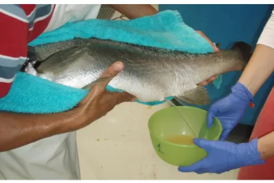
technologies require artificial reproduction. Polyploidy production, already done in other sciaenid species, is one example (Ballarin et al., 2004). It leads to the formation of fish that, being sterile, have less incidence of diseases or higher growth rate (Piferrer et al., 2009). Lastly, these techniques allow selective breeding, which consists in crossing the genetic material of specific individuals (Dupont-Nivet, Vandeputte, Haffray & Chevassus, 2006). Meagre gametes may be collected for artificial reproduction (Poli et al, 2003), particularly when spawning is not occurring naturally (Marino et al., 2000). Semen can be collected by stripping, which might not be effective in every individual (Mylonas et al., 2013b), whereas eggs can also be collected by stripping after hormonal treatment, as seen in Figure 1 (DIVERSIFY, n.d.).
Figure 1: The process of stripping eggs from a female meagre (DIVERSIFY, n.d.).
Sperm and eggs are usually mixed in a certain ratio that maximizes the gamete usage. In general, to ensure genetic variability when doing artificial reproduction, a pool of semen from several males is used (Mylonas, Fostier & Zanuy, 2010). Nevertheless, for selective breeding, the use of only one specific male is also possible.
6. Gamete storage
When conducting artificial reproduction male and female gametes are not always available at the same time. In these cases gametes have to be stored in advance. In this context, gamete storage is an essential tool for artificial fertilization and breeding programs for several reasons. Fertilization can be synchronized according to gametes’ availability (Cabrita et al., 2010; Suquet, 2000). Furthermore, it also allows for the gametes’ transportation between locations such as collection site and hatchery, making easier and cheaper genetic exchanges (Cabrita et al., 2010; Kopeika et al., 2007; Suquet, 2000). The total volume of the gametes can
be efficiently used, which is particularly important when low volumes are available (Cabrita et al., 2010). Finally, in the case of semen cryopreservation, genetic material of valuable males can be theoretically preserved indefinitely (Cabrita et al., 2010; Suquet, 2000).
After collection, gametes start to suffer degradation processes. Their quality decreases, causing a reduced fertilizing ability. In some species, gametes may be stored unaltered from hours to days, at room temperature (Mylonas et al., 2010). Alternatively, the storage conditions can be manipulated to delay the degradation processes (Mishra, Patra, Dash, Verma & Routray, 2016; Yasui et al., 2015). Some examples of parameters that can be manipulated are temperature, oxygen supply, gas exchange, the storage medium (extender) and its dilution ratio, as well as the replenishment of the extender during storage (Kowalski et al., 2014; Mishra et al., 2016). For both female and male gametes of several fish species there are different storage protocols, which involve the manipulation of one or more of these factors.
Regarding storage temperatures, refrigeration is done above 0oC (Yasui et al., 2015). This can be achieved in a simple way, with the use of a refrigerator or a cooler with ice packs for both oocytes and semen, either undiluted or diluted (Komrakova & Holtz, 2009; Wayman, Tiersch & Thomas, 1998). Depending on the species, the gametes may be refrigerated for several days while keeping some quality.
Alternatively, gametes can be stored at temperatures below 0oC. In fishes, only semen can be stored at subzero temperatures (Asturiano, Cabrita & Horváth, 2016). The larger size of the oocytes, the yolk content and the different permeability of the oocytes’ membranes causes some difficulties in their storage, namely the penetration of cryoprotectants and lack of uniformity in the cooling process (Asturiano et al., 2016; Chao & Liao, 2001). On the other hand, it is relatively easier to preserve semen of most fish species, either through short term cooling or long term cryopreservation in liquid nitrogen, at -196oC (Agarwal, 2011; Gwo, Chen & Cheng, 2002; Stoss, 1983). Although cryopreservation allows the cells’ preservation for a longer period of time, and several cryopreservation protocols have been developed for different fish species, there are not many cases of practical applications outside of the research environment (Cabrita et al., 2010). Indeed, from the aquaculture perspective semen refrigeration remains the best alternative (Viveiros, Isaú, Figueiredo, Leite & Maria, 2010a). Extenders are the substances in which gametes are diluted and preserved. Oocytes are generally stored in ovarian fluid (Bellard, 1988), since the usage of dilution media has shown no advantages (Stoss, 1983). In the case of semen storage the usage of extenders is more common. Although it is only essential for cryopreservation, for the incorporation of
cryoprotectants (Agarwal, 2011; Kopeika, Kopeika & Zhang, 2007), they are an important complement for refrigeration as well, extending the spermatozoa’s viability (Lahnsteiner, Berger, Weismann & Patzner, 1996; Maria, 2014). An ideal extender, according to Mann (1964), should not activate semen motility, should be isotonic and have a good buffering capacity and should include nutrients, antioxidants, antibacterial substances and stabilizing colloids. Keeping these characteristics in consideration, the extender’s composition may vary. A simple and effective extender is a solution of NaCl (Fabbrocini et al., 2000; Maria, Viveiros, Freitas & Oliveira, 2006; Oliveira, Viveiros, Maria, Freitas, & Izaú, 2007). The addition of other substances to this extender may be proven beneficial, such as glucose (Viveiros, Orfão, Maria & Allaman, 2009). He and Woods III (2003) also proved that the addition of some aminoacids to a NaCl solution, such as glycine, improved the quality of stored semen. There are also extenders with more complex compositions, usually mimicking the seminal plasma composition. As an example, for the European sea bass it is usually recommended a non-activating medium, with different salts, glucose and BSA (Fauvel, Suquet, Dreanno, Zonno, & Menu, 1998). Additionally, there are commercially available extenders that may be used, such as BTS™ and ACP® (Nascimento, Maria, Pessoa, Carvalho, & Viveiros, 2010; Viveiros, Maria, Orfão, Carvalho, & Nunes, 2008).
The atmosphere with which the gametes are stored, excluding cryopreservation, affects their quality. Generally, normal air is used, but oxygen can be added (Billard, 1981). During storage, gametes consume oxygen (Robitaille, Mumford & Brown, 1987), and thus aerobic conditions should be provided. For this reason, addition of oxygen to the containers results in a longer storage time (Bellard, 1988; Komrakova & Holtz, 2011) for both oocytes (Billard, 1981) and semen (Bellard, 1988). Nevertheless, the presence of oxygen can also be the cause of lipids’ peroxidation, resulting in damage to the cells with deterioration of quality (Chen et al., 2010). Some factors must also be taken in consideration, such as frequency of renewal of atmosphere (Marques & Godinho, 2004), which depends on the amount of oxygen consumed. This varies with the species, semen dilution and ratio of volume of semen to atmosphere. Diluting semen or stocking it with more air allows a reduction in this frequency (Bellard, 1988).
Other aspect to take into consideration in the refrigeration protocols is, in the case of semen, the sedimentation over time, which can be avoided by slightly shaking the containers periodically (Stoss, Büyükhatipoglu & Holtz, 1978). In the case of oocytes their storage is affected by the weight of eggs and the number of layers (Komrakova & Holtz, 2009). In most
cases, these protocols' optimization is done empirically, testing different parameters and evaluating the gametes’ quality over time.
Semen storage protocols are far more diverse and more frequently used than oocytes’ storage protocols. Artificial reproduction techniques normally involve semen storage until collection of oocytes (Rurangwa, Kime, Ollevier & Nash, 2004; Stoss, 1983). In the case of the Sciaenidae family, there are already semen storage protocols for different species, testing different extenders, osmolalities and dilution ratios (Leclercq et al., 2014; Wayman et al. 1998; Wayman, Thomas & Tiersch, 1997). More recently, research done under the DIVERSIFY-EU project, in 2014, found that in meagre sperm quality decreases rapidly after collection, if not diluted in an extender.
Meagre is a species in the early phases of adaptation to aquaculture, which will benefit from the improvement of artificial reproduction techniques that could assist in the management of broodstocks. Preliminary results regarding the refrigeration of meagre semen, done under the DIVERSIFY-EU project, gave promising results for the development of artificial reproduction. The usage of European sea bass extenders efficiently sustained the spermatozoa’s motility for more than 24 hours. However, there are no protocols for semen storage in this species.
7. General considerations on semen and spermatozoa
The ejaculate semen is composed of spermatozoa and seminal plasma. Spermatozoa are the cells responsible to deliver the male genetic information to the egg, and have the capacity to become motile (Miller, Brinkworth & Iles, 2010). The seminal plasma is an enriched medium responsible for maintaining spermatozoa in a quiescent state until ejaculation and providing energy resources (Alavi & Cosson, 2006).
In teleost fish, the spermatozoa structure is divided in head, mid-piece and flagellum (Ginzburg, 1968; Islam & Akhter, 2011). The head is occupied in the most part by the nucleus, containing the genetic material (Ginzburg, 1968; Islam & Akhter, 2011). In the case of the teleosts, the head does not contain an acrosome (Dzyuba & Cosson, 2014), and in meagre it is oval-shaped (Schiavone et al., 2012). The mid-piece is linked to the head (Ginzburg, 1968; Islam & Akhter, 2011) and contains the mitochondria (Berois et al., 2011; Ginzburg, 1968). The flagellum is the apparatus responsible for the movement (Islam & Akhter, 2011), and originates from the posterior part of the nucleus (Cosson et al., 2008). It produces waves that propagate from proximal to distal tip (Boryshpolets, 2011), resulting in cell motility. ATP is the major source of energy for the flagellar beating, produced by the
mitochondria present in the mid-piece (Boryshpolets, 2011; Dzyuba et al., 2016). Nevertheless, during the fast motility period there is a very high ATP consumption that is not compensated by the mitochondria production (Ingermann, 2008). As a result, the energy stores are depleted during this time leading to the spermatozoa immobilization in a short period (Cosson, 2010; Dreanno, Seguin, Cosson, Suquet & Billard, 1999b). This means that motility depends on the endogenous ATP and other energy molecules’ stores present before activation (Cabrita et al., 2010). In most external fertilizing fishes spermatozoa are immotile when inside the testes (Cosson et al., 2008; Ginzburg, 1968). After ejaculation motility is triggered by changes in the osmotic or ionic environment, which in the case of most marine species is caused by seawater (Cosson et al., 2008).
8. Sperm quality and its evaluation
Sperm quality can be defined as the ability to successfully fertilize an egg and develop into a normal embryo (Bobe & Labbé, 2010; Rurangwa et al., 2004). The measurement of different sperm quality parameters has several applications in aquaculture. They can be used as predictors of the fertilizing ability (Bobe & Labbé, 2010; Rurangwa et al., 2004), and thus their evaluation is important to increase artificial reproduction efficiency (Zilli, Schiavone, Zonno, Storelli & Vilella, 2004). When there is a reduced amount of eggs (Rurangwa et al., 2004), the risk of a low fertilization rate and loss of egg batches is prevented (Bobe & Labbé, 2010). Furthermore, cryopreservation should be only done to sperm of proven high quality (Zilli et al., 2004). Sperm quality evaluation also helps to determine the optimal time for semen collection, when its quality is variable along the season (Babiak, Ottesen, Rudolfsen & Johnsen, 2006; Fabbrocini et al., 2000). The occurrence of contamination during collection (Dreanno et al., 1998) and the effect of repeated stripping (Aas, Refstie & Gierde, 1991) may also be assessed. Finally, it helps to improve handling and storage protocols (Fabbrocini et al., 2000; Linhart et al., 2004), as is the case of the present thesis.
Fertilization success is the most integrative parameter and the best indicator of sperm quality (Bobe & Labbé, 2010). Nonetheless, for practical reasons, there is the need to use other parameters, which do not require the usage of eggs. The most commonly used are motility parameters, such as percentage of motile cells and motility duration or spermatozoa’s swimming speed (Beirão et al., 2015, 2011b; Schiavone et al., 2012). Other parameters less frequently used are sperm concentration, sperm cell membrane integrity or viability (Beirão, Pérez-Cerezales, Martínez-Páramo & Herráez, 2010; Cabrita et al., 2011), ATP content (Montgomery, Brown, Gendelman, Ota & Clotfelter, 2014), lipid peroxidation
(Martínez-Páramo, Diogo, Beirão, Dinis & Cabrita, 2012) or bacteriology (Jenkins & Tiersch, 1997; Viveiros et al., 2010b), to mention a few.
In most cases it is difficult to correlate one specific parameter with fertilization rate, since they evaluate specific cellular functions, and fertilization depends on many (Bobe & Labbé, 2010). Furthermore, they show the mean value for the whole spermatozoa population, whereas only a small number of spermatozoa is needed to ensure fertilization (Bobe & Labbé, 2010). Sperm quality parameters are, then, partial descriptors of fertilization ability (Bobe & Labbé, 2010). In this context, sperm quality schemes should be built based on several parameters at the same time. These parameters should be investigated and improved, to increase the number of spermatozoa which meet the requirements for fertilization to occur (Bobe & Labbé, 2010).
According to Bobe & Labbé (2010) there are several factors that can influence the sperm quality, most of them related to broodstock management (Billard, Cosson, Perchec & Linhart, 1995). One example is stress, which can be caused by fish handling or environmental conditions. Nutrition is another factor with impact in sperm quality, as feed should have an adequate formula, especially regarding lipid composition and vitamins (Asturiano et al., 2001; Dabrowski & Ciereszko, 1996). Sperm handling during and after collection can also affect its quality, such as adequate storage conditions as temperature, medium and duration (Babiak et al., 2006; Peñaranda, Pérez, Fakriadis, Mylonas & Asturiano, 2008; Suquet et al., 1998). In the case of meagre, the sperm quality has already been evaluated for concentration, for aspects of motility such as percentage of progressive motile cells and duration of progressive forward movement and morphology (Mylonas et al., 2013b; Mylonas et al., 2016; Schiavone et al., 2012). While Mylonas et al. (2013b) state that sperm quality does not significantly change during the reproductive season., Schiavone et al. (2012) verified a variation during the reproductive season, which goes in accordance to what occurs in other species, and may be due to the aging of spermatozoa (Babiak et al., 2006; Peñaranda et al., 2008).
9. Sperm quality parameters
9.1. Concentration
Sperm concentration is usually presented as the number of cells per milliliter (Junior et al., 2008; Wirtz & Steinmann, 2006). In teleost semen the cell concentration is highly variable, due to factors such as species, individual and time in the reproductive season (Butts, Litvak & Trippel, 2010; Piironen & Hyvärinen, 1983). In several species studied by Piironen and
Hyvärinen (1983) and Poole and Diilane (1998), the concentration ranged from 2.2 to 127.4 x 10⁹ ml-1. Results obtained by Mylonas et al. (2013b) in meagre semen obtained a mean cell concentration between 19 and 32 x 109 ml-1. This parameter can be correlated, in some instances, with other parameters such as fertilization rate and motility (Ciereszko & Dabrowski, 1994; Hwang & Idler, 1969). Nevertheless, its main importance involves the optimization of the usage of semen when doing artificial reproduction, and the sperm to egg ratio needs to be adjusted (Bombardelli, Mörschbächer, Campagnolo & Syperreck, 2006; Sanches et al., 2011).
Several methods can be used to assess sperm concentration of fish semen. The most common involves counting the number of spermatozoa with a microscope and a cell counting chamber, such as a Neubauer chamber (Junior et al., 2008; Wirtz & Steinmann, 2006). These methods, despite cheaper, are time consuming (Fauvel, Suquet & Cosson, 2010). Spermatocrit determination, with hematocrit capillary tubes, is faster and simpler, giving a result in percentage equivalent to sperm concentration (Ciereszko & Dabrowski, 1993). However, this technique is not as effective in marine fish because spermatozoa do not sediment efficiently, which may be due to their density being similar to that of seminal plasma (Fauvel et al., 2010). Alternatively, the value of absorbance, obtained by spectrophotometry, can be used if a correlation with concentration has been previously established (Ciereszko & Dabrowski, 1993) Furthermore, some authors also suggest the use of flow cytometry or coulter counter, which are highly precise but expensive methods (Fauvel et al., 2010). Lastly, a modern image analysis software connected to a microscope is also frequently used to determine concentration fast and efficiently (Fauvel et al., 2010). Depending on the species, the correlation coefficient between these three methods may be high (Ciereszko & Dabrowski, 1993).
9.2. Motility
Sperm motility is an integrative parameter that combines different cellular structures (Bobe & Labbé, 2010). It is the most commonly used parameter to compare different experimental conditions such as sperm collection procedures, semen extenders, or sperm storage conditions (Bobe & Labbé, 2010).
In general, the initiation, duration and the patterns of motility differ between species (Rurangwa et al., 2004). In teleosts with external fertilization, such as meagre, the motility tends to be brief (Coward, Bromage, Hibbitt & Parrington, 2002) and spermatozoa usually swim at high speed and frequency, in rectilinear motion (Cosson et al., 2008; Cosson, 2010).
In meagre the percentage of motile spermatozoa varies between 53% and 74%, with a duration of 34 to 80 seconds (Schiavone et al., 2012). In most species, fertilization usually occurs between 5 and 20 seconds after spermatozoa’s activation, thus the most useful motility data is obtained during this period (Kime et al., 2001).
In several species a correlation between motility and ability to fertilize the eggs has been found (e.g. Cosson et al., 2008; Ottesen, Babiak & Dahle, 2009), including refrigerated and cryopreserved sperm (e.g. Beirão et al., 2011a; Ciereszko & Dabrowski, 1994). Some examples are the correlations between sperm motility parameters and fertilization or hatching rate, in cryopreserved semen in African catfish (Clarias glariepinus) (Rurangwa et al. 2001), gilthead seabream (Beirão et al. 2011a), common carp (Cyprinus carpio) (Leveroni, Calvi, Zoccarato, Gasco & Andrione, 1993; Leveroni, Calvi, Zoccarato, Gasco & Andrione, 1994) and Alburnus alburnus (Lahnsteiner et al., 1996), or in refrigerated rainbow trout (Oncorhynchus mykiss) semen (Ciereszko & Dabrowski, 1994).
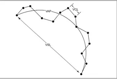
Motility can be assessed subjectively, by direct observation on a microscope (Kime et al., 2001). In these cases, the researcher measures the motility duration and estimates the percentage of motile sperm (Fauvel et al., 2010). To increase the accuracy of these measurements, sperm motility may be videotaped and posteriorly analyzed by different researchers (Kime et al., 2001). Alternatively, nowadays, more laboratories are using computer-assisted sperm analysis (CASA) systems (Fauvel et al., 2010; Kime et al., 2001) to objectively assess motility. The CASA systems analyze sperm movement of every single spermatozoa in a short video file, which is recorded with a camera attached to a microscope and connected to a computer (Rurangwa et al., 2004). This system measures several motility and velocity parameters (Fauvel et al., 2010). The parameters most commonly used for fish sperm analysis are described in Table 1. The differences between the three velocity parameters are represented in Figure 2. The large data sets obtained can be used to compare different sperm samples and thus reveal differences in experimental protocols, individuals and species, to mention a few (Linhart, Rodina, Gela, Kocour & Vandeputte, 2005; Rurangwa et al., 2001).
Table 1: Sperm motility parameters, their abbreviation, unit, and description (Rurangwa et al., 2004).
PARAMETER
(unit) DESCRIPTION
Duration of Motility
(s) time interval in which the cells remain motile MOT • Percentage of Motile Cells
PROG • Percentage of Progressive Spermatozoa
(%) percentage of cells with forward progressive movement MOC • Concentration of Motile Cells
(spz/ml) concentration of cells that exhibit some motility VCL • Curvilinear Velocity
(µm/s) considering the entire distance travelled VSL • Straight Line Velocity
(µm/s) considering a straight line between initial and final point VAP • Average Path Velocity
(µm/s) considering the distance travelled as a smoothed path LIN • Linearity
(%) obtained by VSL/VAP • a measure of path curvature STR • Straightness
(%) obtained by VAP/VCL • a measure of side to side movement
Figure 2: Schematic representation of some motility patterns measured by a CASA system. VCL, curvilinear velocity, in the entire distance travelled; VSL, velocity in a straight line; VAP, velocity of an average path. Black circles represent the position of the cell in each frame. Adapted from Rurangwa et al. (2004).
9.3. Viability
Sperm viability refers to the percentage of live spermatozoa (Garner & Johnson, 1995). Although viability should be attributed to the capacity of sperm to fertilize an egg, this parameter evaluates the percentage of spermatozoa with an integral plasma membrane (Fauvel et al., 2010; Rurangwa et al., 2004).
Different authors have found a correlation between sperm viability and other semen quality parameters, such as sperm velocity parameters in common carp semen and hatching rate in African catfish (Linhart et al., 2005; Rurangwa et al., 2001).
Viability tests are based on staining protocols, which confer different coloration to viable and dead cells (Rurangwa et al., 2004). Each stain has a different cell permeation ability,
depending on the plasma membrane’s integrity (Fauvel et al., 2010). Protocols may only use one dye, such as tryptan blue (Lubzens et al., 1997; Rurangwa, Biegniewska, Slominska, Skorkowski & Ollevier, 2002), or a combination of two, such as eosin and nigrosine (Maria et al., 2006). Alternatively, fluorescent dyes can also be used, to facilitate cell distinction. A commonly used combination of these is propidium iodide (PI) with SYBR-14, for dead and viable cells respectively (Rurangwa et al., 2004), but other combinations have been proposed, such as PI with YO-PRO 1 (Beirão et al., 2010). These stains usually target nucleic acids of the spermatozoa. In the case of the PI/SYBR-14 combination, PI cannot cross the intact plasma membrane, whereas SYBR-14 leaks out when the plasma membrane is deteriorated (Nagy, Jansen, Topper & Gadella, 2003; Rurangwa et al., 2004). The percentage of viable cells can be assessed under a microscope, by direct counting or using an image analysis system (De Baulny, Labbé & Maisse, 1999). Other ways to do so are through flow cytometry (Beirão et al., 2010), with an automatic counter (Fauvel et al., 2010) or a fluorometer (McNiven, Gallant & Richardson, 1993).
9.4. ATP
ATP, together with adenosine diphosphate (ADP) and adenosine monophosphate (AMP), compose the Adenylate Energy Charge (AEC). The AEC can be considered the intracellular content of available energy, for it is composed of high energy nucleotides. Since the values of ATP and AEC are highly correlated, it is reasonable to evaluate just one of these parameters (Fauvel et al., 2010), such as ATP, which has simple and precise protocols (Perchec, Jeulin, Cosson, Andre & Billard, 1995). As previously mentioned, the ATP present in the spermatozoa is the major energy source for flagellar beating and, as demonstrated by Dreanno et al. (1999b) and Cabrita et al. (2005), it can be used as a sperm quality estimator. Indeed, a positive correlation between ATP content and percentage of motile cells was found in some species such as turbot (De Baulny et al., 1996) and steelhead trout (Oncorhynchus mykiss) (Bencic, Krisfalusi, Cloud & Ingermann, 1999b). Furthermore, Bencic et al. (1999b) also observed a positive correlation between ATP and fertility rate in steelhead trout.
ATP determination can be done with a bioluminescence technique that uses firefly luciferin-luciferase, to which there are commercial assay kits (Fauvel et al., 2010; Perchec et al., 2005). The reaction between ATP, luciferin and luciferase leads to the production of luminescence, which can be detected with a luminometer or scintillation counter (Rieger, 1997; Yang, Ho, Chen & Hu, 2002). There are also methods which enable the quantification of not only ATP but also the other components of AEC, such as high pressure liquid chromatography
(Dziewulska, Rzemieniecki & Domagala, 2010; Mukai & Okuno, 2004) and nuclear magnetic resonance spectroscopy (van den Thillart, van Waarde, Muller, Erkelens & Lugtenburg, 1990).
9.5. Lipid Peroxidation
During the aerobic metabolic processes, cells produce reactive oxygen species (ROS). These have cytotoxic properties, causing damages to the desoxyribonucleic acid (Cabrita et al., 2011; Potts, Notarianni & Jefferies, 2000), ATP depletion (Aramli, Kalbassi, Nazari & Aramli, 2013), abnormalities in morphology (Ball, 2008), and lipid peroxidation (Zhou et al., 2006), which leads to a decrease in motility and cell viability (Aramli et al., 2013; Ball, 2008), and a decrease in sperm quality (Apel & Hirt, 2004). The cell membrane’s high content in polyunsaturated fatty acids (PUFAs) makes fish spermatozoa quite susceptible to lipid peroxidation (Labbe et al., 1995; Zhou et al., 2006). To counteract the ROS effects semen has antioxidant mechanisms (Lahnsteiner & Mansour, 2010), which include different antioxidant substances present in the seminal plasma (Cabrita et al., 2011; Potts et al., 2000). However, in vitro storage of the semen increases ROS formation (Ball, 2008; Zhou et al., 2006) namely because dilution in the extenders reduces the antioxidants’ concentration of seminal plasma (Cabrita et al., 2011; Martínez-Páramo, Martínez-Pastor, Martínez-Rodríguez, Herráez & Cabrita, 2009). Nevertheless, antioxidants could be added to stored semen to prevent cellular injuries caused by ROS (Aramli et al., 2013).
During lipid peroxidation, several by-products are formed. Their measure is frequently used to quantify lipid peroxidation (Devasagayam, Boloor & Ramasama, 2003). The thiobarbituric acid (TBA) assay is the most used to evaluate lipid peroxidation in vitro (Buege & Aust, 1978; Devasagayam et al., 2003). It measures the by-products which react with thiobarbituric acid – thiobarbituric acid reactive substances (TBARS) -, such as malondialdehyde (MDA) (Potts et al., 2000; Devasagayam et al., 2003; Yahyavi, Kaykhaii & Hashemi, 2016). This assay is sensitive but non-specific (Devasagayam et al., 2003; Zhou et al., 2006), since TBA might react with other organic components. Alternatively, MDA can be measured using commercially available kits (Beirão et al., 2015; Martínez-Páramo et al., 2012).
9.6. Bacteriology
Bacterial growth can be the cause of sperm quality decrease especially when semen is stored in refrigeration. Bacteria may be present in semen as part of the normal flora, due to contamination or with origin in the extender (Bartha, 2009). They can reduce sperm quality
due to the oxygen consumption, and the production of extracellular enzymes and metabolic by-products that affect spermatozoa (Jenkins & Tiersch, 1997). The growth of bacteria has been associated to decreased fertility (Saad, Billard, Theron & Hollebecq, 1988; Stoss & Refstie, 1983), low motility (Christensen & Tiersch, 1996; Isaú, 2014) and viability (Stoss et al., 1978), as well as increased morphological changes (Jenkins & Tiersch, 1997). Indeed, Jenkins & Tiersch (1997) observed that the usage of a sterile extender, as opposed to a non-sterile one, extended the number of days in which there was sperm motility. Moreover, some authors have suggested the use of antibiotics to inhibit bacterial growth and improve sperm survival and fertility (Billard et al., 2004; Viveiros et al., 2010b).
Bacteriological evaluation can be done quantitatively or qualitatively. The quantitative analysis is the estimation of the number of bacteria per unit of volume by counting the number of colony-forming units (CFUs) (Elain et al., 2015; Jenkins & Tiersch, 1997). The qualitative analysis involves determining the presence of bacteria and their identification (Plusquellec, Beucher, Le Lay, Le Gal & Cleret, 1991) by means of various techniques that evaluate different phenotypical characteristics of the bacteria. Examples of these are observation of morphology and motility, Gram staining, catalase or oxidase test, and glucose fermentation (Salanitro, Blake & Muirhed, 1977).
9.7. Fertilization success
Fertilization success is the most integrative estimator of sperm quality, since it requires the interaction of all sperm cellular functions, rather than one or some of them (Bobe & Labbé, 2010). In fact, in numerous occasions there is no correlation between other sperm quality parameters, such as motility parameters or viability, and fertilization (Bobe & Labbé, 2010; Cabrita et al., 2010). It should also be kept in mind that only a small fraction of spermatozoa are needed to fertilize the eggs, whereas most of the sperm quality parameters take into account the average of overall sperm population (Bobe & Labbé, 2010).
In general, to evaluate sperm fertilization success, sperm and eggs are mixed at a certain ratio to observe the percentage of ova fertilized per amount of sperm cells (Beirão et al. 2011a). Usually, only the amount of fertilized eggs is evaluated, without contemplating further developmental success (Shields, Brown & Bromage, 1997). This is easy to observe in species with transparent eggs, such as meagre (Gamsiz & Neke, 2008). Nonetheless, the hatching rate might also be assessed, incubating the eggs (Cabrita et al., 2010). It should be taken in consideration that there may be significant differences between fertility and hatching rate
(Cabrita et al., 2009; Pérez-Cerezales et al., 2010). Furthermore, the impact of egg quality must also be taken in consideration when evaluating the results (Bobe & Labbé, 2010).