Cop
yright
© ABE&M t
odos os dir
eit
os r
eser
vados
.
Decreased astrocytic GFAP
expression in
streptozotocin-induced diabetes after gliotoxic
lesion in the rat brainstem
Expressão astrocitária diminuída de GFAP no diabetes induzido por estreptozotocina após lesão gliotóxica no tronco encefálico de ratos
Eduardo Fernandes Bondan1,2, Maria de Fátima Monteiro Martins1,2,
Flávio Cesar Viani2
ABSTRACT
Objective: The aim of this study was to evaluate the effect of diabetic hyperglycemia on as-trocyte function, estimated by means of glial ibrillary acidic protein – GFAP – immunohisto-chemical expression. Materials and methods: Adult male rats received a single intravenous injection of streptozotocin (50 mg/kg) and were submitted 10 days later to a single injection of 10 microlitres 0.1% EB solution or 0.9% saline solution into the cisterna pontis. Ten microliters of 0.1% EB or 0.9% saline solution were also injected in non-diabetic rats. Animals were anesthe-tized and perfused through the heart 15 and 31 days after EB or saline injection, and brainstem sections were collected for ultrastructural analysis and GFAP immunohistochemical staining. Results: The GFAP brown-stained areas were evaluated by colorimetry using a computerized image analysis system and the results have shown that diabetes hindered the increase of GFAP astrocyte expression in the EB-injected group compared to non-diabetic animals. However, dia-betes did not affect GFAP response in the saline-injected group or in control animals. Conclu-sion: Streptozotocin-induced diabetic condition reduced astrocytic GFAP expression following gliotoxic injury. Arq Bras Endocrinol Metab. 2013;57(6):431-6
Keywords
Astrocytes; GFAP; central nervous system; diabetes mellitus; ethidium bromide
RESUMO
Objetivo: O objetivo deste estudo foi avaliar o efeito da hiperglicemia na função astrocitária, estimada pela expressão imuno-histoquímica da proteína glial ibrilar ácida – GFAP. Materiais e métodos: Ratos machos adultos receberam uma injeção intravenosa única de estreptozoto-cina (50 mg/kg) e foram submetidos, 10 dias após, à injeção de 10 microlitros de solução de BE 0,1% ou de salina 0,9% na cisterna pontina. Dez microlitros de BE 0,1% ou salina 0,9% foram também injetados em ratos não diabéticos. Os animais foram anestesiados e perfundidos por via intracardíaca aos 15 e 31 dias pós-injeção de BE ou salina, e amostras de tronco encefálico foram coletadas para estudo ultraestrutural e análise imuno-histoquímica para a GFAP. Resul-tados: Utilizando um sistema computadorizado de análise de imagens, os resultados das áreas coradas em marrom pela GFAP, medidas por colorimetria, mostram que o diabetes reduziu o aumento de expressão dessa proteína no grupo injetado com BE em comparação aos animais não diabéticos, mas não alterou a resposta no grupo injetado com salina ou nos controles diabéticos. Conclusão: O estado diabético induzido pela estreptozotocina reduziu a expressão astrocitária de GFAP após dano gliotóxico. Arq Bras Endocrinol Metab. 2013;57(6):431-6
Descritores
Astrócitos; GFAP; sistema nervoso central; diabetes melito; brometo de etídio
1 Post-Graduate Program in Environmental and Experimental Pathology, Universidade Paulista, São Paulo, SP, Brazil 2 Veterinary Medicine Department, Universidade Cruzeiro do Sul, São Paulo, SP, Brazil
Correspondence to:
Eduardo Fernandes Bondan Rua Caconde, 125/51 01425-011 – São Paulo, SP, Brasil [email protected]
Cop
yright
© ABE&M t
odos os dir
eit
os r
eser
vados
.
INTRODUCTION
I
t is widely described that ethidium bromide (EB) injection in the white matter of the central nervous system (CNS) acts like a gliotoxin, causing local oli-godendroglial and astrocytic death, with consequent demyelination (although naked axons remain preser-ved), blood-brain barrier disruption, and Schwann cell invasion due to the glia limitans breakdown (1-5). Surviving astrocytes present vigorous reaction around the injury site with increased immunorreactivity to the speciic cell marker, glial ibrillary acidic protein (GFAP), and reexpression of vimentin (VIM) (5). Hyperglycemia found in diabetes mellitus is known to cause well-characterized morphological and functional changes in peripheral neurons and Schwann cells (6). Much less is known about the effects of hyperglycemia on CNS cells, mainly on glia. It is recognized that dia-betes exacerbates astrocytic (7,8) and neuronal (9,10) damage induced by ischemia and reperfusion. On the other hand, insulin treatment prevents diabetes-indu-ced alterations in astrocyte glutamate uptake and re-verts the decreased GFAP expression in rats at 4 and 8 weeks of diabetes duration (11). Glial modiications were clearly pointed out in some studies (12,13) using streptozotocin-diabetic rats after the injection of EB, with marked delay on macrophagic scavenging activi-ty of myelin debris, on oligodendrocyte and Schwann cell remyelination (12), as well as on blood-brain bar-rier repair (13), although astrocytic response was not properly investigated and compared between diabetic and non-diabetic animals. In such context, the aim of the present investigation was to evaluate the effect of diabetic hyperglycemia on astrocyte function (estima-ted by means of GFAP immunohistochemical expres-sion) in rats injected or not with EB in the brainstem, serving as normal homeostatic regulators in the neural microenvironment or as reactive and repairing cells af-ter injury.MATERIALS AND METHODS
This experiment was approved by the Ethics Com-mission of Universidade Paulista (protocol number 002/09). Adult male Wistar rats, 3 to 4 months old, were used, from which some received, after 12 hours of fasting, a single injection of streptozotocin (50 mg/kg, Sigma) in 0.01M citrate buffer (pH 4.5) into the tail vein. Ten days after that, blood glucose was measured
and animals with levels of 300 mg/dL or more were considered diabetic. At this time, they were submitted to a local injection of 10 microlitres of 0.1% EB (group I) or 0.9% saline (group II) solution into the cisterna pontis. All rats were anaesthetized with ketamine and xylazine (5:1; 0.1 ml/100 g) and a burr hole was made on the right side of the skull, 8 mm rostral to the fron-toparietal suture.
Injections were performed freehand using a Hamil-ton Syringe, itted with a 35o angled polished 26-gauge needle into the cisterna pontis, an enlarged subarach-noid space below the ventral surface of the pons. Non-diabetic rats also received 10 microlitres of 0.1% EB solution (group III) or 0.9% saline solution (group IV). Diabetic (group V) and non-diabetic rats (group VI) were also used without receiving any intracisternal in-jection (control groups).
Body weight and blood glucose levels (Dextrostix, Ames) were recorded at 3 different times – at the mo-ment of the streptozotocin injection, 10 days after and at the time of euthanasia. Water and food were given ad libitum during the experimental period. All rats were anaesthetized; some were submitted to intracardiac perfusion with buffered 10% formaldehyde (for im-munohistochemical purpose), and some with 4% glu-taraldehyde in 0.1 M Sorensen phosphate buffer (pH 7.4) (for transmission electron microscopy study) 15 and 31 days after intracisternal injection or not. Thin slices of the brainstem (pons and mesencephalon) were collected and post-ixed in 0.1% osmium tetroxide, dehydrated with graded acetones and embedded in Araldite 502 resin, following transitional stages in ace-tone. Thick sections were stained with 0.25% alkaline toluidine blue. Selected areas were trimmed, and thin sections were stained with 2% uranyl and lead acetate and viewed in a JEM -1200 EX2 JEOL transmission electron microscope.
Sec-Cop
yright
© ABE&M t
odos os dir
eit
os r
eser
vados
.
ondary goat anti-rabbit antibody (E0433, Dako) was used as the binding antibody diluted at 1:100 for 30 minutes. The material was revealed with DAB (diami-nobenzidine) for 5 minutes and counterstained with Harris hematoxylin 1:2.
Astrocytic evaluation was done at the 31st day in groups I, II, III and IV using a computerized image analysis system (Image-Pro-Plus 4.5, Media Cybernet-ics, Silver Spring, USA), measuring by colorimetry the area stained in brown in a total area of 302,952.5 µm2, chosen from the lesion edge, where astrocytic reaction occurred. Negative controls for immunostaining (sec-tions lacking primary antibody application) were done. Data were analyzed by t test and statistical signiicance was set at p < 0.05.
RESULTS
The EB-induced lesions were similar to those previou-sly described in the brainstem of diabetic (12,13) and non-diabetic rats (2,5). In general terms, they were characterized by demyelinated areas in the ventral sur-face of the pons and mesencephalon containing, in the central region, phagocytic cells, some myelin-derived membranes in a distended extracellular space, as well as naked axons. At the periphery, the presence of oli-godendrocytes and Schwann cells was noted, the latter occurring in areas of an enlarged extracellular space devoid of astrocytic prolongments, notably around blood vessels and in subpial areas. Astrocyte processes
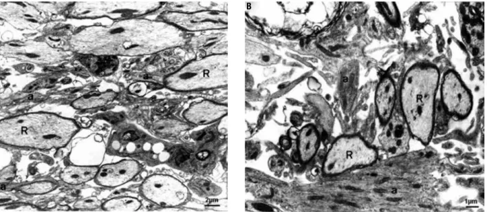
were invariably seen near the incipient, but preponde-rant, oligodendroglial remyelination (Figure 1A and B), and Schwann cells also appeared to contribute to myelin repair. Ultrastructural analysis apparently sho-wed that astrocytic processes among oligodendrocyte remyelinated axons were slightly thinner in diabetic animals compared with non-diabetic ones. Although oligodendroglia prevailed in the brainstem myelin re-pair from the 15th to the 31st day, sheaths formed by Schwann cells in astrocyte-free areas were thicker than those produced by oligodendrocytes during the same period. Lymphocytes and iniltrating pial cells were also observed, the irst contacting phagocytic cells and myelin debris.
All rats submitted to streptozotocin injection pre-sented hyperglycemia (levels from 300 to 650 mg/ dL) at the 10th day and at perfusion day. During the experimental period they developed characteristic polyuria, polydipsia, and weight loss (body weight data are shown in Table 1). As previously described (12,13), diabetic rats from group I presented delayed macrophage activity at the 15th and 31st day after EB injection, as shown by the inding of huge amounts of myelin-derived membranes in the extracellular space, and a lesser extent of remyelination by both oligoden-drocytes and Schwann cells at the edges of the lesions in comparison with non-diabetic rats from group III. A greater proportion of axons persisted without myelin and remyelinated ones clearly presented thinner my-elin sheaths.
Figure 1. EB-induced lesions in groups I (A, diabetic) and III (B, non-diabetic) 15 days post-injection. Note the presence of axons in initial oligodendroglial remyelination (R) among astrocytic processes (a). Note in B some hypertrophic astrocyte prolongments (a) with greater bundles of intermediate ilaments. Electron micrographs - A) Bar = 2 µm; B) Bar = 1 µm.
Cop
yright
© ABE&M t
odos os dir
eit
os r
eser
vados
.
In saline-injected rats from groups II (diabetic) and IV (non-diabetic), mild lesions circumscribed to the pons and along the needle track were detected in just one animal from each group at day 15 post-injec-tion, probably due to the surgical procedure, and no difference was noted between them. Ultrastructural analysis of these two lesions showed a small and focal expansion of the extracellular space, containing some loose lamellae and few phagocytic macrophages. No evidence of primary demyelination or loss of neuroglia was found.
EB-induced lesions presented increased astrocyte re-action close to the edges of the injury site, expressed by the inding of thickened and strongly brown-stained as-trocytic processes 15 and 31 days post-injection (Figu re 2A and B), although no astrocytes were observed in the central areas of the lesions. Astrocytic immunoreactivity following saline injection was very discrete.
In both diabetic and non-diabetic groups, GFAP-stained areas at 31 days were signiicantly greater in EB-injected rats than in saline-EB-injected or control animals (Table 2), but were smaller in the diabetic rats (group I, 41669.63 ± 7204.08) in comparison with non-diabetic
A B
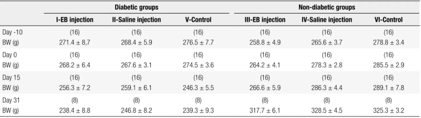
Table 1. Body weight of the animals in the experimental groups
Diabetic groups Non-diabetic groups
I-EB injection II-Saline injection V-Control III-EB injection IV-Saline injection VI-Control
Day -10 BW (g)
(16) 271.4 ± 8,7
(16) 268.4 ± 5.9
(16) 276.5 ± 7.7
(16) 258.8 ± 4.9
(16) 265.6 ± 3.7
(16) 278.8 ± 3.4 Day 0
BW (g)
(16) 268.2 ± 6.4
(16) 267.6 ± 3.1
(16) 274.5 ± 3.6
(16) 264.2 ± 4.1
(16) 278.3 ± 2.8
(16) 285.5 ± 2.9 Day 15
BW (g)
(16) 256.3 ± 7.2
(16) 259.1 ± 6.1
(16) 246.3 ± 5.5
(16) 266.6 ± 5.9
(16) 286.3 ± 4.4
(16) 289.1 ± 7.8 Day 31
BW (g)
(8) 238.4 ± 8.8
(8) 246.8 ± 8.2
(8) 239.3 ± 9.3
(8) 317.7 ± 6.1
(8) 328.5 ± 4.5
(8) 325.3 ± 3.2
BW: body weight; Day-10: day of streptozotocin injection in groups I, II and III; Day 0: day of EB administration in groups I and III or day of saline administration in groups II and IV. Data are presented as means ± standard deviations (SD) for the number of rats given in parenthesis.
Figure 2. GFAP immunohistochemical expression at 31 days in EB-induced lesions from diabetic (A) and non-diabetic rats (B). A and B) Bar = 50 µm.
ones (III, 55354.38 ± 5825.37; p = 0.001). As for dia-betic rats compared with non-diadia-betic, no difference was found between control animals (groups V and VI) and saline-injected rats (groups II and IV), nor for dia-betic groups II and V and non-diadia-betic groups IV and VI, with rats injected with saline solution or not.
DISCUSSION
Astrocytes play a key role in CNS homeostasis, inclu-ding maintenance of the blood-brain barrier, neuropro-tection from reactive oxygen species, regulation of neu-ronal activity and synaptic transmission, energy supply, as well as control of extracellular pH and ion and neu-rotransmitter concentrations, among many other func-tions (14,15).
Cop
yright
© ABE&M t
odos os dir
eit
os r
eser
vados
.
(about 6 nm) and microtubules (about 23 nm). In contrast to microtubules and actin ilaments, the com-position of intermediate ilaments changes among cell types, their developmental stages, and functional sta-tus (16). Astrocyte precursors and immature astrocytes present principally nestin and vimentin and, as astro-cytes mature, nestin expression disappears, GFAP be-comes increasingly expressed, and vimentin decreases to undetectable levels (15).
Insults to the CNS, such as trauma, ischemia, tu-mors, neuroinlammation, and neurodegenerative disorders lead to astrocytic activation, also known as reactive gliosis or astrogliosis, increasing the produc-tion of intermediate ilaments. In such condiproduc-tions, re-active astrocytes become highly positive for GFAP and vimentin, also reexpressing the third ilament protein, nestin (14-16). Reexpression of vimentin and strong astrocytic immunoreactivity to GFAP were clearly seen by Bondan and cols. (5) following EB injection in the rat brainstem from the 3rd to the 31st day post-gliotoxic injection.
This increased GFAP expression around the EB-in-duced lesions was also conirmed in the present study, but it was noted in the streptozotocin-diabetic rats that somehow diabetes hindered the increase in this expres-sion, although the same was not observed after saline solution injection. Besides, diabetic rats that did not re-ceive any intracisternal injection (EB or saline; control group) had no signiicant difference in GFAP expres-sion from non-diabetic ones, suggesting that such dif-ference caused by the diabetic status was only detected when a strong glial response to injury was induced (5).
Meanwhile, decreased astrocyte GFAP expression in type 1 diabetic rats was also found by other researchers with no additional harmful condition beyond diabetes (11,18-22). On the other hand, insulin treatment has shown to prevent diabetes-induced decreases in astro-cytic GFAP content (11)
Although astrocytes were not individually counted in our study, the decrease in GFAP content seen in diabetic rats apparently relected a decrease in GFAP expression rather than a decrease in the number of as-trocytes. This observation is similar to that of Coleman and cols. (11), and differs from Lechuga-Sancho and cols. (21),who reported a decrease in rat hypothalamic astrocyte numbers after 6 weeks of diabetes onset. It is recognized that astrocyte counts based on quantiica-tions of GFAP-positive cells are not really representa-tive, as diminution of GFAP immunoreactivity could lead to undercounting.
While it was initially thought that astrocyte pro-liferation was a major component of glial scar, it has been demonstrated repeatedly that there are actually few astrocytes undergoing cell division during gliosis (17). To corroborate this afirmation it is important to notice that no astrocyte in mitotic activity was seen in our studies, with astrocytic response following gliotoxic lesions (4,5).
The association of reactive astrocytes with enhanced GFAP and cellular hypertrophy, coupled with the in vi-tro observations that mature astrocytes do not represent a supportive environment for axon growth, has led to a widespread concept that reactive astrocytes are always detrimental to regeneration in the CNS. However, it
Table 2. Areas with GFAP staining in µm2 in a total area of 302,952.5 µm2 at 31 days, in rats diabetic or not, injected with EB or not
Animal
Diabetic groups Non-diabetic groups
I-EB injection (µm2)
II-Saline injection (µm2)
V-Control (µm2)
III-EB injection (µm2)
IV-Saline injection (µm2)
VI-Control (µm2)
1 50,241 5,834 9,313 47,281 6,132 6,249
2 60,312 10,105 8,131 39,522 5,041 8,833
3 48,154 4,581 5,121 51,430 4,190 5,214
4 53,826 6,912 4,297 40,265 7,013 5,575
5 57,115 6,134 5,642 34,127 7,264 7,266
6 61,232 7,283 6,715 36,112 6,145 4,127
7 62,841 8,145 4,237 50,717 4,827 4,483
8 49,114 4,905 8,541 33,903 8,549 9,028
Mean 55,354.38a 6,737.38c 6,499.6c 41,669.63b 6,145.13c 6,346.88c
standard deviation (SD) ±5,825.36 ±1,806.27 ±1,978.4 ±7,204.08 ±1,442.37 ±1,871.01
Cop
yright
© ABE&M t
odos os dir
eit
os r
eser
vados
.
has been proved in several models of brain and spinal cord injury that not all reactive astrocytes produce non-permissive molecules for neural regeneration, such as tenascin and condroitin sulfate family of proteoglycans, which are inhibitory for neurite outgrowth (17). In op-position to this proteoglycan up-regulation associated with regenerative failure in vivo, reactive astrocytes also produce molecules that can support regeneration, such as laminin (17). In addition, astrocytes are recognized to support oligodendrocytes during myelination and remyelination (15), and this role is particularly impor-tant after myelin loss due to naturally occurring or ex-perimentally induced demyelinating processes, such as the EB gliotoxic model.
The reaction of astrocytes following trauma form-ing a glial scar surroundform-ing the area of lesion could wall off the damaged area in an attempt to isolate the injury site and prevent any further damage to the nearby tis-sue, which is still viable.
Decreases in GFAP expression are invariably asso-ciated with detrimental conditions in the CNS (23) and this was also expected in relation to diabetes. Due to the complex scenario of CNS repair, the ambiguous roles of astrocytes in nervous tissue remodeling after injury, and as diabetes apparently negatively affects as-trocyte reaction, it is dificult to precisely infer if this impaired astrocytic response would play a beneicial or a deleterious inluence on the restoration of mor-phological and functional tissue integrity after neural damage.
Acknowledgements: this study was supported by São Paulo Rese-arch Foundation (Fapesp – 2008/58696-2) and National Coun-sel of Technological and Scientiic Development (CNPq).
Disclosure: no potential conlict of interest relevant to this article was reported.
REFERENCES
1. Graça DL, Bondan EF, Pereira LAVD, Fernandes CG, Maiorka PC. Behaviour of oligodendrocytes and Schwann cells in an experi-mental model of toxic demyelination of the central nervous sys-tem. Arq Neuropsiquiatr. 2001;59:358-61.
2. Pereira LAVD, Dertkigill MS, Graça DL, Cruz-Höfling MA. Dyna-mics of remyelination in the brain of adult rats after exposure to ethidium bromide. J Submicrosc Cytol Pathol. 1998;30:341-8. 3. Bondan EF, Lallo MA, Sinhorini IL, Pereira LAVD, Graça DL. The
effect of cyclophosphamide on the rat brainstem remyelination following local ethidium bromide injection in Wistar rats. J Sub-microsc Cytol Pathol. 2000;32:603-12.
4. Bondan EF, Lallo MA, Dagli MLZ, Pereira LAVD, Graça DL. Blood--brain barrier breakdown following gliotoxic drug injection in the brainstem of Wistar rats. Arq Neuropsiquiatr. 2002;60:582-9.
5. Bondan EF, Lallo MA, Dagli MLZ, Sanchez M, Graça DL. Investiga-tion into the astrocytic immunoreactivity to GFAP and vimentin in the brainstem of Wistar rats submitted to the ethidium bromide gliotoxic model. Arq Neuropsiquiatr. 2003;61:642-9.
6. Vincent AM, Kato K, McLean LL, Soules ME, Feldman EL. Sensory neurons and Schwann cells respond to oxidative stress by incre-asing antioxidant defense mechanisms. Antioxid Redox Signal. 2009;11:425-38.
7. Li P, Ding C, Muranyi M, He Q, Lin Y. Diabetes mellitus causes astrocyte damage after ischemia and reperfusion injury. J Cereb Flow Metab. 2005;25:S430.
8. Muranyi M, Ding C, He Q, Lin Y, Li P. Streptozotocin-induced dia-betes causes astrocyte death after ischemia and reperfusion in-jury. Diabetes. 2006;55:349-55.
9. Li PA, Siesjo BK. Role of hyperglycaemia-related acidosis in ischa-emic brain damage. Acta Physiol Scand. 1997;161:567-80. 10. Li PA, Gisselsson L, Keuker J, Vogel J, Smith MI, Kuschinsky W, et
al. Hyperglycemia exaggerated ischemic brain damage following 30 min of middle cerebral artery occlusion due to capillary obs-truction. Brain Res. 1998;804:36-44.
11. Coleman ES, Dennis JC, Braden TD, Judd RL, Posner P. Insulin treatment prevents diabetes-induced alterations in astrocyte glu-tamate uptake and GFAP content in rats at 4 and 8 weeks of dia-betes duration. Brain Res. 2010;8:131-41.
12. Bondan EF, Lallo MA, Trigueiro AH, Ribeiro CP, Sinhorini IL, Gra-ça DL. Delayed Schwann cell and oligodendrocyte remyelination after ethidium bromide injection in the brainstem of Wistar rats submitted to streptozotocin diabetogenic treatment. Braz J Med Biol Res. 2006;39:637-46.
13. Bondan EF, Martins MFM. Blood-brain barrier breakdown and re-pair following gliotoxic drug injection in the brainstem of strepto-zotocin-diabetic rats. Arq Neuropsiquiatr. 2012;70:221-5. 14. Ramos AT, Viott AM, Machado GF. Astrócitos. In: Graça DL,
Bon-dan EF, Pereira LAV, Maiorka PC (eds.). Biologia da desmielini-zação e da remielinidesmielini-zação: a base da esclerose múltipla. Santa Maria: Editora UFSM; 2011. p. 35-59.
15. Sofroniew MV, Vinters HV. Astrocytes: biology and pathology. Acta Neuropathol. 2010;119:7-35.
16. Pekny M. Astrocytic intermediate ilaments: lessons from GFAP and vimentin knock-out mice. In: Castellano-López B, Nieto-Sam-pedro M. Glial cell function. Amsterdam: Elsevier; 2001. p. 22-30. 17. Fitch MT, Silver J. Astrocytes are dynamic participants in central
nervous system development and injury responses. In: Jessen KR, Richardson WD. Glial cell development. Oxford: Oxford Uni-versity Press; 2001. p. 263-77.
18. Barber AJ, Antonetti DA, Gardnes TW. Altered expression of re-tinal occludin and glial ibrillary acidic protein in experimental diabetes. Invest Ofthalmol Vis Sci. 2000;41:3561-8.
19. Coleman ES, Judd R, Hoe L, Dennis J, Posner P. Effects of dia-betes on astrocyte GFAP and glutamate transporters in the CNS. Glia. 2004;48:166-78.
20. Dennis JC, Coleman ES, Swyers SE, Moody SW, Wright JC, Judd R, et al. Changes in mitotic rate and GFAP expression in the pri-mary olfactory axis of streptozotocin-induced diabetic rats. J Neurocytol. 2005;34:3-10.
21. Lechuga-Sancho AM, Arroba AI, Frago LM, Garcia-Cáceres C, de Célix AD, Argente J, et al. Reduction in the number of astrocytes and their projections in association with increased synaptic pro-tein density in the rat hypothalamus of poorly controlled diabetic rats. Endrocrinol. 2006;147:5314-24.
22. Afsari ZH, Renno WM, Abd-El Basset E. Alterations of glial ibrilla-ry acidic proteins immunoreactivity in astrocyte of the spinal cord diabetic rats. Anat Rec. 2008;291:390-9.