Cop
yright
© ABE&M t
odos os dir
eit
os r
eser
vados
.
Surgical management of pediatric
Cushing’s disease: an analysis
of 15 consecutive cases at a
specialized neurosurgical center
Estratégia cirúrgica na doença de Cushing em pacientes pediátricos: análise de 15 casos consecutivos operados em centro neurocirúrgico especializado
Ricardo Santos de Oliveira1, Margaret de Castro2, Sonir Roberto Rauber Antonini2,
Carlos Eduardo Martinelli Júnior2, Ayrton Custódio Moreira2, Helio Rubens Machado1
AbstrAct
Objective: The aim of this study was to review the results of surgery for pediatric patients with Cushing’s disease who were less than 18 years old and underwent transsphenoidal surgery in a specialized center during a 25-year period. Subjects and methods: Retrospective study, in which the medical records, histology and pituitary imaging of 15 consecutive pediatric patients with Cushing’s disease (mean age: 13 years) were evaluated by the same team of endocrinolo-gists and a neurosurgeon from 1982 to 2006. Patients were considered cured when there was clinical adrenal insuficiency and serum cortisol levels were below 1.8 μg/dL or 50 nmol/L after one, two, three, or seven days following surgery; they therefore required cortisone replacement therapy. Follow-up was for a median time of 11.5 years (range: 2 to 25 years). Results: Clinical and biochemical cure was achieved in 9/15 patients (60%) exclusively after transsphenoidal surgery. Hypopituitarism was observed in four patients; growth hormone deiciency, in two; permanent diabetes insipidus, in one case. Conclusions: Cushing’s disease is rare in children and adolescents. Transsphenoidal surgery is an effective and safe treatment in most of these patients. Plasma cortisol level < 1.8 μg/dL following surgery is the treatment goal and is a good predictive factor for long-term cure of Cushing’s disease. Arq Bras Endocrinol Metab. 2010;54(1):17-23
Keywords
Cushing’s disease; pituitary tumor; transsphenoidal surgery; pediatric neurosurgery
resumo
Objetivo: O objetivo deste estudo foi avaliar os resultados cirúrgicos em pacientes pediátricos com doença de Cushing com idade inferior a 18 anos, submetidos à cirurgia transfenoidal num centro especializado, durante um período de acompanhamento de 25 anos. Sujeitos e métodos: Estudo retrospectivo dos prontuários médicos de 15 pacientes pediátricos com doença de Cushing (ida-de média (ida-de 13 anos), sendo avaliados aspectos clínicos, laboratoriais, histológicos e radiológicos. Todos os pacientes foram avaliados pela mesma equipe de endocrinologistas e operados por um mesmo neurocirurgião, entre 1982 e 2006. O tempo médio de seguimento foi 11,5 anos (2 a 25 anos). Os pacientes foram considerados curados quando houve insuiciência adrenal e níveis de cortisol plasmático inferiores a 1,8 μg/dL ou 50 nmol/L no pós-operatório um, dois, três ou sete dias após a cirurgia; estes pacientes necessitaram de reposição de corticosteroide. Resultados: Cura clínica e bioquímica foi alcançada em 9/15 pacientes (60%) após a cirurgia transfenoidal. Hipopituitarismo foi observado em quatro pacientes; déicit de hormônio de crescimento, em dois; diabetes insípido per-manente, em um. Conclusões: A doença de Cushing é rara na infância e na adolescência. A cirurgia transfenoidal é um tratamento efetivo e seguro para a maioria dos pacientes. Uma concentração de cortisol plasmático < 1,8 μg/dL nos primeiros dias pós-cirurgia transfenoidal é o objetivo do tratamen-to e um fatratamen-tor preditivo tardio para a cura da doença de Cushing. Arq Bras Endocrinol Metab. 2010;54(1):17-23
Descritores
Doença de Cushing; tumor hipoisário; cirurgia transfenoidal; neurocirurgia pediátrica
1 Divisão de Neurocirurgia
Pediátrica, Departamento de Cirurgia e Anatomia, Faculdade de Medicina de Ribeirão Preto (FMRP), Universidade de São Paulo (USP), Ribeirão Preto, SP, Brasil
2 Divisão de Endocrinologia,
FMRP-USP, Ribeirão Preto, SP, Brasil
Correspondence to: Ricardo Santos de Oliveira Divisão de Neurocirurgia Pediátrica, Departamento de Cirurgia e Anatomia, FMRP-USP, Campus Universitário 14049-900 – Ribeirão Preto, SP, Brasil
Cop
yright
© ABE&M t
odos os dir
eit
os r
eser
vados
.
IntroDuctIon
C
ushing’s disease (CD) is a life-threatening con-dition in children. It is characterized by hyperse-cretion of an adrenocorticotropic hormone (ACTH) secreting tumor, which causes chronic adrenal overpro-duction of cortisol (1). The main clinical features and some aspects of the management of pediatric patients with CD differ somewhat from those seen in adults. They include growth impairment, weight gain, hyper-tension and pubertal delay or arrest. Transsphenoidal surgery remains the mainstay of therapy; it allows re-moval of an ACTH-producing adenoma without the need for long-term replacement therapy, though cure rates vary (2-6). Surgical technical dificulties and post-therapy hypopituitarism can be more damaging for children than adults because growth and puberty are not yet complete (4,7). We report on surgical management and outcome of the treatment of 15 pediatric CD pa-tients, who had only been operated on by the same neu-rosurgeon over the last 25 years at a specialized center.subjects AnD methoDs
Patients
This study was approved by the Ethics Committee of the Hospital das Clínicas of the Faculdade de Medici-na de Ribeirão Preto (FMRP) of Universidade de São Paulo (USP).
Medical records, imaging indings, and operative notes of 15 children younger than 18 years (7 males and 8 females), who were admitted consecutively to our service with CD from December 1982 to Decem-ber 2006, were retrospectively reviewed. Patient age (mean ± standard deviation, SD) at diagnosis was 13 ± 2.9 years (range: 6 to 18). The mean duration of CD was 3 years (range: 0.5 to 10). The main presenting features were weight gain (14/15), impaired growth (11/15), and hypertension (10/15). Abdominal striae, hirsutism, acne and amenorrhea were observed in 8, 7, 5 and 3 of the 15 patients, respectively.
All patients were evaluated by a multidisciplinary team, in which a neuroendocrinologist, a pediatric endo-crinologist, and a pediatric neurosurgeon were included.
Diagnosis of CD was established by suggestive clini-cal indings, loss of plasma or salivary cortisol circadian rhythm and no suppression of cortisol levels after an overnight dexamethasone suppression test (20 μg/kg up to 1 mg) (8,9). The pituitary etiology of
hypercorti-solism (CD) was determined by measurement of plasma ACTH levels, high-dose and very high-dose dexameth-asone suppression tests in all patients, and the cortico-trophin releasing hormone (CRH) test (8). All patients had some preoperative imaging. Computed tomogra-phy (CT) was performed in ten patients and magnetic resonance (MR), in ive cases. Five out of 15 patients had normal preoperative imaging; three of them per-formed CT. Bilateral inferior petrosal sinus sampling (BIPSS) for ACTH measure was performed in 2 of the 15 patients. Diagnosis of CD was conirmed by surgery and positive pituitary tissue pathology. Histological indings were divided into two groups: (i) obvious pi-tuitary adenomas with ACTH staining in 12 cases and (ii) biopsies with high numbers of ACTH-staining cells without evidence of adenoma in 3 patients.
surgical approach and dificulties
All operations were performed by the same neurosur-geon. The surgical technique and strategy described in detail by Hardy, in 1969, was followed (10). Briely, resection of all presumed adenomas was performed via a sublabial, paraseptal, transsphenoidal approach. Af-ter exposition of the sellar region and incision in the dura the entire pituitary gland was carefully examined, and all abnormal tissue was excised. In patients with no clearly abnormal tissue, depending on age, history, and severity of disease, hemihypophysectomy, subtotal hypophysectomy, or, rarely, total hypophysectomy was performed. Aggressiveness of the surgery was individu-alized, considering patient characteristics, preoperative morbidity, severity of clinical symptoms, and recur-rence of disease.
Fluoroscopy was especially helpful to reduce the risk of straying from the midline and injuring the carotid artery and cavernous sinus. An airpower drill (Midas Rex®, Medtronic Neurological) was used to open the sphenoid sinus and reach the pituitary sella in three cases, because the sphenoid sinus aeration was absent.
outcome evaluation
Cop
yright
© ABE&M t
odos os dir
eit
os r
eser
vados
.
resuLts
During the 25-year period, 15 children and adolescents with CD were treated with transsphenoidal surgery and followed up in the Hospital of Clinics of Ribeirão Pre-to, University of São Paulo (Table 1).
In this series, imaging correctly localized the ade-noma within the pituitary fossa in ten patients (66.6%), as conirmed by surgery. Eight of the 15 patients had a microadenoma; 2 had a macroadenoma; and 5 patients were normal in preoperative images. One patient (case 11) presented with a bilateral microadenoma in
preope-rative MR. Two of the 15 patients (cases 12 and 14) underwent BIPSS, which successfully lateralized the le-sion in both, and surgical cure was achieved after TSS.
In most patients a distinct tumor was visualized and excised. Adenomas were found in 12/15 patients based on histology (ACTH staining +); 3 patients pre-sented high numbers of ACTH staining cells (hyper-plasia) without evidence of adenoma.
Transsphenoidal excision of the corticotroph adeno-mas was performed in all patients. Clinical and/or bio-chemical cure was achieved in nine patients (60%) exclu-sively after TSS; eight of them achieved both criteria and
table 1. Surgical management of 15 consecutive pediatric patients with Cushing’s disease
case no.
Age (yrs) at surgery/
sex
Length of history
neuroradiological Features
bIPss Initial operative approach
resec-tion
histology Post-op serum cortisol
surgical cure
complemen-tary therapy
rt ADX remission outcome FW (yrs)
1 12/M 8 mos normal CT NA TSS partial adenoma ACTH+
7.2 no 2nd TSS† yes yes yes DM
hypert 25
2 13/M 1 yr micro (CT) NA TSS‡ total adenoma
ACTH+
< 1.2 yes NA no no NA normal 23
3 18/F 15 mos micro (CT) NA TSS total adenoma ACTH+
< 1.2 yes NA no no NA normal 20
4 12/M 5 yrs normal (CT) NA TSS total adenoma ACTH+
7.0 no 2nd TSS
(Nelson’s Syndrome)
yes yes no AP in reposition
20
5 14/M 4 yrs normal (CT) NA TSS total adenoma ACTH+
< 1.2 yes NA no no NA GH deficit 20
6 15/F 8 mos micro (CT) NA TSS total adenoma ACTH+
22.4 no 2nd TSS,
3rd TSS
(Nelson’s Syndrome)
no yes no death§ 3
7 14/F 10 mos micro (CT) NA TSS* total adenoma ACTH+, GH+
11.2 no 2nd TSS,
3rd TSS
(Nelson’s Syndrome)
yes yes no ketoconazole 15
8 12/F 3 yrs macro (CT) CS invasion
NA TSS partial adenoma ACTH+
4.2 no NA yes no yes AP in
reposition 15
9 6/F 2.5 yrs micro (CT) NA TSS* total adenoma ACTH+
< 1.2 yes NA no no NA normal 10
10 16/F 6 mos macro (CT) CS invasion
NA TSS total adenoma ACTH+
< 1.2 yes NA no no NA AP in reposition,
DDAVP 9
11 11/F 5 yrs bilateral micro (RM)
NA TSS total hyperplasia, ACTH+
< 1.2 yes NA no no NA normal 4
12 9/M 1 yr normal (RM) + (R) TSS total hyperplasia, ACTH+
< 1.2 yes NA no no NA normal 4
13 13/M 4 yrs micro (RM) NA TSS total adenoma ACTH+
< 1.2 yes NA no no NA normal 3
14 15/M 6 yrs normal (RM) + (L) TSS* total hyperplasia, ACTH+
2.7 no NA no no yes normal 2
15 16/F 10 yrs micro (RM) NA TSS total adenoma ACTH+
4.8 no NA no no no ketoconazole 1
M: male; F: female; CT: computed tomography; MR: magnetic resonance; TSS: transsphenoidal surgery; RT: radiotherapy; ADX: adrenalectomy; FW: follow-up; NA: not applicable; L: left-side gradient; R: right-side gradient; BIPSS: bilateral inferior petrosal sinus; Hyper: hypertension; DM: diabetes mellitus; Micro: microadenoma; Macro: macroadenoma; AP: anterior hypopituitarism; GH: growth hormone; CS: cavernous sinus.
† Hemorrhage secondary to an anomaly in the venous sinus during sella dissection; ‡ Bleeding during the surgical approach; § Meningitis three years after the 3rd TSS.
Cop
yright
© ABE&M t
odos os dir
eit
os r
eser
vados
.
one presented only clinical remission (case 14). It is im-portant to point out that case number 14 presented postop-erative plasma cortisol levels of 2.7 μg/dL. All other patients presented clinical and biochemical cure after TSS and complementary treatment. As this series of patients has been collected in a long period of time (24 years), during which there have been several improvements in diagnosis, surgical and radiotherapic techniques, the patients were divided into two groups: operated before and after 1998. There was a remarkable improvement of percentage of surgical cure (37.5% versus 85.7%) in the latter ten years.
All nine patients have remained cured and have re-quired no further treatment to date (median follow-up period: 12.1 years; range: 2 to 23 years). The six remain-ing patients had persisted CD, with a cortisol level in ex-cess of 300 nmol/L. A second TSS was performed in four patients. Three of them, who had persistent hypercorti-solemia, were treated with postoperative external-beam direct pituitary irradiation, using a 6-MV linear accelera-tor, with 45 Gy in 25 fractions over a period of 35 days after surgery. Bilateral adrenalectomy was performed in four patients (cases 1, 4, 6 and 7). Three patients devel-oped Nelson’s syndrome and TSS was performed in two patients (cases 6 and 7) for the third time. Postoperative radiotherapy was performed as the only complementary treatment in one patient (case 8) (Figure 1).
Hypopituitarism was observed in four patients, two of them with macroadenoma (case 8 and 10), one pa-tient (case 4) after radiotherapy to treat Nelson’s syn-drome, and one patient after TSS (case 5); permanent diabetes insipidus was observed in only one patient (case 10) and two patients developed growth hormone dei-ciency (case 5 and 14). One patient (case 6) developed meningitis after the third TSS and died.
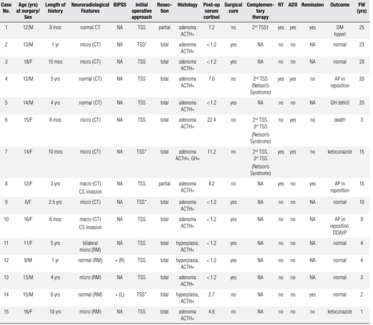
Figure 1. Case 10: (A) Sagittal and coronal (B) contrast-enhanced MR images obtained in an adolescent with Cushing’s disease, demonstrating a macroadenoma (asterisk); (C) contrast-enhanced sagittal MR imaging scan of the sella four years after transsphenoidal surgery and radiotherapy, demonstrating no evidence of residual tumor.
DIscussIon
CD is rare in the pediatric age group (6,11,12). The disease remains a complex diagnostic challenge in child-hood, because many of the diagnosis criteria and deini-tion of cure after transsphenoidal surgery are based on results obtained in adults that have been extrapolated to the pediatric population (13). In this study, the mean prediagnosis duration of symptoms was 36 months – approximately half of that reported in adults (14,15). This inding was consistent with other reports of pe-diatric CD and has been attributed to the additional symptoms of growth delay and pubertal arrest, which prompt earlier medical evaluation (13). Children with CD present with obesity, stunted growth, and mental and behavioral problems. More than one-third of pa-tients also have hypertension. The frequencies of the symptoms found were similar to those of other pub-lished series (5,16). Increased cortisol and ACTH lev-els, absence of diurnal variation, cortisol and ACTH re-sponse to CRH test, poor suppression of serum cortisol levels with a low dose of dexamethasone, and suppres-sion with a high dose are considered to be key evidence for biochemical diagnosis of CD (11,17).
CD is often due to microadenomas that may not be detectable in routine contrast-enhanced CT and MR images. The correct detection rate of corticotroph ad-enoma for pediatric CD ranges from 52% to 75% for CT and/or MR scans in the literature (5). Most of the corticotrophin adenomas are small (< 4 mm), and their images have similar intensity to those of normal pitu-itary tissue, making accurate identiication of the tumor site dificult (18,19). Five out of these 15 patients had normal preoperative imaging; 3 of them performed only
Cop
yright
© ABE&M t
odos os dir
eit
os r
eser
vados
.
CT, previously MR availability. Recently, BIPSS was performed successfully in two patients with normal MR scan. This procedure proved to be a reliable method of tumor localization when CT or MR were normal, as also found by other researchers who have reported accuracy ranges of 71% to 83% for BIPSS lateralization (5,14,16). Transsphenoidal surgery is a safe and effective irst-choice treatment for the management of CD (4-6,20); it was the preferred approach for excising the adeno-ma in the series of the present study. However, some peculiarities are observed in the pediatric population. Since children have smaller nasal apertures, the subla-bial route provided a wider corridor to access the sellar region than the direct transnasal rhinoseptal approach; this allowed the researchers to use the same nasal specu-lums utilized in adult patients (4). Care should be taken not to open the blades of Hardy’s nasal speculum too wide within the sphenoid sinus, because the chances of injuring the carotid artery are greater in children, due to the thin surrounding bones and the narrower sphenoid sinus (10). During sellar dural opening, es-pecially in microadenomas, care should be taken not to injure the intercavernous sinuses at the anterior and posterior sellar dural limits or the cavernous sinuses lat-erally, because this may cause excessive venous bleeding (13). In two cases, severe hemorrhage was observed – which was secondary to an anomaly of the venous sinus observed during sella dissection in one case. In more
table 2. Overview of previously reports on the surgical outcome of the TSS in pediatric Cushing’s disease
series number of cases surgical cure (%) recurrence reoperation remission mortality Follow-up
Styner and cols. (11) 15 14 (93.3) 0 2 1 0 3.6 years (10 months-8.2 years)
Buchfelder and Falbusch (21) 15 13 (86.6) 0 0 1 1 3.1 years (1.2-11 years)
Haddad and cols. (23) 5 5 (100) 0 0 0 0 4.6 years (0.5 to 9 years)
Partington and cols. (27) 15 12 (80)a 3 0 3 0 4.5 years (0 to 13.5 years)
Magiakou and cols. (6) 37b 35 (94.5) 2 2 2 2 22 months (5 to 60 months)
Dyer and cols. (22) 36 23 (64) 5 5 5 1 6 months to 21 years
Weber and cols. (12) 9 5 (55.5) 0 0 1 0 1.6 to 10.7 years
Mathivon and cols. (25) 16 9 (56.2) 5 2 3 0 40 ± 35 months
Devoe and cols. (20) 26 19 (73) 7 6 5 0 7.2 years (1.5 to 13.6 years)
Massoud and cols. (24) 12 9 (75) 3 3 2 0 6.8 (1 to 14 years)
Joshi and cols. (5) 25 14 (56) 10 0 11 0 6.9 years (1.3 to 12 years)
Kanter and cols. (13) 33c 22 (66.6) 3 3 8 0 3.6 years (0 to 9 years)
Storr and cols. (28) 27 16 (59) 0 0 0 0 7.1 ± 5.3 years (0.5 to 17.8 years)
Mehrazin (26) 8 6 (75) 2 2 0 0 13.4 (1 to 23 years)
Das and cols. (4) 10 4 (40) 6 1 3 0 5.3 years (1 to 10 years)
Current series 15 8 (53.3) 2 4 2 1 11 years (1 to 25 years)
a In the group of 12 patients with initial remission, 3 patients had a late recurrence; b excluded cases operated on in another service; c ten patients were submitted on TSS surgery in another service4.
recent cases, the endonasal route and neuronavigation have been employed with success.
In this series, even when a tumor was identiied during surgery, surgical cure was not achieved in all patients. The surgical cure rate over 25-year period of this study was 60%. However, there was a remark-able improvement of surgical cure (85.7%) in the lat-ter ten years. In the lilat-terature, surgical cure rates range from 40% to 100% (mean: 70.5%), according to table 2 (4-6,12,13,20-28). In the present study, plasma cor-tisol levels considered were < 1.8 μg/dL during the irst days after surgery for cure deinition – this is a very strict criterion. Notably, the criteria for cure vary sig-niicantly in the studies from the last 25 years, justifying the variable surgical cure rates observed (29,30). In ad-dition, the experience of the neurosurgeon is another important factor. Finally, there have been several im-provements in surgical techniques, such as endoscopy and neuronavigation. All these aspects have improved surgical treatment of the CD, but also have rendered very dificult to compare patients operated 20 years ago with those operated more recently.
morpho-Cop
yright
© ABE&M t
odos os dir
eit
os r
eser
vados
.
logic study. Among then, one patient did not achieved cure. The others have been cured in a follow-up period of four years. Pituitary hyperplasia can be deined as a non-neoplastic increase in one or more functionally dis-tinct types of pituitary cells (31). Kovacs and cols. (32) reported a patient who present corticotroph hyper-plasia and had a long-lasting remission (14 years), but CD recurred. The authors suggest that corticotroph hyperplasia may cause CD and the elimination of the negative inhibitory feedback effect by corticosteroids after adrenalectomy plays a role in adenoma initiation (32). Therefore, ultimate outcome analysis is critically dependent on the criteria adopted to deine cure and may only be made with careful and long-term follow-up. Despite a clear need for uniformity in the deinition of postoperative cure in CD, there is still considerable variation in deinitions between centers, making mean-ingful comparison of data dificult (33,34).
As advocated by Trainer and cols. (19), a postop-erative cortisol level of < 1.8 μg/dL or 50 nmol/L has been adopted as criterion of cure, based on the physi-ological principle that high cortisol levels will suppress normal corticotroph function, so that complete remov-al of a corticotroph adenoma will render the patient ACTH deicient, with low or undetectable cortisol lev-els. Some authors (34) have proposed that the presence of an intrasellar lesion and postoperative serum cortisol < 50 nmol/L are good predictors of cure in long term, with some degree of hypopituitarism. However, Yap and cols. (35) concluded that undetectable postopera-tive cortisol is not always predicpostopera-tive of long-term cure in an adult series.
Pituitary radiotherapy is effective for the treatment of CD, but it is only used on patients with persisting disease after surgery; growth hormone deiciency seems to be an unavoidable complication after treatment with radiotherapy (5,36). In this series, radiotherapy was per-formed as complementary treatment in four patients af-ter TSS, with two of them achieving cure of CD. Recent-ly, it has been reported that high-precision stereotactic radiosurgery (37) and gamma knife surgery (38) can ef-fectively treat persistent or recurrent CD following TSS. Bilateral adrenalectomy has long been considered the treatment of choice for CD in childhood. Nowa-days, it still has a role, but should be reserved for pa-tients in whom surgery and radiotherapy fail to stop the secretion of ACTH from the pituitary adenoma (25). Although adrenalectomy is the only treatment that offers an immediate control of hypercortisolism
with 100% certainty, it is necessary to consider its side effects, including potential adrenal insuficiency crisis. Therefore, lifelong need for glucocorticoid and min-eralocorticoid replacement therapy hyperpigmentation, elevated ACTH levels, and an enlarged sella turcica at-tributable to Nelson’s syndrome have been described in 12 to 67% of cases (38-41). In addition, there is a continued need for glucocorticoid and mineralocorti-coid replacement therapy. Bilateral adrenalectomy was performed in four patients; three of them developed Nelson’s syndrome and were treated with further sur-gery and radiotherapy. Pituitary radiotherapy at the time of adrenalectomy seems to reduce the risk of Nel-son’s syndrome development (41).
In conclusion, TSS remains a safe and effective pri-mary treatment of pediatric CD, with minimal morbid-ity and mortalmorbid-ity and with a cure rate comparable to those reported in published series. However, interpre-tation of the results of surgery depends on the criteria adopted to deine postoperative cure. Our data support the proposals of others that the goal of surgery for CD should be to render postoperative cortisol levels low or undetectable, while maintaining normal pituitary func-tion. A neurosurgeon, who is a specialist in pituitary disease, is indicated.
Disclosure: no potential conlict of interest relevant to this article was reported.
reFerences
1. Savage MO, Storr HL, Chan LF, Grossman AB. Diagnosis and treat-ment of pediatric Cushing’s disease. Pituitary. 2007;10(4):365-71. 2. Bigos ST, Somma M, Rasio E, Eastman RC, Lanthier A, Johnston
HH, et al. Cushing’s disease: management by transsphenoidal pi-tuitary microsurgery. J Clin Endocrinol Metab. 1980;50(2):348-54. 3. Chee GH, Mathias DB, James RA, Kendall-Taylor P.
Transsphenoi-dal pituitary surgery in Cushing’s disease: can we predict outco-me? Clinical Endocrinol (Oxf). 2001;54(5):617-26.
4. Das NK, Lyngdoh BT, Bhakri BK, Behari S, Bhatia V, Jain VK, et al. Surgical management of pediatric Cushing’s disease. Surg Neu-rol. 2007;67(3):251-7.
5. Joshi SM, Hewitt RJ, Storr HL, Rezajooi K, Ellamushi H, Gross-man AB, et al. Cushing’s disease in children and adolescents: 20 years of experience in a single neurosurgical center. Neurosurg. 2005;57(2):281-5.
6. Magiakou MA, Chrousos GP. Cushing’s syndrome in children and adolescents: current diagnostic and therapeutic strategies. J En-docrinol Invest. 2002;25(2):181-94.
7. Liddle GW. Tests of pituitary-adrenal suppressibility in the diagnosis of Cushing’s syndrome. J Clin Endocrinol Metab. 1960;20:1539-60.
Cop
yright
© ABE&M t
odos os dir
eit
os r
eser
vados
.
9. Martinelli CE Jr, Sader SL, Oliveira EB, Daneluzzi JC, Moreira AC. Salivary cortisol for screening of Cushing’s syndrome in children. Clin Endocrinol (Oxf). 1999;51(1):67-71.
10. Hardy J. Transphenoidal microsurgery of the normal and patholo-gical pituitary. Clin Neurosur. 1969;16:185-217.
11. Styne DM, Grumbach MM, Kaplan SL, Wilson CB, Conte FA. Treat-ment of Cushing’s disease in childhood and adolescence by transs-phenoidal microadenomectomy. N Engl J Med. 1984;310(14):889-93. 12. Weber A, Trainer PJ, Grossman AB, Afshar F, Medbak S, Perry LA, et al. Investigation, management and therapeutic outcome in 12 cases of childhood and adolescent Cushing’s syndrome. Clin En-docrinol (Oxf). 1995;43(1):19-28.
13. Kanter AS, Diallo AO, Jane JA Jr, Sheehan JP, Asthagiri AR, Oskouian RJ, et al. Single-center experience with pediatric Cushing’s disease. J Neurosurg. 2005;103(5 Suppl):413-20. 14. Boggan JE, Tyrrell JB, Wilson CB. Transsphenoidal microsurgical
management of Cushing’s disease. Report of 100 cases. J Neuro-surg. 1983;59(2):195-200.
15. Mampalam TJ, Tyrrell JB, Wilson CB. Transsphenoidal microsur-gery for Cushing disease. A report of 216 cases. Ann Intern Med. 1988;109(6):487-93.
16. Fahlbusch R, Honegger J, Buchfelder M. Neurosurgical manage-ment of Cushing’s disease in children. In: Savage MO, Bourgui-non JP, Grossman AB, editors. Frontiers in pediatric endocrinolo-gy. Oxford: Blackwell Scientiic Publications; 1994. p. 68-72. 17. Streeten DH, Faas FH, Elders MJ, Dalakos TG, Voorhess M.
Hyper-cortisolism in childhood: shortcomings of conventional diagnos-tic criteria. Pediatrics. 1975;56(5):797-803.
18. Marcovitz S, Wee R, Chan J, Hardy J. Diagnostic accuracy of pre-operative CT scanning of pituitary prolactinomas. AJNR Am J Neuroradiol. 1988;9(1):13-7.
19. Trainer PJ, Lawrie HS, Verhelst J, Howlett TA, Lowe DG, Gross-man AB. Transsphenoidal resection in Cushing’s disease: unde-tectable serum cortisol as the deinition of successful treatment. Clin Endocrinol (Oxf). 1993;38(1):73-8.
20. Devoe DJ, Miller WL, Conte FA, Kaplan SL, Grumbach MM, Ro-senthal SM, et al. Long-term outcome in children and adolescents after transsphenoidal surgery for Cushing’s disease. J Clin Endo-crinol Metab. 1997;82(10):3196-202.
21. Buchfelder M, Fahlbusch R. Neurosurgical treatment of Cushing’s disease in children and adolescents. Acta Neurochir Suppl (Wien). 1985;35:101-5.
22. Dyer EH, Civit T, Visot A, Delalande O, Derome P. Transsphenoi-dal surgery for pituitary adenomas in children. Neurosurgery. 1994;34(2):207-12.
23. Haddad SF, VanGilder JC, Menezes AH. Pediatric pituitary tumors. Neurosurgery. 1991;29(4):509-14.
24. Massoud AF, Powell M, Williams RA, Hindmarsh PC, Brook CG. Transsphenoidal surgery for pituitary tumours. Arch Dis Child. 1997;76(5):398-404.
25. Mathivon L, Carel JC, Coutant R, Derome P, Adamsbaum C, Boug-nères P, et al. [Cushing disease in children and in adolescents. Therapeutic results]. Arch Pediatr. 1997;4(6):521-8.
26. Mehrazin M. Pituitary tumors in children: clinical analysis of 21 cases. Childs Nerv Syst. 2007;23(4):391-8.
27. Partington MD, Davis DH, Laws ER Jr, Scheithauer BW. Pituitary adenomas in childhood and adolescence. Results of transsphe-noidal surgery. J Neurosurg. 1994;80(2):209-16.
28. Storr HL, Afshar F, Matson M, Sabin I, Davies KM, Evanson J, et al. Factors inluencing cure by transsphenoidal selective ade-nomectomy in paediatric Cushing’s disease. Eur J Endocrinol. 2005;152(6):825-33.
29. Czepielewski MA, Rollin GA, Casagrande A, Ferreira NP. Criteria of cure and remission in Cushing’s disease: an update. Arq Bras Endocrinol Metab. 2007;51(2):1362-72.
30. Patil CG, Prevedello DM, Lad SP, Vance ML, Thorner MO, Katz-nelson L, et al. Late recurrences of Cushing’s disease after initial successful transsphenoidal surgery. J Clin Endocrinol Metab. 2008;93(2):358-62.
31. Al-Gahtany M, Horvath E, Kovacs K. Pituitary hyperplasia. Hor-mones. 2003;2(3):149-58.
32. Kovacs K, Horvath E, Coire C, Cusimano M, Smyth H, Scheithauer BW, et al. Pituitary corticotroph hyperplasia preceding adenoma in a patient with Nelson’s syndrome. Clin Neuropathol. 2006;25(2):74-80. 33. Newell-Price J. Transsphenoidal surgery for Cushing’s dise-ase: deining cure and following outcome. Clin Endocrinol. 2002;56(1):19-21.
34. Rees DA, Hanna FW, Davies JS, Mills RG, Vaidis J, Scanlon MF. Long-term follow-up results of transsphenoidal surgery for Cushing’s disease in a single centre using strict criteria for remis-sion. Clin Endocrinol (Oxf). 2002;56(4):541-51.
35. Yap LB, Turner HE, Adams CB, Wass JA. Undetectable posto-perative cortisol does not always predict long-term remission in Cushing’s disease: a single centre audit. Clin Endocrinol. 2002;56(1):25-31.
36. Mahmoud-Ahmed AS, Suh JH. Radiation therapy for Cushing’s disease: a review. Pituitary. 2002;5(3):175-8.
37. Jalali R, Brada M. Radiosurgery for pituitary adenoma. Crit Rev Neurosurg. 1999;9(3):167-73.
38. Weiss MH, Couldwell WT. Gamma Knife surgery for Cushing dise-ase. J Neurosurg. 2007;106(6):976-7.
39. Hopwood NJ, Kenny FM. Incidence of Nelson’s syndrome after adrenalectomy for Cushing’s disease in children: results of a na-tionwide survey. Am J Dis Child. 1977;131(12):1353-6.