Michelle Mikhael AMMARI(a)
Luiz Flávio Martins MOLITERNO(b)
Raphael HIRATA JÚNIOR(c)
Mariana Canano SÉLLOS(d)
Vera Mendes SOVIERO(b)
Wagner Pereira COuTINHO FILHO(e)
(a)Department of Specific Training, School of
Dentistry, Universidade Federal Fluminense - UFF, Nova Friburgo, RJ, Brazil.
(b)Department of Preventive and Community
Dentistry, School of Dentistry, Universidade do Estado do Rio de Janeiro - UERJ, Rio de Janeiro, RJ, Brazil.
(c)Department of Microbiology, Immunology
and Parasitology, School of Medicine, Universidade do Estado do Rio de Janeiro - UERJ, Rio de Janeiro, RJ, Brazil.
(d)School of Dentistry, Universidade do Estado
do Rio de Janeiro - UERJ, Rio de Janeiro, RJ, Brazil.
(e)Department of Microbiology, School of
Dentistry, Centro Universitário Serra dos Órgãos - UNIFESO, Teresópolis, RJ, Brazil.
Efficacy of chemomechanical caries
removal in reducing cariogenic
microbiota: a randomized clinical trial
Abstract: The aim of this study was to compare the eficacy of che -mochemical methods (Carisolv™ and Papacárie®) versus the manual method (excavators) in reducing the cariogenic microbiota in dentine caries of primary teeth. Forty-six healthy children (5 to 9 years old) having at least one primary tooth with a cavitated dentine carious lesion were included in the study. The teeth presented no clinical or radiographic signs of pulpal involvement. The sample of 74 teeth was randomly divided into three different groups: Papacárie® (n = 25), Ca-risolv™ (n = 27) and Manual (n = 22). Samples of carious and sound dentine were collected with sterile excavators before and after caries removal in the three groups. The dentine samples were transferred to glass tubes containing a 1mL thioglycollate medium used as a car-rier and enriched for microbiological detection of mutans strepto-cocci and Lactobacillus spp, after incubation for 6h at room tempera-ture. The minimum detection value for colony forming units (CFU) was 3.3 x 10² CFU/ml, and the results were converted into scores from 0 to 4. A signiicant difference was observed in relation to the microbio -logical scores before and after caries removal for all methods (Wilcoxon test; p < 0.001). The use of chemomechanical methods for caries removal did not improve the reduction of cariogenic microorganisms in dentine caries lesions, in comparison with manual excavation.
Keywords: Dental Caries; Papain; Microbiology; Tooth, Deciduous.
Introduction
The therapeutic approach to carious dentine lesions has been recon-sidered by the scientiic community over time as a way to preserve as much tooth structure as possible, increase the longevity of teeth and prevent the repetitive restorative cycle.1,2,3 Based on the minimal inva-sive dentistry concept, more conservative approaches have been recom-mended,4,5 such as partial caries removal,6 atraumatic restorative treat-ment7 and chemomechanical methods,8,9 instead of conventional caries removal by drilling and manual excavation.
Although the chemomechanical system was developed over 30 years ago, it only started to gain attention in the late 90s, when Carisolv™ was released on the market and began to be used widely. This system involves application of a gel composed of an amino acid solution and sodium hypochlorite, which are able to dissolve collagen ibers degraded Declaration of Interests: The authors
certify that they have no commercial or associative interest that represents a conflict of interest in connection with the manuscript.
Corresponding Author: Michelle Mikhael Ammari E-mail: [email protected]
DOI: 10.1590/1807-3107BOR-2014.vol28.0031 Epub XXX XX, 2014
Submitted: Nov 14, 2012
by caries, facilitating their removal by appropriated manual instruments.8,10 In order to disseminate the large-scale use of the chemomechanical method to remove carious dentine, and considering the high cost of Carisolv™, a proteolytic gel named Papacá-rie® was released in Brazil. Papacárie® combines the collagen degradation effect of papain (a natural pro-tease) and the bactericide effect of chloramines.9,11
A certain number of residual microorganisms, 101 to 103 of colony forming units (CFU), in the dentine after cavity preparation is considered acceptable and not harmful to teeth.12,13,14 Clinical research compar-ing the conventional method (drillcompar-ing) with the Cari-solv™ chemomechanical method has concluded that both methods are similar in terms of CFU reduction in residual dentine.13,14,15,16
The purpose of the present study was to compare the eficacy of the chemochemical methods (Cari -solv™ and Papacárie®) versus the manual method (excavators) in reducing the cariogenic microbiota in dentine caries of primary teeth.
Methodology
Study design:This study was designed as a ran-domized, controlled clinical trial following the CON-SORT statement17. Three hundred and eight children (5 to 9 years old) were screened at the Pediatric Den-tal Clinic of Universidade Estadual do Rio de Janeiro. Children who presented at least one tooth with den-tine caries accessible (cavitated) for excavation in the occlusal or buccal surfaces were considered eligible for the study. Informed consent was obtained from parents and the study was approved by the Research Ethics Committee (1007-CEP-HUPE) at the Univer-sidade Estadual do Rio de Janeiro (UERJ). An experi-enced clinician detected carious lesions visually and in radiographs. Bite-wing radiographs were used to assess the depth of the occlusal lesions. All radio-graphs were performed using pediatric ilm-holders (Indusbello®, Londrina, Brazil). Dental examinations were carried out in a dental chair under standard-ized conditions after prophylaxis.
Forty-eight children (mean age of 6 to 9 years) were included in the study and 95 primary teeth with dentine carious lesions accessible for excava-tion were selected. Twenty-one teeth were excluded
due to extensive lesion depth (in the inner third of the dentine) or due to clinical or radiographic signs of pulpal involvement. The inal sample (74 primary teeth, 40 anterior teeth with buccal caries lesion and 34 posterior teeth with occlusal lesions) was randomly allocated to one of the three methods using a ran-dom numbers table. When more than one tooth was selected in the same child, the teeth were included in the table following the examination sequence from the upper right to the lower right quadrants. After randomization, the inal sample was allocated as fol -lows: Carisolv™ (Medi Team Dental AB, Savedalen, Sweden) (n = 27); Papacárie® (Fórmula & Ação, São Paulo, Brazil) (n = 25); Manual excavation (n = 22).
Calibration: A trained operator performed the
clinical interventions. Theoretical and practical train-ing with 17 teeth were performed durtrain-ing a pilot study carried out under the same conditions as the present study. During training, it was emphasized that consistence was more important than the color of the dentine, as a clinical sign of the level of infec-tion. The criteria were discussed exhaustively and good interexaminer agreement was achieved regard-ing the decision about stoppregard-ing caries removal. A kappa coeficient of 0.89 was obtained.
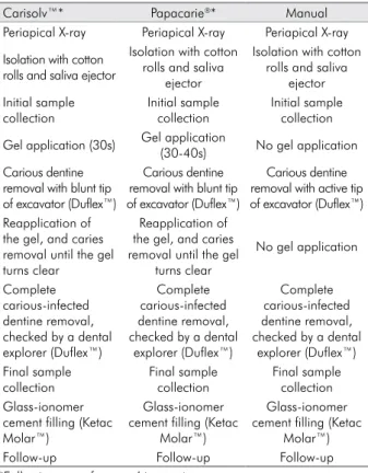
Intervention: The sequence of caries removal for each method is described in Table 1. The clinical decision to stop caries removal was based on tactile criteria1,12. Caries removal was stopped when the dentine showed slight resistance to excavation and no tug-back sensation was felt when the tip of a blunt explorer was pressed into the dentine. If the dentine was reasonably irm, caries removal was stopped. The color of the dentine was not used as an indica-tor of the moment to stop excavation. Apart from this clinical criterion, Papacárie® and Carisolv™ were employed according to the manufacturers’ instruc-tions, which establish that caries removal is complete when the gel appears clear and without debris. After caries removal, teeth from the three groups were restored with conventional glass-ionomer cement (Ketac Molar/ 3M-ESPE™, Monrovia, USA).
collected before caries removal and the second, after caries removal. The active part of the same dentine excavator (11½ Dulex™) used in the irst and the sec -ond dentine removal procedures was used to mea-sure the dentine samples. Microbiological procedures were based on experiments performed elsewhere15. The dentine samples were transferred to glass tubes containing 1mL thioglycollate medium used as a carrier and enriched for microbiological detection of mutans streptococci and Lactobacillus spp, after incubation for 6h at room temperature. The dentine samples were subsequently stirred vigorously in a vortex blender to produce a suspension. Serial decimal dilutions were prepared in phosphate buffered saline (PBS, 0.01M, pH 7.2). Aliquots of 10µL were inocu-lated, in triplicate, in selective mediums for mutans streptococci (Mitis-Salivarius Agar - Difco Labora-tories, Detroit, MI, containing 20% sucrose and 0.25 IU bacitracin/mL) and for Lactobacillus spp. (Rogosa Agar, BBL - BD, Basingstoke, United Kingdom). The agar plates were incubated at 37 ºC for 48 h under an anaerobic atmosphere (Anaerogen, Oxoid
Bas-ingstoke, United Kingdom), and the colonies were counted and corrected for the dilution factor. The minimum detection count value of colony forming units (CFU) was 3.3 X 10² CFU/ml, and the results were converted into scores from 0 to 4 (Table 2) to facilitate the description of the results. The microbi-ological analysis was performed blindly in relation to the caries removal method.
Statistical analysis:Data were analyzed by means of the Kruskal-Wallis and Wilcoxon tests to verify if there were any differences in the reduction of cario-genic lora among the methods used. The statistical level of signiicance was set at 5%.
Results
Ten (13.5%) microbiological samples were excluded due to contamination by fungus or no bacterial growth (5 teeth from the CarisolvTM group and 5 from the Manual group). Table 3 shows the distribution of CFU scores before and after caries removal for the three groups (n = 64). CFU scores did not differ signiicantly among the three groups, neither before nor after car-ies dentine removal (Kruskal-Wallis test; p > 0.05).
A signiicant difference was observed in relation to the microbiological scores before and after caries removal for all methods (Wilcoxon test; p < 0.001). CFU scores after caries removal were signiicantly lower than the initial scores, regardless of the method.
Table 3. Reduction of CFU (colony forming units) scores after treatment with CarisolvTM, Papacárie® and Manual excavation.
Reduction of CFU score (Difference between initial and final score)
Treatment
CarisolvTM Papacarie® Manual
4 2 1 5
3 7 9 3
2 10 5 2
1 3 10 7
No reduction 0 0 0
Total 22 25 17
Table 1. Description of the caries removal technique for each group
Carisolv™* Papacarie®* Manual
Periapical X-ray Periapical X-ray Periapical X-ray
Isolation with cotton rolls and saliva ejector
Isolation with cotton rolls and saliva
ejector
Isolation with cotton rolls and saliva
ejector Initial sample collection Initial sample collection Initial sample collection
Gel application (30s) Gel application
(30-40s) No gel application Carious dentine
removal with blunt tip of excavator (Duflex™)
Carious dentine removal with blunt tip of excavator (Duflex™)
Carious dentine removal with active tip of excavator (Duflex™) Reapplication of
the gel, and caries removal until the gel turns clear
Reapplication of the gel, and caries removal until the gel
turns clear
No gel application
Complete carious-infected dentine removal, checked by a dental explorer (Duflex™)
Complete carious-infected dentine removal, checked by a dental
explorer (Duflex™)
Complete carious-infected dentine removal, checked by a dental
explorer (Duflex™) Final sample collection Final sample collection Final sample collection Glass-ionomer
cement filling (Ketac Molar™)
Glass-ionomer cement filling (Ketac
Molar™)
Glass-ionomer cement filling (Ketac
Molar™)
Follow-up Follow-up Follow-up
*Following manufacturers’ instructions.
Table 2. Score description of CFU (colony forming units)
Score CFU values
0 ≤ 3.3 x 10²
1 3.4 x 10² – 3.3 x 10³
2 3.4 x 10³ – 3.3 x 10⁴
3 3.4 x 10⁴ – 3.3 x 10⁵
Discussion
Chemomechanical methods for caries removal can be expected to display more extensive antimicrobial properties in both carious and clinical sound dentin tissues, by virtue of the presence of substances hav-ing antimicrobial properties. In the present study, a signiicant reduction in the cariogenic microbiota in dentine was observed after caries removal, regardless of the method used. The results of the two chemo-mechanical methods used were comparable to those obtained after caries removal by manual excavation. Therefore, the two chemomechanical methods evalu-ated in this study were not more effective in reducing the cariogenic microbiota in dentine carious lesions, in comparison with the manual method.
Previous studies reported similar results with pri-mary teeth, showing that Carisolv™ did not improve the reduction of cariogenic microbiota (mutans strep-tococci and Lactobacillus spp.) in dentine caries, com-pared with manual excavation,14,15 or drilling.13,15 Con-versely, in a clinical study, the reduction of cariogenic lora after caries removal was attributed to the anti -bacterial properties of Carisolv™, which has amino acids and sodium hypochlorite in its composition.16
Differences in selection criteria, sampling proce-dures and bacterial culture techniques could explain the controversial results in studies assessing antimicrobial eficacy of the chemomechanical methods.13,14,15,16 Some studies used a speciic size of sterile bur to collect the dentine samples,12,13,16 whereas others used a manual sterile excavator14,15 or the speciic Carisolv™ excavator.16 In the present study, dentine samples were collected with a sterile excavator (111/2 Dulex™). The use of burs was avoided because of the higher risk of causing acci-dental pulp exposure. Additionally, their high speed could cause a negative effect on the children’s behavior. In addition, some molecular methods, including qPCR, may render different results, because of their sensitivity, especially when dealing with fastidious organisms such as mycobacteria.18 Nevertheless, molecular methods may detect DNA of non-viable organisms, as observed in an attempt to detect DNA of Yersinia pestis from skeletons whose patients had deceased from plague in the 14th century.19As reported recently, integrated approaches use both culture methods and molecular detection of other fastidious, dificult-to-grow organisms in dental
caries to provide supplementary results for the progres-sion of dental caries.20
Unlike this clinical trial, other clinical studies have been conducted under local anesthesia and rubber dam,13,15,16 and burs were used on the enamel to facilitate access to the carious dentine.15 In the present study, the manufacturers’ instructions (Papacárie® and Carisolv™) for chemomechanical caries removal were followed strictly. Therefore, only caries lesions with direct access to the dentine were selected for this clinical trial, and hand instruments were used for undermined enamel. Clinical procedures were carried out with cotton rolls and suction, under proper moisture control.
According to Subramaniam et al.,16, the reduction in microbiota lora could be attributed to the anti -bacterial properties of Carisolv™, which contains amino acids and sodium hypochlorite. During the dentine caries removal in the present clinical trial, it was clear that the proteolytic agents, like the amino acids and hypochlorite in the Carisolv™ gel, and the papain and chloramines in the Papacárie® gel, facili-tated carious dentine excavation. Despite the action of these agents, the chemomechanical methods did not promote antibacterial action in the present study, considering that the Manual group reduced the CFU to a similar extent, even without any chemical agent. The results of the present study showed no signiicant difference in the ability of the caries removal meth-ods to reduce cariogenic bacteria among the groups. Several studies have been carried out over the years to make the concept of dentine caries removal more con-servative.1,4,6,12,21 In pediatric dentistry, chemomechani-cal methods (Carisolv™ and Papacárie®) and ART aim to remove only softened and infected dentine, avoid-ing over-preparation of the cavity, eliminatavoid-ing pain and discomfort, and reducing both the need for anesthesia and the level of dental anxiety. Satisfactory results with chemomechanical methods have been observed in clini-cal studies, in regard to preserving tooth structure and promoting patient acceptability.15,16, 22,23,24
this is the most commonly used method in similar clinical trials.10,13,14,22,24 Moreover, it must be borne in mind that, regardless of the method used to excavate caries, there is little evidence to support the concept of complete caries removal. Actually, it is not even possible to remove all the infected dentine.21 The relevant bacterial reduction observed in the present study, regardless of the method used to remove caries, indicates that the necrotic and highly infected dentine was removed by excavation. Although the clinical cri-teria of dental irmness are subjective, it seemed to be enough to guide the operator to decide when to stop removing the caries. In addition, it has been demon-strated that the remaining bacteria left in the bottom of the cavity are not harmful to the dentin-pulp com-plex and do not lead to further lesion progression or pulp reactions, since the cavity is appropriately sealed with a restorative material.20 Prior to the study, good interexaminer agreement was achieved between the operator and a more experienced clinician regarding the moment to stop removing the caries.
Many clinicians still ind it hard to accept the con -cept that leaving infected dentine in the bottom of the cavity is not deleterious, because they were taught to stop cleaning the cavity only when an undoubtedly sound dentine was reached. However, the dentin is invaded by microorganisms very early during the
caries process and even a so-called complete caries removal does not guarantee complete elimination of the bacteria21.After partial caries removal, some bac-teria still remain within the affected dentine tissue, but this bacteriological content is compatible with health.12Moreover, caries recurrence cannot be attrib-uted exclusively to the residual bacterial counts in den-tine, since other factors may represent a more relevant inluence on the recurrence of secondary caries, like marginal failure and presence of gap on remaining restored/sealed carious dentin, leading to leakage and iniltration of bacteria and carbohydrates.14 The irrel-evance of the remaining carious dentine is accepted, in accordance with the concept that caries progression is driven by the metabolically active bioilm stagnated on the tooth surface, and not by the infected dentine left prior to sealing the cavity.6,21
Conclusion
In accordance with the results obtained in the present study, the reduction in cariogenic micro-biota in dentine after caries removal by chemome-chanical means was comparable with the reduction by conventional manual excavation. Thus the use of chemomechanical gels does not make caries removal more effective than manual excavation in reducing cariogenic microbiota in the remaining dentine.
1. Ericson D, Kidd E, McComb D, Mjör I, Noack MJ. Minimally invasive dentistry concepts and techniques in cariology. Oral Health Prev Dent. 2003;1(1):59-72.
2. Borges BC, Campos GB, da Silveira AD, Lima KC, Pinheiro IV. Efficacy of a pit and fissure sealant in arresting dentin non-cavitated caries: a 1-year follow-up, randomized single-blind, controlled clinical trial. Am J Dent. 2010 Dec;23(6):311-6. 3. Borges BC, De Souza Bezerra Araújo RF, Dantas RF, De
Araújo Lucena A, De Assunção Pinheiro IV. Efficacy of a non-drilling approach to manage non-cavitated dentin occlusal caries in primary molars: a 12-month randomizd controlled clinical trial. Int J Paediatr Dent. 2012 Jan;22(1):44-51.
4. Banerjee A, Watson TF, Kidd EA. Dentine caries excava-tion: a review of current clinical techniques. Br Dent J. 2000 May;188(9):476-82.
5. Ricketts DN, Pitts NB. Novel operative treatment options. Monogr Oral Sci. 2009 Jun;21:174-187.
6. Bjørndal L, Larsen T. Changes in the cultivable flora in deep carious lesions following a stepwise excavation procedure. Caries Res. 2000 Nov-Dec;34(6):502-8.
7. Frencken JE, Makoni F, Sithole WD, Hackenitz E. Three-year survival of one-surface ART restourations and glass-ionomer sealants in a school oral health programme in Zimbabwe.” Caries Res. 1998;32(2):119-26.
8. Ericson D, Zimmerman M, Raber H, Götrick B, Bornstein R, Thorell J. Clinical evaluation of efficacy and safety of a new method for chemomechanical removal of caries. A multi-centre study. Caries Res. 1999 May-Jun;33(3):171-7.
9. Bussadori SK, Guedes CC, Bachiega JC, Santis TO, Mota LJ. Clinical and radiographic study of chemical-mechanical re-moval of caries usig Papacárie: 24 month follow up “ J Clin Pediatr Dent. 2011 Spring;35(3):251-254.
10. Fure S, Lingström P. Evaluation of the chemomechanical removal of dentin caries in vivo with a new modified Cari-solv™ gel. Clin Oral Invest. 2004 Sep;8(3): 139-44.
11. Ammari MM, Moliterno L. Remoção químico-mecânica da cárie: evidências atuais. Rev Bras Odontol. 2005;62(1/2)125-7. 12. Kidd EA, Joyston-Bechal S, Beighton D. Microbiological
validation of assessments of caries activity during cavity preparation. Caries Res. 1993;27(5):402-8.
13. Lager A, Thornqvist E, Ericson D. Cultivatable bacteria in dentin after caries excavation using rose-bur or carisolv. Car-ies Res. 2003 May-Jun;37(3):206-11.
14. Azrak B, Callaway A, Grundheber A, Stender E, Willershau-sen B. Comparison of the efficaccy of chemomechanical caries removal (Carisolv) with that of conventional excava-tion in reducing the cariogenic flora. Int J Paediatr Dent. 2004 May;14(3):182-91.
15. Lima GQ, Oliveira EG, Souza JI, Monteiro Neto V. Com-parison of the efficacy of chemomechanical and mechani-cal methods of caries removal in the reduction of Strepto-coccus mutans and Lactobacillus spp in carious dentine of primary teeth. J Appl Oral Sci. 2005 Dec;13(4):399-405. 16. Subramaniam P, Babu KL, Neeraja G. Comparison of the
antimicrobial efficacy of chemomechanical caries removal (carisolv) with that of conventional drilling in reducing car-iogenic flora. J Clin Pediatr Dent. 2008 Spring;32(3):215-9. 17. Needleman I, Worthington H, Moher D, Schulz K, Altman
DG. Improving the completeness and transparency of
re-ports of randomized trials in oral health: The CONSORT Statement. Am J Dent. 2008 Feb;21(1):7-12.
18. Räsänen NH, Rintala H, Miettinen IT, Torvinen E. Compari-son of culture and qPCR methods in detection of mycobac-teria from drinking waters. Can J Microbiol. 2013;59:280-286 19. Gilbert MT, Cuccui J, White W, Lynnerup N, Titball RW, Coo-per A, Prentice MB. Absence of Yersinia pestis-specific DNA in human teeth from five European excavations of putative plague victims. Microbiology. 2004; 150: 341-354.
20. Nyvad B, Crielaard W, Mira A, Takahashi N, Beighton D. Dental caries from a molecular microbiological perspective. Caries Res. 2013;47: 89-102.
21. Kidd EA. How ‘clean’ must a cavity be before restoration?. Caries Res. 2004 May-Jun;38(3):305-13.
22. Maragakis GM, Hahn P, Hellwig E. Clinical evaluation of chemomechanical caries removal in primary molars and its acceptance by patients. Caries Res. 2001 May-Jun;35(3):205-10. 23. Nadanovsky P, Cohen Carneiro F, Souza de Mello F. Removal
of caries using only hand instruments: a comparison of me-chanical and cheo-meme-chanical methods. Caries Res. 2001 Sep-Oct;35(3):384-9.