TEMPORAL MUSCLE HAEMATOMA AS A CAUSE OF
SUBOPTIMAL HAEMICRANIECTOMY
Case report
Charles André
1, Marco de O. Py
1, Paulo Niemeyer-Filho
2ABSTRACT - Objective: To call attention to an unusual complication of decompressive haemicraniectomy in the treatment of malignant haemispheric infarction. Method: We describe a case in which partial decompression occurred despite large craniectomy. Complete decompression followed resection of the temporal muscle. Pertinent literature is briefly reviewed. Case description: A 55-year old woman developed massive right middle cerebral artery infarction evolving to cerebral haerniation in 40 hours. Decompressive haemicraniectomy without cortical excision was unable to revert coma and decerebrate posturing because of a massive temporal muscle haemorrhage with persistent contralateral deviation of midline structures. Muscle resection was followed by adequate external haerniation of the affected haemisphere and fast recovery. Cranioplasty was succesfully performed 22 days later, following gradual regression of cerebral oedema. Conclusion: There is an increasing perception of the need to operate patients with massive middle cerebral or internal carotid artery territory infarctions before the development of coma and cerebral haerniation. The most common factor leading to inadequate surgical decompression is small size craniectomy. The case reported calls attention to temporal muscle bleeding as an additional complication of craniectomy.
KEY WORDS: ischaemic stroke, acute cerebral infarct, craniectomy.
Hematoma de músculo temporal como causa de inadequada descompressão após hemicraniectomia descompressiva: relato de caso
RESUMO - Objetivo: Alertar para o risco de descompressão cirúrgica subótima após hemicraniectomia descompressiva. Método: Revisão da literatura pertinente após descrição de caso clínico exemplificador. Resultados - Descrição do caso: Mulher de 55 anos, com instalação rápida de infarto cerebral no território da artéria cerebral média à direita. Evolução em 40 horas para coma e síndrome de herniação uncal. Tratada com salina hipertônica, hiperventilação e hemicraniectomia descompressiva (sem excisão de tecido cerebral), mantendo, porém, coma e decerebração persistentes. Exame de controle demonstrando volumoso hematoma de músculo temporal, levando a persistente desvio contralateral do segmento anterior do hemisfério afetado. Reoperação após 72 horas (exérese de músculo temporal), com rápida recuperação do nível de consciência e extubação. Controles neurorradiológicos mostrando adequada herniação externa do hemisfério, seguida de lenta regressão do edema cerebral, permitindo cranioplastia após 22 dias da primeira cirurgia. Conclusão: Progressivamente torna-se clara a importância potencial da cirurgia descompressiva. Estudos recentes sugerem maior benefício em casos de cirurgia precoce (de preferência antes da instalação de herniação cerebral) e de grandes craniectomias com descompressão completa para a evolução favorável de pacientes com infarto cerebral maciço. Hematoma de músculo temporal pode constituir causa não rara de descompressão incompleta.
PALAVRAS-CHAVE: acidente vascular cerebral agudo, craniectomia descompressiva, infarto cerebral.
1Neurology Service, Hospital Universitário Clementino Fraga Filho, Universidade Federal do Rio de Janeiro and 2Centro de Neurocirurgia,
Clínica São Vicente, Rio de Janeiro RJ, Brazil.
Received 28 January 2003, received in final form 20 March 2003. Accepted 3 April 2003.
Dr. Charles André - Serviço de Neurologia - Hospital Universitário Clementino Fraga Filho - Avenida. Brigadeiro Trompowsky, s/no, sala
10E36 - 21941-590 Rio de Janeiro RJ - Brasil. E-mail: [email protected] Decompressive hemicraniectomy is being
increasingly performed all over the world in an attempt to save the life of patients with intracranial hypertension and internal cerebral herniation associated with complete middle cerebral artery
CASE
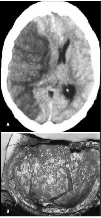
A 55-year old woman with acute cerebral infarction was admitted 12 hours after the onset of a massive MCA infarct (Fig 1a). Cardiovascular risk factors included type II diabetes mellitus, systemic arterial hypertension and cur-rent smoking. The patient had already suffered a myocar-dial infarction and been subjected to coronary bypass sur-gery 10 years before. At admission, she was alert and exhi-bited mild confusion, complete left haemiplegia and hae-mianestesia and difficulty moving her eyes leftwards. Des-pite conventional management including vital sign moni-toring, oxigen supplementation, subcutaneous low mole-cular weight heparin (enoxaparine) 40 mg/day, intravenous hydration with isotonic saline solution and fixed doses of mannitol (1.2 g/Kg/day), the patient developed coma and signs of uncal haerniation with respiratory insufficiency 24 hours later. Controlled hyperventilation and emergency treatment with hypertonic saline were followed by decom-pressive haemicraniectomy performed 4 hours after dete-rioration. A large (14 cm diametre) bone flap was removed (Fig 1b) with no cerebral tissue resected.
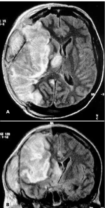
The patient, however, was still comatose and exhibited bilateral decerebrate reaction to painful stimuli 48 hours after surgery. A magnetic resonance exam of the brain (MR) disclosed persistent mass effect with marked deviation of anterior midline structures related to a large epidural mass thought to represent a temporal muscle haemorrhagic suffusion (fig 2). After resection of the temporal muscle (fig 3), the patient awakened, was extubated and had a subsequent uneventful clinical course with progressive partial recovery (feeding and walking independently but still with severe arm paresis and haemianopia 6 months later). The bone flap was reimplanted 22 days later.
DISCUSSION
Clinically significant cerebral oedema develops after at least 10% of large MCA infarcts, probably more frequently in ICA infarcts. Mortality among tho-se in which this progrestho-ses to the point of cerebral haerniation – the so called malignant MCA infarcton – reaches 80%1. A number of current approachs to treat this complication are of dubious value. These include widely used drugs such as mannitol or bar-biturates2-4. Vigorous sustained mechanical hyper-ventilation may even be harmful in similar contexts of uncontrolled intracranial hypertension5. Hypo-ther)mia is being actively investigated in the treat-ment of massive brain infarction and is possibly asso-ciated with delayed evolution of infarction6,7. It is at least doubtfull however that it could reduce definite infarction size or improve neurological outcome. Hypothermia is probably less effective than decom-pressive craniectomy, but it may have an addictive role to surgery8. Although feasible, blanket cooling is associated with a number of severe complications
including bleeding diathesis, arritmias and cardiac failure and uncontrolled intracranial hypertension during rewarming7. Alternative methods for mini-mally invasive endovascular cooling in acute stroke are being investigated9, but the associated risks are not negligible and their clinical role is still undefined.
The technique for decompressive haemicraniec-tomy is already well established10,11. It includes the Fig 1. A) Brain computerised tomography at admission: there is marked deviation of midline structures (over 12 mm) caused by an extensive middle cerebral artery infarct. B) Surgical view of a stellate durotomy following a large (14 cm diametre) craniectomy.
A
remotion of a large (≥ 12 cm) bone flap with a circu-lar or oval shape including the frontal, parietal, tem-poral and parts of the occipital squamae (with special care to avoid producing sharp bone edges); fixation of the dura at the edge of the craniectomy to prevent epidural bleeding; opening of the dura (usually one longitudinal and three radial incisions almost rea-ching the osseous rim) and placement of a dural patch made of lyophilised cadaver dura or more re-cently microporic poliester-uretane (Neuro-Patch) in the incision. The lenght (15 to 20 cm) and width
(2.5 to 3.5 cm) of the patch are somewhat variable. Extensive beveling is avoided as it may lead to profuse venous bleeding. Also, the midline should be spared by 1 cm because opening of bridging veins also pro-motes bleeding. The bone flap is inserted in the abdo-minal wall and reimplanted between 3 and 12 weeks later (alternatively an artificial flap may be made at that time).
In a recent retrospective study of 60 operated patients, Wagner and coleagues12 found a surprising 70% incidence of ischaemic (28.4%) or haemorrhagic lesions (41.6%) directly related to haemicraniectomy. Most lesions were small and probably of no major clinical impact. Small epidural or subgaleal hae-morrhages occurred in 10% of all patients, and small subdural haematomas in 5%. Only one patient deve-loped a large epidural haematoma needing reope-ration. The authors found a statistically significant inverse correlation between the frequency of paren-chimal bleeding caused by craniectomy (but not of ischaemic lesions or epidural bleeding) and the size of the bone defect. Also, sharp bone edges were associated with haemicraniectomy-related lesions of any type. Most importantly, patients with haemicra-niectomy-associated parenchimal haemorrhages (but not those with other lesions) exhibited a higher mor-tality rate – 45% compared with 20% in patients without haemorrhages.
Fig 2. Magnetic resonance in the first post-operative day: adequate posterior decompression of the brain and persisting mass effect and deviation of anterior midline structures caused by a large volume temporal muscle haematoma. A) Axial view; B) Coronal view.
Fig 3. Control CT following excision of the temporal muscle and adequate decompression. The ipsilateral haemisphere is now externally haerniated, with minimal contralateral deviation of midline structures.
A
Craniectomy-associated parenchimal bleeding and ischaemic lesions are probably due to mushro-om-like haerniation associated with an increase in shear forces acting and distorting the brain. Epidural haemorrhages are probably caused by the wound surface. We alert here to haemorrhagic suffusion of the temporal muscle leading to inadequate decom-pression and persisting harm to the herniating brain. We speculate that this bleeding complication may be more common in acute stroke patients receiveing platelet antiaggregants and anticoagulants in the pre-operative phase. In these patients, consideration should be given to the exeresis of large temporal muscles with haemorrhagic suffusion.
The importance of the early indication of hae-micraniectomy in patients with massive MCA infarcts has been emphasised. In an open study11 of 63 pa-tients with complete MCA territoy strokes – with or without additional anterior or posterior artery territory infarction – those operated early (mean time from onset 21 hours, range 8 to 42 hours), had a 16%mortality rate. Only 4 of these 31 patients had signs of uncal haerniation with a unilaterally fixed and dilated pupil. The results compared favourably with those attained in patients previously studied by the same group and operated later (mean 39 hours, range 6 to 112 hours), who exhibited a 34.4% mortality rate13. Also, functional evolution (Barthel index and Rankin scale) was better and lenght of stay in the intensive care unit was shorter in the early-operation group.
We are now partially able to predict the patients with maximum risk of developing haerniation from MCA infarctions. Many authors emphasised the pre-dictive role of large hypodensities as seen in admis-sion CT exam. Total or large (> 50%) hypodensities in the MCA territory are associated with a 85% death risk14. The sensitivity for prediction of the syndrome seems somewhat low, however.
A number of published reports tried to find out important clinical predictors of fatal brain swelling. Nausea/vomiting was suggested as an important marker in one model15 but not in other16. Kasner and cols16 reviewed clinical and radiological criteria in 201 patients with extensive MCA infarctions, 94 (47%) of which died from cerebral oedema. A number of factors independently increased the risk of develo-ping fatal cerebral oedema (Table). Risk was correla-ted with increasing numbers of factors present, being greater than 85% in patients with 4 or 5 factors. The model is not perfect and also has a relatively low sensitivity but is able to predict 70% of the fatal events related to cerebral oedema.
The role of routine monitoring of intracranial pressure and a number of other parameters in guiding surgical indication in patients initially treated conservatively is still a matter of debate. Costs and the possible delay of urgent surgery must be wheighed against the importance of the obtained information, especially avoidance of unneccessary interventions and adequate identification of the pa-tients who could most profit from surgery. Although haerniation is usually associated with increasing intracranial pressure, the degree of displacement of brainstem structures may be greater than its increase as measured by a peripheraly located parenchymal device17. Isolated intracranial pressure monitoring is not an adequate standard in monitoring of
antioe-dema treatment18, which should also include
measurements of cerebral perfusion pressure, oxigen consumption (jugular bulb venous oxigen saturation) and osmolality.
REFERENCES
1. Hacke W, Schwab S, Horn M, Spranger M, De Georgia M, von Kummer R. Malignant middle cerebral artery territory infarction: clinical course and prognostic signs. Arch Neurol 1996;53:309-315.
2. Kaufmann AM, Cardoso ER. Aggravation of vasogenic cerebral edema by multiple-dose mannitol. J Neurosurg 1992;77:584-589.
Table. Factors independently increasing the risk of fatal brain oedema in extensive middle cerebral artery (MCA) infarction*
Risk factor Odds ratio (CI 95%)
History of hypertension 3.0 (1.2-7.6)
History of cardiac failure 2.1 (1.5-3.0)
Increased white cell count 1.08 for every 1000 cells/microL (1.01-1.14) Hypodensity involving > 50% of the MCA territory 6.3 (3.5-11.6)
Extension to other arterial territories (posterior or anterior cerebral artery) 3.3 (1.2-9.4)
3. Bereczki D, Liu M, do Prado GF, Fekete I. Cochrane report: a systematic review of mannitol therapy for acute ischemic stroke AMD cerebral parenchymal hemorrhage. Stroke 2000;31:2719-22.
4. Schwab S, Spranger M, Schwarz S, Hacke W. Barbiturate coma in severe hemispheric stroke: useful or obsolete? Neurology 1997;48:1608-1613. 5. Muizelaar JP, Marmarou A, Ward JD, et al. Adverse effects of prolonged hyperventilation in patients with severe head injury: a randomized clinical trial. J Neurosurg 1991;75:731-739.
6. Schwab S, Schwarz S, Spranger M, Keller E, Bertram M, Hacke W. Moderate hypothermia in the treatment of patients with severe middle cerebral artery infarction. Stroke 1998;29:2461-2466.
7. Schwab S, Georgiadis D, Berrouschot J, Schellinger PD, Graffagnino C, Mayer SA. Feasibility and safety of moderate hypothermia after massive hemispheric infarction. Stroke 2001;32:2033-2035.
8. Doerfler A, Schwab S, Hoffmann TT, Engelhorn T, Forsting M. Combination of decompressive craniectomy and mild hypothermia ameliorates infarction volume after permanent focal ischemia in rats. Stroke 2001;32:2675-2681.
9. Georgiadis D, Schwarz S, Kollmar R, Schwab S. Endovascular cooling for moderate hypothermia in patients with acute stroke: first results of a novel approach. Stroke 2001;32:2550-2553.
10. Delashaw JB, Broaddus WC, Kassell NF, et al.Treatment of right hemispheric cerebral infarction by hemicraniectomy. Stroke 1990;21:874-881. 11. Schwab S, Steiner T, Aschoff A, et al. Early hemicraniectomy in patients
with complete middle cerebral artery infarction. Stroke1998;29:1888.1893 12. Wagner S, Schnippering H, Aschoff A, Koziol JA, Schwab S, Steiner T.
Suboptimum hemicraniectomy as a cause of additional cerebral lesions in patients with malignant infarction of the middle cerebral artery. J Neurosurg 2001;94:693-696.
13. Rieke K, Schwab S, Krieger D, von Kummer R, Aschoff A, Hacke W. Decompressive surgery in space-occupying hemispheric infarction: results of an open, prospective study. Crit Care Med 1995;23:1576-1587. 14. von Kümmer R, Meyding-Lamadé U, Forsting M, et al. Sensitivity and prognostic value of early computed tomography in middle cerebral artery trink occlusion. AJNR 1994;15:9-15.
15. Krieger DW, Demchuk AM, Kasner SE, Jauss M, Hanton L. Early clinical and radiological predictors of fatal brain swelling in ischemic stroke. Stroke 1999;30:287-292.
16. Kasner SE, Demchuk AM, Berruschot J, et al. Predictors of fatal brain edema in massive hemispheric ischemic stroke. Stroke2001;32:2117-2113. 17. Schwab S, Aschoff A, Spranger M, et al. The value of intracranial pressure
monitoring in acute hemispheric stroke. Neurology 1996;47:393-398. 18. Steiner T, Pilz J, Schellinger P, et al. Multimodal online monitoring in