Diagnostic Value of Myocardial Radionuclide Imaging in Patients
with Multivessel Coronary Disease
Maria Eduarda Menezes de Siqueira, Erly Medeiros Vieira Segundo Neto, Juliana Fernandes Kelendjian, Paola
Emanuela Pogio Smanio
Instituto Dante Pazzanese de Cardiologia, São Paulo, SP - Brazil
Abstract
Background: Myocardial perfusion radionuclide imaging (MPRI) is a noninvasive method extremely useful for evaluating ischemia in patients with coronary disease. However, the idea that this method would not be effective for patients with multivessel coronary disease is still entertained.
Objective: Assess the value of MPRI with gated-SPECT for tracing pharmacological stress-induced ischemia in patients with multivessel obstructive coronary disease.
Methods: We evaluated 68 patients with multivessel obstructive coronary disease applying coronary angiography for performing MPRI under pharmacologic stress with dipyridamole. The tests were analyzed by two nuclear medicine physicians without prior knowledge of the result of the coronary angiography.
Results: Gated-SPECT revealed that, out of the patients, 64 (92.8%) showed images of perfusion abnormalities and 4 (7.2%) showed normal perfusion, given that three of the latter showed functional changes.
Conclusion: MPRI has shown high value for identifying patients with multivessel coronary disease, since most patients had perfusion abnormalities indicative of ischemia. (Arq Bras Cardiol 2011; 97(3) : 194-198)
Keywords: Myocardial reperfusion; coronary artery disease / radionuclide imaging; radionuclide imaging / diagnostic use; dipyridamole.
Mailing Adress: Maria Eduarda Menezes de Siqueira •
Alameda dos Jurupis, 856 – Moema – 04088-002 – São Paulo, SP – Brazil E-mail: [email protected]
Manuscript received December 21, 2010; revised manuscript received December 21, 2010; accepted April 13, 2011.
Introduction
Cardiovascular diseases are the leading cause of death worldwide, mainly the coronary artery disease (CAD)1, the
early detection of which allows prompt treatment and change in the prognosis.
According to the physiological principles of ischemic cascades, changes in myocardial perfusion are the first events in episodes of ischemia. This fact explains the relevance of methods for assessing perfusion abnormalities (blood flow and flow reserve), using information from noninvasive methods. Among the patients with CAD, those with lesions of the left coronary artery trunk and multivessel disease undergo the highest risk of adverse events2,3.
The main objective of noninvasive diagnostic methods is to identify the higher risk group, where coronary artery bypass grafting improves symptoms and survival4. The myocardial
perfusion radionuclide imaging (MPRI) is important since it detects changes to myocardial perfusion and left ventricular function, helping in CAD diagnosis, thus showing great value in clinical decisions5,6.
Studies in the 80s already showed that the extent of perfusion abnormalities via MPRI holds an important association with
adverse events (cardiac death and nonfatal acute myocardial infarction). In effect, the risk of cardiac death or AMI in patients with chest pain and suspected CAD increases with the number of segments showing reversible hypoperfusion indicative of ischemia7. This concept applies not only to exercise stress MPRI,
but also to the wide spectrum of nuclear medicine procedures with pharmacological stress8.
Earlier, when some method delivered only myocardial perfusion information, which included relative assessment of the perfusion in one wall compared to the other walls, the idea that MPRI would underestimate or fail to identify perfusion abnormalities was usually accepted when multiple artery irrigation territories were compromised (the so-called balanced uptake)9. In the cases of triple vessel disease of equal severity,
homogeneous hypoperfusion was believed to happen, leading to wrong negative results in tests.
Nonetheless, this fact is unusual, as finding multiple coronary lesions with the same degree of stenosis is extremely rare. If existing, stenosis is not expected to lead to the same regional flow reduction. Differences of regional flow in vessels with stenosis of similar severity are connected to collateral circulation, compression forces and other mechanisms not identified yet10. The world literature makes few references to
balanced uptake5.
Inclusion criteria:
• Patients with obstructive coronary lesions higher than 50% in three coronary arteries confirmed by cardiac catheterization.
Exclusion criteria:
• Patients with clinical or hemodynamic instability (chest pain over the last 48 hours, complex arrhythmias).
• Patients who underwent treatment of vessel(s) affected by surgery or angioplasty before radionuclide imaging.
• Contraindication to dipyridamole.
• Atrial fibrillation.
• Asthma/bronchitis.
• Use of caffeine or xanthine over the 24 hours before the test.
All patients were informed about the study and signed informed consent before the work has been started. The study began after submission and approval of the ethics committee and local research.
The results of MPRI’s relating to perfusion abnormalities, such as ischemia, and its magnitude (intensity and extent) in a qualitative fashion (mild, moderate and severe) were analyzed. In addition to this, functional changes were observed, such as LV cavity dilation after stress (considering the more quantitative visual analysis, TID > 1.23), drop in LV ejection fraction after stress compared to rest higher than 5%, presence of pulmonary uptake of radiopharmaceutical drug after stress and segmental changes to systolic contractility and thickening, even in absence of myocardial perfusion abnormalities.
The statistical analysis was performed using Pearson’s chi-square tests, Fisher’s exact test, Student’s T-test and the nonparametric Mann-Whitney test. The only continuous variable compared was the age, which did not reject the Shapiro normality test.A significance level of 5% (p = 0.05) was considered for inferential analysis. All tests had conclusion with two-tailed hypothesis.
The specificity was not calculated as there was no control group of patients without coronary lesion.
Results
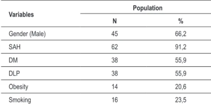
Regarding the clinical and epidemiological characteristics of the population, most participants out of the 68 patients were male (66.2%). The demographic characteristics are shown in Table 1.
Referring to the gated-SPECT radionuclide imaging findings, 64 out of the 68 patients (92.8%; IC 84.86 - 98.10) showed myocardial perfusion abnormalities and 4 (7.2%; IC 1.90 – 15.13) showed normal perfusion, and, among the latter, three showed functional changes (we refer to it as poor prognosis criteria) in the gated-SPECT analysis). 61 out of the 64 patients with perfusion abnormality (95.3%) also had poor prognosis criteria (Figure 1).
Only one (1.5%; IC 1.13 - 13.18) patient had normal perfusion and functional parameters.
When analyzing poor prognosis criteria, 25 patients (39.1%) were found with a drop in LV ejection fraction (LVEF) higher than 5%, 57 (82.6%) with abnormal systolic thickening in at may evaluate left ventricle (LV) function and detect regional
changes in systolic contractility and thickening, increasing the accuracy in the detection of high risk cardiovascular disease11-13.
Our hypothesis is that gated-SPECT MPRI has great sensitivity in detecting patients with multiple vessel territories.
Objectives
This study primarily targets at assessing the value of gated-SPECT MPRI for tracing pharmacological stress-induced ischemia in patients with dipyridamole in a group of patients diagnosed with multivessel obstructive coronary disease through cardiac catheterization.
Secondarily, we intend to ascertain the additional value of functional variables employing gated-SPECT for identifying patients with multivessel obstruction who may have normal myocardial perfusion.
Methods
It is a cross-sectional study including a group of 68 patients subject to cardiac catheterization at Instituto Dante Pazzanese de Cardiologia, from December 2009 to September 2010, with diagnosis of obstructive coronary disease higher than 50% in three epicardial vessels of different vessel territories, including the following three items: 1) anterior descending artery or one of its diagonal branches, provided they have great caliper and anatomic importance, 2) circumflex artery or one of the marginal branches, provided they have great caliper and anatomic importance, 3) right coronary artery.
After the patients were selected, they underwent gated-SPECT MPRI using the radiopharmaceutical drug 99mTc-sestamibi
associated with pharmacological stress with dipyridamole over the standard protocol of two days. The administered dose of dipyridamole was 0.56 mg/kg/min over 4 minutes, and 0.31 mCi/kg of MIBI-99mTc injected in the second minute after the
end of intravenous infusion of dipyridamole.
The images were acquired on gamma camera Millennium VG (GE Medical Systems, Milwaukee, USA), equipped with two scintillation detectors, angled 90 degrees, with high resolution and low energy parallel-hole collimators. Images were acquired 60 minutes after the injection of radiopharmaceutical drug on the two phases (baseline and after dipyridamole). The adverse effects during administration of dipyridamole were promptly reversed with intravenous aminophylline at a dose of 1.0 mg/ kg/min. The radionuclide images were processed by software QGS for perfusion and QPS for analysis of ventricular function. The analysis of perfusion images employed standard qualitative evaluation.
All patients underwent MPRI up to 60 days after cardiac catheterization, before treatment of coronary lesions by surgery or angioplasty, and the therapeutic procedure was not postponed due to the performance of the test. Patients were referred to the test as suggested by assistant physician. The images were evaluated by two nuclear medicine expert physicians without prior knowledge of the results of catheterization.
least one the LV walls, 45 (65.2%) with LV transient dilation after stress and three (4.3%) with pulmonary uptake of radiopharmaceutical drug.
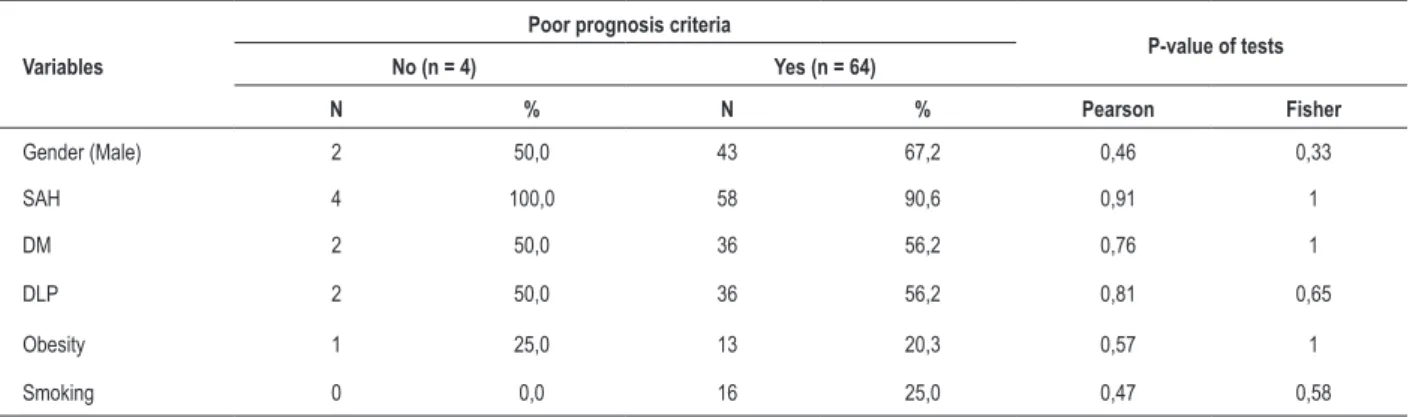
Regarding the variables which could influence the presence of poor prognosis criteria, statistical association with risk factors for cardiovascular disease was not found to explain these findings (Table 2). Moreover, these functional findings with gated-SPECT were not associated with important changes to coronary anatomy such as, for instance, presence of severe lesion in the proximal 1/3 of the anterior descending artery or on the left coronary artery trunk.
The assessment of presence relating to perfusion findings considered only “present” or “absent,” and there was no analysis per artery territory. Out of the total, 27 patients showed multiple perfusion defects.
The sensitivity of the method to detect any abnormality on gated-SPECT was 88.40% (IC 95%; 78.4 - 94.90) when perfusion and functional defects were evaluated.
Discussion
MPRI is a noninvasive diagnostic method well established in the diagnosis of CAD and cardiovascular risk stratification. One of its objectives is to identify the presence of myocardial ischemia, either directly, by the presence of perfusion changes after stress, or indirectly through the functional findings indicative of stress-induced ischemia.
The main information delivered by this test is the relative uptake of the radiopharmaceutical after stress on one of the myocardium walls relating to the other and the baseline study. When present, this finding suggests the existence of myocardial ischemia on that wall14.
However, the doubt on whether, in multivessel CAD affecting all vascular beds, the degree of ischemia could be underestimated due to the balanced hypoperfusion (since all myocardial walls would receive less blood labeled with the radiopharmaceutical drug) persists. In this situation, there would be no normal reference segments in radionuclide imaging to be compared with the segments fed by stenosed vessels. And all the segments would be found in similar status.
Nonetheless, since 1997, with the technical development
brought by gated-SPECT, the perfusion study has become synchronized to electrocardiogram, also delivering functional information, such as the analysis of global and regional systolic contractility and thickening at baseline and post-stress stages9.
Other important information added was baseline and post-stress LVEF. If there is drop in LVEF higher than 5% after stress compared to baseline, stress-induced ischemia may also be evoked14.
Shirai et al13 showed that the change in contractility increases
sensitivity for detection for patients with multivessel coronary disease compared to perfusion abnormality only, underpinning the data of this study13. Also in this study, the method was found
safe for multivessel CAD patients, as there were no AMI or death events.
In this scenario, there were significant findings where, even normal perfusion images may indicate multivessel disease and poor prognosis. Previous studies show that gated-SPECT MPRI may add prognostic and diagnostic value to myocardial perfusion images only5,15-18.
Functional findings with gated-SPECT include pulmonary uptake and transient ischemic dilation of the LV during stress.
The pulmonary uptake means that a substantial amount of the radiopharmaceutical drug is apparent in the pulmonary branches during stress, and absent during rest. It shows the possibility of multivessel disease in patients, besides high of pulmonary arterial pressure during the stress.
Transient LV dilation refers to a pattern of image in which this cavity seems larger during stress compared to rest. In patients where the whole LV seems more dilated in stress, there is probable association with extensive ischemia and systolic dysfunction after prolonged ischemia, resulting in left ventricular dysfunction after stress, without presence of it in baseline phase. It is worth to note the possibility of epicardial silhouette to look similar both in stress and at rest, associated with LV dilation. This finding may reveal the presence of diffuse subendocardial ischemia, which would also be associated with more extensive CAD, and, therefore, more severe. The presence of these two findings shows that the patient has high risk for cardiovascular events19,20.
In this study, only one out of the 68 patients analyzed showed no perfusion or ventricular function abnormalities applying gated-SPECT technique. When checking the patient’s coronary anatomy, we realize that the lesions caused to three vessels led to stenosis between 50-70% of its light.
Lima et al5 studies 143 patients with three-vessel diseases
with 112 controls with no coronary lesions, whether unilateral or bilateral, and concluded that the combination of perfusion abnormality and functional analysis increases the sensitivity (88%) to detect severe coronary disease. This data confirm the sensitivity found in our study.
One of the limitations of this study is the fact that only one patient had a completely normal MPRI (for both perfusion and functional abnormalities). The other patients who presented normal perfusion and that suffered from multivessel disease were identified by the presence of functional changes. Besides this, there was the number of 68 patients.
The main difficulty in the inclusion of patients was the failure to attend on the scheduled date. Other difficulties were: the MPRI
SAH - Systemic arterial hypertension; DM - Diabetes mellittus; DLP - Dyslipidemia.
Table 1 - Demographic characteristics of the population (n = 68)
Variables Population
N %
Gender (Male) 45 66,2
SAH 62 91,2
DM 38 55,9
DLP 38 55,9
Obesity 14 20,6
analysis was only qualitative, in accordance with service routine by the time the work was performed, and, thus, the percentage of the area affected was not evaluated; only the presence or absence of ischemia was evaluated, with no correlation to the artery territory, although literature data show few (13-50%) multivessel coronary disease patients with perfusion abnormality in multiple territories13,21,22.
We hope to prove with this study that, as we observe the patients studied, the gated-SPECT myocardial radionuclide imaging is able to satisfactorily identify perfusion abnormalities in patients with multivessel involvement, particularly by associating perfusion and functional information.
Conclusion
The results suggest that MPRI is a noninvasive diagnostic method of great value in identifying patients with multivessel disease, who were mostly detected by the
presence of perfusion abnormalities. Those with normal perfusion were identified by the findings of poor prognosis ascertained by functional analysis employing gated-SPECT technique.
Potential Conflict of Interest
No potential conflict of interest relevant to this article was reported.
Sources of Funding
There were no external funding sources for this study.
Study Association
This study is not associated with any post-graduation program.
Figure 1 - Correlation between perfusion abnormality and poor prognosis criteria.
SAH - Systemic arterial hypertension; DM - Diabetes mellittus; DLP - Dyslipidemia.
Table 2 - Association between cardiovascular risk factors and poor prognosis criteria
Variables
Poor prognosis criteria
P-value of tests
No (n = 4) Yes (n = 64)
N % N % Pearson Fisher
Gender (Male) 2 50,0 43 67,2 0,46 0,33
SAH 4 100,0 58 90,6 0,91 1
DM 2 50,0 36 56,2 0,76 1
DLP 2 50,0 36 56,2 0,81 0,65
Obesity 1 25,0 13 20,3 0,57 1
References
1. Godoy MF, de Lucena JM, Miquelin AR, Paiva FF, Oliveira AL, Augustin JL Jr, et al. Mortalidade por doenças cardiovasculares e níveis socioeconômicos na população de São José do Rio Preto, estado de São Paulo, Brasil. Arq Bras Cardiol. 2007;88(2):200-6.
2. Myocardial infarction and mortality in coronary artery surgery study (CASS) randomized trial. N Engl J Med. 1984;22:310(12):750-8.
3. Jones RH, Hannan EL, Hammermeister KE, Delong ER, O’Connor GT, Luepker RV, et al; for the Working Group Panel on the Cooperative CABG Database Project. Identification of preoperative variables needed for risk adjustment of short-term mortality after coronary artery bypass graft surgery. J Am Coll Cardiol. 1996;28(6):1478-87.
4. Shaw LJ, Berman DS, Maron DJ, Mancini GB, Hayes SW, Hartigan PM, et al; COURAGE Investigators. Optimal medical therapy with or without percutaneous coronary intervention to reduce ischemic burden: results from the Clinical Outcomes Utilizing Revascularization and Aggressive Drug Evaluation (COURAGE) trial nuclear substudy. Circulation. 2008;17(10):1283-91.
5. Lima RS, Watson DD, Goode AR, Siadaty MS, Ragosta M, Beller GA, et al. Incremental value of combined perfusion and function over perfusion alone by gated SPECT myocardial perfusion imaging for detection of severe three-vessel coronary artery disease. J Am Coll Cardiol. 2003;42(1):64-70. 6. Loong CY, Anagnostopoulos C. Diagnosis of coronary artery disease by
radionuclide myocardial perfusion imaging. Heart. 2004;90(Suppl 5):v2-9. 7. Ladenheim ML, Pollock BH, Rozanski A, Berman DS, Staniloff HM, Forrester
JS, et al. Extent and severity of myocardial hypoperfusion as predictors of prognosis in patients with suspected coronary artery disease. J Am Coll Cardiol. 1986;7(3):464-71.
8. Iskandrian AS. Single-photon emission computed tomographic thalliumim aging with adenosine, dipyridamole and exercise. Am Heart J. 1991;122(1 Pt 1):279-84.
9. Kostacos EJ, Araujo LI. Incidence of balanced ischemia in patients with dipyridamole perfusion imaging [abstract]. J Nucl Cardiol. 2004;11(4):S5-S6. 10. Madias JE, Knez K, Win MT. True-positive exercise electrocardiogram /
false-negative thallium-20 1scintigram: a proposal of a mechanism for the paradox. Clin Cardiol. 2000;23(8):625-9.
11. Sharir T, Germano G, Kavanagh PB, Lai S, Cohen I, Lewin HC, et al. Incremental prognostic value of post-stress left ventricular ejection fraction and volume by gated myocardial perfusion single photon emission computed tomography. Circulation. 1999;100(10):1035-42.
12. Hida S, Chikamori T, Tanaka H, Usui Y, Igarashi Y, Nagao T, et al. Diagnostic value of left ventricular function after stress and at rest in the detection of multivessel coronary artery disease as assessed by electrocardiogram-gated SPECT. J Nucl Cardiol. 2007;14(1):68-74.
13. Shirai N, Yamagishi H, Yoshiyama M, Teragaki M, Akioka K, Takeuchi K, et al. Incremental value of assessment of regional wall motion for detection of multivessel coronary artery disease in exercise (201) TI gated myocardial perfusion imaging. J Nucl Med. 2002;43(4):443-450.
14. Desai D, Kozeski G, Akinboboye O. Detection of multivessel coronary artery disease: looking beyond the extent of perfusion abnormalities. J Nucl Cardiol. 2009;16(1):4-5.
15. Yamagishi H, Shirai N, Yoshiyama M, Teragaki M, Akioka K, Takeuchi K, et al. Incremental value of left ventricular ejection fraction for detection of multivessel coronary artery disease in exercise (201)Tl gated myocardial perfusion imaging. J Nucl Med. 2002;43(2):131-9.
16. De Winter O, Velghe A, Van de Veire N, De Bondt P, De Buyzere M, Van De Wielle C, et al. Incremental prognostic value of combined perfusion and function assessment during myocardial gated SPECT in patients aged 75 years or older. J Nucl Cardiol. 2005;12(6):662-70.
17. Matsuo S, Matsumoto T, Nakae I, Koh T, Masuda D, Takada M, et al. Prognostic value of ECG-gated thallium-201 single-photon emission tomography in patients with coronary artery disease. Ann Nucl Med. 2004;18(7):617-22.
18. Gimelli A, Rossi G, Landi P, Marzullo P, Tervasi G, L’abbate A, et al. Stress/rest myocardial perfusion abnormalities by gated SPECT: still the best predictor of cardiac events in stable ischemic heart disease. J Nucl Med. 2009;50(4):546-53. 19. Duarte PS, Smanio PE, Oliveira CA, Martins LR, Mastrocolla LE, Pereira JC, et al. O significado clínico da dilatação transitória do ventrículo esquerdo avaliada pela cintilografia do miocárdio com Tc99m-Sestamibi. Arq Bras Cardiol. 2003;81(5):474-8.
20. Smanio PE, Watson DD, Segalla DL, Vinson EL, Smith WH, Beller GA. Value of gating of technetium-99m sestamibi single-photon emission computed tomographic imaging. J Am Coll Cardiol. 1997;30(7):1687-92.
21. Christian TF, Miller TD, Bailey KR, Gibbons RJ. Noninvasive identification of severe coronary artery disease using exercise tomographic thallium-201 imaging. Am J Cardiol. 1992;70(1):14-20.