Reduction of Central Sleep Apnea in Heart Failure Patients with
Beta-Blockers Therapy
Christiano Pereira Silva, Geraldo Lorenzi-Filho, Bianca Marcondes, Gilmar Osmundo Junior, Sandrigo Mangini,
Aguinaldo Figueiredo Freitas Junior, Phillipe Vieira Pires, Edimar Alcides Bocchi, Fernando Bacal
Instituto do Coração - Faculdade de Medicina da Universidade de São Paulo, São Paulo, SP - Brazil
Abstract
Background: Sleep apneas are frequent in patients with heart failure (HF). Estimate of the pre-beta blocker age (BB) point out to 45% of central apneas in these patients.
Objective: Assess the influence of BB in central apneas and their interference in the quality of sleep and life of patients with heart failure.
Methods: 65 patients with heart failure underwent diagnostic polysomnography. Polysomnography have been assessed according to the use or not of BB. On the day of examination, the patients answered the Minessota questionnaire for quality of life with HF. After 6 and 12 months from the polysomnography date, all patients were contacted by phone, in order to repeat the Minessota questionnaire.
Results: The prevalence of sleep apnea (IAH > 15/h) hit 46.1% in the total population, however, central sleep apnea was identified in 18.4% of patients. The use of BB, in a multivariate analysis, was the only predictor of a minor index of central apnea and hypopnea (IAH) (p=0.002), greater saturation (p=0.02) and smaller average desaturation of oxygen (p=0.03). Additionally, the use of BB could predict a better quality of life after 6 and 12 months (p=0.002 and 0.001 respectively) and a smaller number admissions in these periods (p=0.001 and p=0.05 respectively).
Conclusion: The use BB reduced the rate of central sleep apnea in total population, if we compare to literature data. Additionally, the BB improved parameters of quality of sleep and life of patients with heart failure. (Arq Bras Cardiol 2010;94(2): 223-229)
Key Words: Sleep apnea syndromes; heart failure; adrenergic beta-antagonists.
Mailing address: Christiano Pereira Silva •
Rua Charles Spencer Chaplin, 85 / 21 - Vila Andrade - 05642-010 - São Paulo, SP - Brazil
E-mail: [email protected], [email protected]
Manuscript received February 07, 2009; revised manuscript received May 23, 2009; manuscript accepted July 09, 2009.
Introduction
Heart failure (HF) is one of the cardiovascular diseases that most lead to morbidity and mortality, causing considerable economic and social impacts1. Nearly 5 million Americans
currently have heart failure. In Europe, studies have reported prevalence from 0.5% (in younger people) to 16.1% in those older than 752. Decompensated heart failure (DHF) causes
at least 20% of total hospital admissions among people of the same age3. Hence, it is important to investigate each variable
that may predict the evolution and prognosis of these patients. Most of these variables are well known, while others are getting quickly popular.
One of these variables is the respiratory sleep disorder (RSD), especially central sleep apnea (CSA). The Cheyne-Stokes (CS) pattern is the most known pattern of such disorder4,5. The CSA
occurs especially with patients with HF, where prevalence is
around 30% and 50%6. The physiopathological mechanism
that explains this high rate of prevalence is the hypocapnia resulting from the tachypnea and hyperpnea deriving from lung congestion. Apnea increases the sympathetic activation and the risk of ventricular arrhythmias, which are likely to cause the increase of mortality, observed in the patients7.
Our study monitored 65 patients with severe HF, optimized drug therapy and patients with symptoms related to CSA. These patients were submitted to night polysomnography and later monitored for one year. The purpose of the study was to assess the impact of beta-blockers on central apneas. We also sought to assess how much the presence of CSA and beta-blocker therapies influenced the treatment of these patients.
Patients and methods
Patients
de São Paulo, were selected to take part in the study. The selection criteria were: Patients with functional HF (NYHA) II or III, dyspnea at rest or rough coughing at night, apnea witnessed by the spouse and left ventricle ejection fraction (LVEF) < 35%. The exclusion criteria included previous cerebrovascular disease, use of central nervous system depressants, body mass index > 30 and chronic respiratory disease.
All patients were submitted to a diagnostic polysomnography test to assess apnea. The drugs prescribed to patients were not changed to do the tests.
The Minnesota Living with Heart Failure Questionnaire of was given to all patients, immediately before the diagnostic test. The entire population was monitored for one year. Six and 12 months after the diagnostic test, the authors called the population under study in order to apply another Minnesota questionnaire and to make questions about re-admissions and death.
The investigation was made according to the principles outlined by the Declaration of Helsinki. The Ethics Commission of Hospital das Clínicas da FMUSP approved the study protocol, and an informed consent was filled by each patient/person responsible before the inception of the follow-up study.
Polysomnography
Night polysomnography incorporated a digital system (EMBLA 17 channels, FLAGA hf. Medical Devices). The investigation consisted in monitoring EEG, electrooculogram, submental electromyogram, ECG, thoracic-abdominal excursions, oronasal flow and arterial saturation of oxygen by pulse oximetry. Central sleep apnea was defined by absence of oronasal flow during > 10 seconds, associated to absence of respiratory stress. Obstructive sleep apnea was defined by absence of oronasal flow for > 10 seconds, however, in the presence of thoracic-abdominal movements (respiratory stress). Hypopnea was characterized by a > 50% reduction in oronasal flow, inasmuch it was > 10 s and associated to > 3% in the drop of arterial oxygenation. The index of apnea-hypopnea (IAH) was calculated considering the average number of apneas and hypopneas per hour of sleep. The indexes of central apneas (ICA) and obstructive apneas (OA) were calculated by the average of these events per hour.
Statistic analysis
The classification variables were displayed in contingency tables with absolute (n) and relative (%) frequencies. The association among them was assessed by Fisher’s exact test. The distribution of quantitative variables was assessed with t-Student test or Wilcoxon test. The statistically significant variables found in the single-variable analysis were used in the logistic regression model. They were considered statistically significant when the p value was < 0.05.
Results
A m o n g t h e p a t i e n t s s u b m i t t e d t o d i a g n o s t i c polysomnography, 55 patients (87.6%) used beta-blocker (Carvedilol) in an average daily dose of 28.8 mg. Hypotension (7.6%), bradycardia (3%) and chronic obstructive pulmonary
disease (1.5%) did not allow the other patients to use the drug. The clinical characteristics of the entire population are described in Table 1.
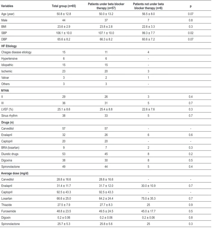
The patients without beta-blocker presented a lower systolic blood pressure (p=0.02), the only significant difference among patients with and without these drugs.
Thirty patients (46.1%) presented IAH > 15/h. Central sleep apnea was found in 12 patients (18.4% of the entire population, corresponding to 40% of patients with IAH > 15/h). Obstructive sleep apnea was found in 3 patients (10%), mixed apnea in 4 (13.3%) and hypopnea in 11 patients (36.6%).
The smaller prevalence of central sleep apnea in this population was surprising, although not statistically compared to literature. The multivariate regression analysis showed that the continuous use of beta-blocker was an independent predictor of absence of central sleep apnea (p<0.002).
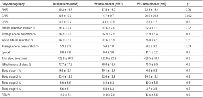
By comparing the results of polysomnographies of patients with and without beta-blocker, we confirmed a smaller index of central sleep apnea in patients taking the drug (p=0.002). Additionally, as shown in Table 2, patients belonging to the beta-blocker group presented greater average night saturation of oxygen, smaller worse saturation during the night and a smaller average of arterial desaturation, significantly different from those of group without beta-blocker (p=0.02, 0.01 and 0.03, respectively).
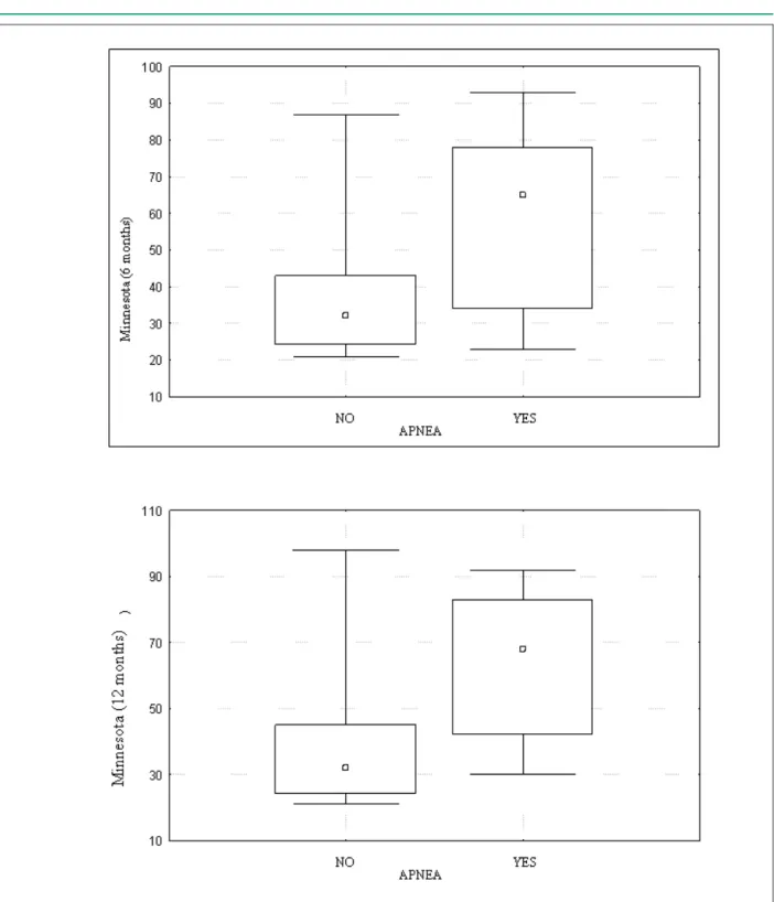
The quality of life, according to the Minnesota questionnaire, was significantly better, after 6 and 12 months, among patients without central sleep apnea (p=0.002 and p=0.001 respectively – Figures 1 and 2). Admissions were more common in the population with central sleep apnea (p=0.001 and p=0.005 to 6 and 12 months, respectively – Figures 3 and 4). There was no difference in the mortality rate in populations with and without central sleep apnea.
Discussion
This study was targeted at assessing the influence of beta-blockers in central sleep apnea, in patients with heart failure patients, as well as of these drugs can interfere in the quality of sleep of these patients. Besides this, it also sought to investigate whether the central sleep apnea would impact the prognosis and/or the quality of life of these patients.
The smaller prevalence of central sleep apnea in patients using beta-blocker, although not statistically compared to literature, if compared to what literature describes, was the main finding of the study. We also confirmed what had already been reported: respiratory disorders, such as central sleep apnea, influence the prognosis and directly affect the quality of life of patients with HF.
Table 1 - Characteristics of the population
Variables Total group (n=65) Patients under beta blocker therapy (n=57)
Patients not under beta
blocker therapy (n=8) p
Age (year) 50.8 ± 12.8 50.0 ± 13.2 56.5 ± 8.0 0.07
Male 44 37 7 0.8
BMI 23.6 ± 2.9 23.8 ± 2.8 22.6 ± 3.3 0.3
SBP 106.1 ± 10.0 107.1 ± 10.0 99.3 ± 7.7 0.02
DBP 65.6 ± 8.2 66.3 ± 8.2 60.6 ± 7.2 0.07
HF Etiology
Chagas disease etiology 15 11 4
Hypertensive 6 6
-Idiopathic 15 15
-Ischemic 23 20 3
Valvar 3 2 1
Others 3 3
-NYHA
II 29 26 3 0.4
III 36 31 5 0.7
LVEF (%) 25.1 ± 8.6 25.4 ± 8.8 22.6 ± 7.6 0.3
Sinus rhythm 38 33 5 0.7
Drugs (n)
Carvedilol 57 57 -
-Enalapril 32 26 6 0.6
Captopril 20 20 -
-BRA (losartan) 9 7 2 0.3
Diuretic drugs 53 45 8 0.2
Digoxina 38 30 8 0.5
Spironolactone 49 44 5 0.4
Average dose (mg/d)
Carvedilol 28.8 ± 16.6 28.8 ± 16.6 -
-Enalapril 31.4 ± 11.7 31.7 ± 12.0 30.0 ± 10.9 0.7
Captopril 92.5 ± 43.3 92.5 ± 43.3 -
-Losartan 66.6 ± 25.0 64.2 ± 24.4 75.0 ± 35.3 0.7
Thiazide 27.5 ± 7.9 27.7 ± 8.3 25 0.9
Furosemide 48.8 ± 23.5 49.5 ± 24.5 45.0 ± 17.7 0.5 Digoxin 0.2 ± 0.06 0.2 ± 0.06 0.2 ± 0.06 0.8 Spironolactone 25.7 ± 5.3 25.8 ± 5.6 25 0.3
BMI - body mass index; SAP - systolic blood pressure; DBP - diastolic blood pressure; LVEF - left ventricle ejection fraction; ACE - inhibitor of angiotensin-converting enzyme; ARB - angiotensin receptor blocker.
mortality risk increased by two to three times8-10. Our study
did not find mortality differences; however, the quality of life and hospital admissions were significantly different among patients with and without CSA, in detriment to patients with apnea.
Few epidemiological studies report prevalence of CSA in patients with HF. The two larger studies involved 450
and 81 patients, reporting prevalence of 33% and 40%, respectively11,12. The main risks to CSA were male sex,
hypocapnia, atrial fibrillation and advanced age.
Table 2 - Polysomnography of patients under and not under beta-blocker therapy
Polysomnography Total patients (n=65) W/ beta-blocker (n=57) W/O beta-blocker (n=8) p*
AHI% 19.3 ± 18.7 17.5 ± 18.2 32.3 ± 18.4 0.06
CAI% 6.6 ± 12.7 4.7 ± 9.7 20.0 ± 21.8 0.002
OAI% 4.2 ± 10.2 4.4 ± 10.9 2.5 ± 1.7 0.2
Arterial saturation awaken % 93.4 ± 2.4 93.6 ± 2.5 92.3 ± 1.1 0.02 Average arterial saturation % 92.8 ± 2.8 92.9 ± 2.9 91.9 ± 1.4 0.1 Worse arterial saturation % 82.9 ± 5.9 83.6 ± 5.9 78.0 ± 4.1 0.01 Average arterial desaturation % 5.9 ± 2.3 5.4 ± 1.8 8.6 ± 3.2 0.03
Epworth 8.8 ± 4.0 8.4 ± 3.6 11.1 ± 6.2 0.2
Total sleep time (min) 422.8 ± 70.2 420.6 ± 72.9 438.5 ± 46.7 0.3 Effectiveness of sleep % 77.7 ± 17.8 78.0 ± 18.7 75.3 ± 9.5 0.5 Sleep stage 1 % 9.6 ± 12.1 10.1 ± 12.7 6.6 ± 5.2 0.1 Sleep stage 2 % 63.4 ± 12.9 62.6 ± 12.8 69.1 ± 13.1 0.2 Sleep stage 3 % 8.6 ± 6.5 8.4 ± 6.3 10.3 ± 8.5 0.6 Sleep stage 4 % 5.6 ± 6.1 5.8 ± 6.3 3.7 ± 3.6 0.2
REM % 14.0 ± 7.1 14.0 ± 7.0 13.6 ± 8.5 0.9
AHI - apnea and hypopnea index; CAI - central apnea index; OAI - obstructive apnea index; REM - rapid eye movement; *p: patients receiving or not beta-blockers.
of beta-blocker therapy – when the use of beta-blockers in patients with ventricular dysfunction affects, at Instituto do Coração, over 90% of patients13. Tamura et al14 have recently
studied 45 patients with HF and reported low prevalence of CSA among chronic beta-blocker users. Additionally, they reported that five patients not using beta-blockers, with IAC > 5 reduced considerably this index after 6 months of Carvedilol therapy (9.5 ± 4.9 to 1.3 ± 2.4, p=0.03). Kohnlein et al15 found similar results by studying 50 patients
with HF with and without the use of beta-blockers15.
Maybe a way to explain the reason for the CSA reduction with beta-blocker is the undisputable improvement that the left ventricular function has with these drugs. Through prevention of ventricular remodeling , prophylaxis of arrhythmias, ischemia, fibrosis and apoptosis, drugs like Carvedilol, Metoprolol and Bisoprolol may increase the ejection fraction of the left ventricle, which is provably associated to a better quality of life and to a reduced mortality of these patients.
Therapy with beta-blocker may also reduce the severity of CSA by restoring the central chemosensitivity to CO2. Such sensitivity may destabilize breathing during sleep, when PaCO2 is below the respiratory threshold, which results in CSA. There is a significant positive correlation between the increase of central chemosensitivity to CO2 and plasmatic norepinephrine, in patients with HF. Inhibiting the sympathetic activity reduces the level of plasmatic norepinephrine and prevents the increase of sensitivity to CO2. That was reported by Takahashi et al16, when they administered endovenous propranolol in
healthy volunteers and promoted a significant depression in this sensitivity16.
Conclusion
In conclusion, this study found that beta-blocker therapy probably reduces the prevalence of central sleep apnea in patients with HF, and improves sleep quality of these patients. Besides this, once again it was proven that patients with CSA have a worse quality of life and are subject to a greater number of admissions. Nevertheless, there was no difference of mortality related to respiratory disease.
Limitations
The number of patients belonging to the group not taking beta-blockers was smaller than we intended. That was due to the huge difficulty in finding patients with HF not taking beta-blockers as a follow-up to heart failure outpatient care and Transplantation at InCor. Even when we investigated patients from other groups in the hospital, it was also hard to select patients without these drugs.
Potential Conflict of Interest
No potential conflict of interest relevant to this article was reported.
Sources of Funding
This study was partially funded by FAPESP.
Study Association
References
1. Sin DD, Man GC. Cheyne–Stokes respiration: a consequence of a broken heart? Chest. 2003; 124: 1627-8.
2. Eriksson H, Svardsudd K, Larsson B, Ohlson LO, Tibblin G, Welin L, et al. Risk factors for heart failure in the general population: the study of men born in 1913. Eur Heart J. 1989; 10: 647-56.
3. Pépin JL, Chouri-Pontarollo N, Tamisier R, Lévy P. Cheyne-Stokes respiration with central sleep apnoea in chronic heart failure: proposals for a diagnostic and therapeutic strategy. Sleep Med Rev. 2006; 10: 33-47.
4. Tobin JM, Snyder JV. Cheyne-Stokes respiration revisited: controversies and implications. Crit Care Med. 1984; 12 (10): 882-7.
5. Lorenzi-Filho G, Genta PR, Figueiredo AC, Inoue D. Cheyne-Stokes respiration in patients with congestive heart failure: causes and consequences. Clinics. 2005; 60 (4): 333-44.
6. Naughton MT, Liu PP, Benard DC, Goldstein RS, Bradley TD. Treatment of congestive heart failure and Cheyne-Stokes respiration during sleep by continuous positive airway pressure. Am J Respir Crit Care Med. 1995; 151: 92-7.
7. Lanfranchi PA, Braghiroli A, Bosimini E, Mazzuero G, Colombo R, Donner CF, et al. Prognostic value of nocturnal Cheyne-Stokes respiration in chronic heart failure. Circulation. 1999; 99: 1435-40.
8. Findley LJ, Zwillich CW, Ancoli-Israel S, Kripke D, Tisi G, Moser KM. Cheyne– Stokes breathing during sleep in patients with left ventricular heart failure. South Med J. 1985; 78: 11-5.
9. Hanly PJ, Zuberi-Khokhar NS. Increased mortality associated with Cheyne–
Stokes respiration in patients with congestive heart failure. Am J Respir Crit Care Med. 1996; 153: 272-6.
10. Sin DD, Logan AG, Fitzgerald FS, Liu PP, Bradley TD. Effects of continuous positive airway pressure on cardiovascular outcomes in heart failure patients with and without Cheyne– Stokes respiration. Circulation. 2000; 102: 61-6.
11. Sin DD, Fitzgerald F, Parker JD, Newton G, Floras JS, Bradley TD. Risk factors for central and obstructive sleep apnea in 450 men and women with congestive heart failure. Am J Respir Crit Care Med. 1999; 160: 1101-6.
12. Javaheri S, Parker TJ, Liming JD, Corbett WS, Nishiyama H, Wexler L, et al. Sleep apnea in 81 ambulatory male patients with stable heart failure: types and their prevalences, consequences, and presentations. Circulation. 1998; 97: 2154-9.
13. Silva CP, Bacal F, Pires PV, Mangini S, Issa VS, Moreira SF, et al. Heart failure treatment profile at the ß-blockers era. Arq Bras Cardiol. 2007; 88 (4): 475-9.
14. Tamura A, Kawano Y, Naono S, Kotoku M, Kadota J. Relationship between ß-blocker treatment and the severity of central sleep apnea in chronic heart failure. Chest. 2007; 131: 130-5.
15. Kohnlein T, Welte T. Does beta-blocker treatment influence central sleep apnoea? Respir Med. 2007; 101: 850-3.