© 2013 Sociedade Brasileira de Hemodinâmica e Cardiologia Intervencionista. Published by Elsevier Editora Ltda. All rights reserved.
Association between Vascular Remodelling and
Necrotic Core in Coronary Arteries: Analysis by
Intracoronary Ultrasound with Virtual Histology
Tammuz Fattah
1, Bruno S. Matte
2, Julise A. Balvedi
3, Juliane Rossato
4, Márcio Mossmann
5,
Xana M. Mendes
6, Taicir C. Salem
7, Paulo V. Crestani
8, Alexandre C. Zago
9ABSTRACT
Background: Anatomopathological studies suggest an as sociation of positive vascular remodeling and vulnerable coronary plaques. The objective of this study was to verify whether there is a correlation between positive vascular remodeling and necrotic core in atherosclerotic coronary lesions. Methods: We studied 270 cross sections obtained by Virtual Histology in 30 patients who had positive re modeling in coronary artery segments with lesions > 50%,
identified by coronary angiography. Seven cross sections were assessed per segment of coronary artery, including the cross section with the highest remodeling index, denominated cross section of interest (cross section 4). Results: Mean age was 60.8 ± 8.8 years, 80% were male and 30% were
diabetic. Unstable angina was the most frequent clinical presentation (56.6%) and the left anterior descending artery was the most analyzed vessel (43%). The vessel reference area was 15.5 ± 4.9 mm² and the remodeling index in
cross section 4 was 1.2 ± 0.1. Repeated measures analysis
of variance showed a higher percentage of necrotic core in the cross section of interest (P < 0.001). We observed
a positive correlation of coronary artery remodeling and necrotic core (r = 0.79; P < 0.001). Conclusions: Positive
coronary artery remodeling is associated to the presence of necrotic core, which characterizes vulnerable athe rosclerotic plaques. The search for positive arterial remo
1 Master. Interventionist cardiologist physician at the Hemodynamics Service of Instituto de Cardiologia do Estado de Santa Catarina. Flo rianópolis, SC, Brazil.
2 Master. Interventionist cardiologist physician at the Hemodynamics Service of Hospital Universitário. Canoas, RS, Brazil.
3 Medical graduation student at Universidade Federal do Rio Grande do Sul. Porto Alegre, RS, Brazil.
4 Master. Biomedical physician at the Interventionist Cardiology Research Group of Hospital de Clínicas de Porto Alegre. Porto Alegre, RS, Brazil.
5 Resident physician at the Hemodynamics Unit of Hospital de Clínicas de Porto Alegre. Porto Alegre, RS, Brazil.
6 Medical graduation student at Universidade Federal do Rio Grande do Sul. Porto Alegre, RS, Brazil.
7 Medical graduation student at Universidade Federal do Rio Grande do Sul. Porto Alegre, RS, Brazil.
8 Medical graduation student at Universidade Federal do Rio Grande do Sul. Porto Alegre, RS, Brazil.
9 Doctor and postdoctorate degree. Cardiologist physician, coordinator of the Interventionist Cardiology Research Group of Hospital de Clínicas de Porto Alegre. Porto Alegre, RS, Brazil.
Correspondence to: Tammuz Fattah. Rua Adolfo Donato da Silva, s/n – Praia Comprida – São José, SC, Brazil – CEP 88103901
Email: [email protected]
Received on: 1/2/2013 • Accepted on: 3/5/2013
Original Article
RESUMO
Associação entre Remodelamento Vascular e Núcleo Necrótico em Artérias Coronárias: Análise por Ultrassom Intracoronário com Histologia Virtual
Introdução: Estudos anatomopatológicos sugerem a associação de remodelamento vascular positivo e placas coronárias vulneráveis. O objetivo deste estudo foi avaliar se existe correlação entre o grau de remodelamento vascular positivo e o porcentual de núcleo necrótico em lesões ateroscleróticas coronárias. Métodos: Foram estudados 270 cortes transversais obtidos pela Histologia Virtual de 30 pacientes, os quais apresentavam remodelamento positivo em segmento de artéria coronária com lesão > 50%,
identiicada pela angiograia coronária. Foram avaliados 7 cor tes transversais por segmento de artéria coronária, incluindo o corte transversal com o maior índice de remodelamento arterial, denominado corte transversal de interesse (corte transversal 4). Resultados: A média de idade foi de 60,8 ± 8,8 anos, 80%
eram do sexo masculino e 30% diabéticos. Angina instável foi a apresentação clínica mais frequente (56,6%) e a artéria descendente anterior foi o vaso mais analisado (43%). A área de referência do vaso foi de 15,5 ± 4,9 mm² e o índice de
remodelamento no corte transversal 4 foi de 1,2 ± 0,1. Análise
de variância de medidas repetidas mostrou maior porcentual de núcleo necrótico no corte transversal de interesse (P < 0,001).
deling may be a useful strategy for detecting vulnerable plaques before rupture.
DESCRIPTORS: Coronary vessels. Coronary stenosis. Ultrasonics.
coronário com o núcleo necrótico (r = 0,79; P < 0,001).
Con-clusões: O remodelamento positivo da artéria coronária está associado à presença de núcleo necrótico, o qual caracteriza placas ateromatosas vulneráveis. A pesquisa de remodelamento arterial positivo pode ser estratégia útil para a detecção de placas vulneráveis antes de sua ruptura.
DESCRITORES: Vasos coronários. Estenose coronária. Ultrassom.
A
cute coronary syndromes present as unstable angina, acute myocardial infarction, or sudden death. Several studies have shown a strong association between these syndromes and atheromatous plaque rupture or erosion, which by exposing the subintimal content, induces the formation of thrombi, and often
results in persistent or transient vessel occlusion.13
The main histological characteristic of vulnerable coronary plaques is the presence of large lipid droplets coated by a thin ibrous cap, iniltrated by inlamma
tory cells.47 Another characteristic of unstable coronary
plaque is the presence of positive remodelling, deined as an increase in vessel diameter at the atheromatous lesion site, which is associated with cardiovascular
events in pathological studies.812
The currently available data regarding the association between the atheromatous plaque characteristics and vascular remodelling pattern has not been suficiently studied. The main theory is that positive remodelling occurs early in order to maintain the vessel lumen
by accommodating the progressive plaque growth.13,14
Studies using histological sections and intravas cular ultrasound images have demonstrated that the physiopathological role of arterial remodelling may be more complex than a mere compensation process, with a strong association between vascular arterial remodelling, local inlammatory response, and plaque
vulnerability.1517
Greyscale intravascular ultrasound is a tool that aids in the diagnosis of coronary artery disease. The images are very useful for the characterization of the extent, distribution, and morphology of atheromatous
plaques in vivo, as well as vessel walls.18 However, this
method does not allow for an adequate differentiation of the atheromatous plaque content.
The Virtual Histology
(Volcano Therapeutics – Rancho Cordova, United States) intravascular ultrasound with radiofrequency backscatter is an auxiliary tool to the intravascular ultrasound, which transforms the captured ultrasound wave signals into colour images
that differentiate the atheromatous plaque components.19
This study aimed to evaluate whether there is a correlation between the degree of positive vascu lar remodelling and the necrotic core percentage of
atheromatous plaque in coronary arteries through
evaluation by Virtual Histology
.
METHODS
Population
Patients of both genders aged 18 years and older with signiicant obstructive lesions in the coronary
artery (> 50%) deined by quantitative angiography
underwent evaluation with intravascular ultrasound and
Virtual Histology
from January 2007 to January 2011 as clinically indicated.
The study was performed in the Haemodynamics Unit of Hospital de Clínicas de Porto Alegre (Porto Alegre, RS, Brazil) and was approved by the Research Ethics Committee of the hospital. The study was per formed in accordance with the Declaration of Helsinki for research in human subjects.
Inclusion criteria
Patients who met the following criteria were included:
– Signiicant obstructive lesion in the coronary
artery (> 50%) at the coronary angiography;
– Presence of positive arterial remodelling in the analysis with greyscale intravascular ultrasound;
– Intravascular ultrasound performance with Virtual
Histology
.
Exclusion criteria
Patients who had one or more of the following criteria were excluded:
– Presence of stent in the evaluated vessel;
– Saphenous vein or internal thoracic artery grafts as target vessel;
– Ostial target lesion in the left main coronary or right coronary artery;
– Presence of lateral branch > 2 mm originating
from the analyzed vessel segment;
– Acute myocardial infarction;
– Images obtained through intravascular ultra
sound and/or with Virtual Histology
inadequate for the analysis; and
– Impossibility to obtain clinical and demographic data from charts or by telephone contact.
Data collection
Upon review of the records of intravascular ul trasound of the Hemodynamics Unit of Hospital de Clínicas de Porto Alegre, 113 patients with coronary artery disease submitted to intravascular ultrasound and
Virtual Histology
evaluation between January 2007 and January 2011 were selected.
The following variables were collected by review ing hospital charts or through telephone calls: age, gender, risk factors, and clinical presentation syndrome. Eligible cases were enrolled in the study, and the pa tients received individual identiication codes. Analysis
with Virtual Histology
was performed blindly by an interventional cardiologist with extensive experience in the method, in accordance with the American College of Cardiology Clinical Expert Consensus Document on Standards for Acquisition, Measurement and Reporting
of Intravascular Ultrasound Studies.20
The ultrasound images and Virtual Histology
patient data were stored on DVD media and were analyzed after opening the image iles in the workstation console
of the Volcano S5
intravascular ultrasound equipment (Volcano Therapeutics).
The sequence of analyzed images was chosen by reconstructing the ultrasound images in the longitudinal axis, outlining the region of interest. The crosssectional analyses were conducted as described:
• The diameter and the reference area of the coro nary artery were determined by calculating the mean between the diameters and the areas of reference proximal and distal to the analyzed segment, respectively. The proximal and distal reference diameters were obtained from the mean between the highest and lowest axis of the external elastic membrane in mm. The proximal and distal reference areas were determined by the outline
of the external elastic membrane in mm2.
• The cross-sections used to determine the vessel
diameter and reference area were selected considering the symmetry criterion, i.e., the ratio between the smal
lest and the largest diameter < 0.7, in order to exclude
crosssections with vascular remodelling.
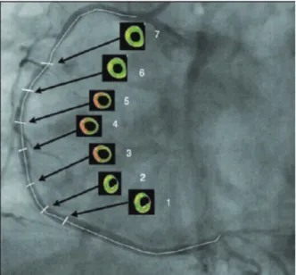
• After locating the cross-section with the highest
rate of arterial remodelling, termed the crosssection of interest (crosssection 4) and the performance of quantitative measures of intravascular ultrasound and
evaluation with Virtual Histology
, the evaluation of six more crosssections was performed with ive frames of
difference; three proximal to and three distal to cross section 4 (Figure 1).
• The following measures were determined by
ultrasound assessment:
– Proximal and distal reference diameters of the coronary artery in mm, for the calculation of reference vessel diameter through simple mean;
– Proximal and distal reference areas of the coro
nary artery in mm2, for the calculation of the reference
vessel area through simple mean;
– Vessel area of crosssections in mm2, for the
calculation of vessel remodelling index;
– Lumen area in crosssections in mm2;
– Atheromatous plaque area of the crosssections
in mm2, calculated by subtracting the area of the vessel
from the lumen area; and
– Plaque burden of crosssections in percentages.
The remodelling index was calculated by the ratio between the vessel area in the crosssection analyzed and reference vessel area, deining as positive remod
elling a ratio > 1.05.11,12
The different compositions of the atheromatous plaque were assessed and characterized by Virtual
Histology
, using the program PcVH 2.2 (Volcano Thera
peutics). For quantitative analysis of Virtual Histology
, tissue corresponding to the atheromatous plaque was colorcoded, which was then divided into four groups according to the main components of the atheromatous plaque: green (ibrotic), yellowgreen (ibrolipidic), red (necrotic core), and white (calcium). Data from the
analysis by Virtual Histology
were reported as absolute values and as percentages relative to the atheromatous plaque area (Figure 2).
Statistical Analysis
The ETCETERA module of the WINPEPI program, version 2.32 was used to calculate the sample size. A correlation coeficient of 0.3 was used, with a power of 90% and a signiicance level of 0.01 to test for the zero difference in the twotailed test. The sample size was calculated at 159 crosssections, that is, 22 patients with seven crosssections each.
The baseline characteristics and the data of the
intravascular ultrasound and Virtual Histology
analysis were tabulated and entered into a database created us ing SPSS software, version 18.0. Quantitative data with Gaussian distribution were described through means and standard deviations. In the presence of asymmetry, median, interquartile range (P25 to P75), and minimum and maximum values were used. Categorical data were described as numbers and percentages.
The variation in the percentage of necrotic core between different crosssections of the coronary artery was studied by analysis of variance for repeated measures; data were logarithmically transformed before analysis. To evaluate the correlation between the remodelling index and the amount of necrotic core, Spearman’s correlation coeficient was used. SPSS software, ver sion 18.0, was used for the analyses, and the level of signiicance was set at < = 0.05.
RESULTS
In total, 113 patients were evaluated, of whom only 30 (26.5%) met the inclusion and exclusion criteria. From the 30 patients included in this study, a total of 270 crosssections were evaluated, derived from pre viously obtained intravascular ultrasound images with
Virtual Histology
; 60 of these crosssections were used to calculate the reference vessel area.
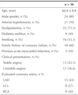
The population’s mean age was 60.8 ± 8.8 years,
most were males (80%), and 30% were diabetics. The most common clinical presentation was unstable angina (56.6%) and the left anterior descending artery was the most often studied vessel (43%) (Table 1).
The measurements performed by intravascular
ultrasound showed proximal reference area of 16.5 ±
5.2 mm2, distal reference area of 14.5 ± 4.9 mm2, ves
sel reference area of 15.5 ± 4.9 mm2, and remodelling
index in the crosssection of interest (crosssection 4)
of 1.2 ± 0.1 (Table 2).
After quantitatively analyzing the percentage of necrotic core in the atheromatous plaque and arranging the results in charts according to the analyzed cross section, the representation of the amount of necrotic core along the studied segment is visualized. Crosssection 1 represents the most distal crosssection of the vessel,
Figure 2 – Images obtained by greyscale intravascular ultrasound (left) and Virtual Histology
(right). The quantiication of the atheromatous plaque components (in mm2 and percentages) are shown above the images.
Table 1
Clinical and angiographic characteristics
n = 30
Age, years 60.8 ± 8.8
Male gender, n (%) 24 (80)
Arterial hypertension, n (%) 21 (70)
Dyslipidaemia, n (%) 22 (73.3)
Diabetes mellitus, n (%) 9 (30)
Smoking, n (%) 16 (53.3)
Family history of coronary failure, n (%) 18 (60)
Previous acute myocardial infarction, n (%) 3 (10)
Clinical presentation, n (%)
Stable angina 13 (43.3)
Unstable angina 17 (56.6)
Evaluated coronary artery, n %
LAD 13 (43)
LCx 8 (27)
RCA 9 (30)
LAD, left anterior descending artery; LCx, left circumlex artery; RCA, right coronary artery.
Component name
Area (mm2)
% of plaque
Fibrosis (FT)
Fibrolipidic (FF)
Lipidic-necrotic (NC)
Calcium dense (DC)
Media 4.01 ...
0.14 1.1
2.31 18.1
1.63 12.8
while crosssection 7 is the most proximal. Crosssection 4, or the crosssection of interest, and crosssections 2, 3, 5, and 6 complement the representation of the chart for the quantitative understanding of the necrotic core in the segment of the coronary artery studied.
The analysis of the variation in the percentage of necrotic core between the seven crosssections of the coronary artery, using analysis of variance for repeated measures, showed higher amounts of necrotic core in the crosssection of interest, which has the highest rate of vascular remodelling (Figure 3).
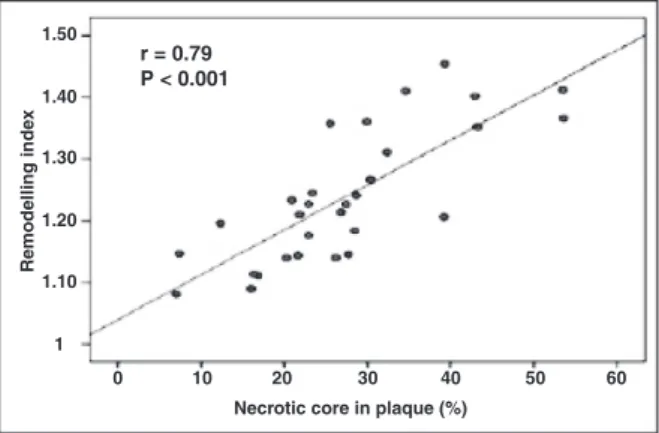
The results obtained when analyzing the linear correlation between the coronary artery remodelling index and the percentage of necrotic core in the ath eromatous plaque show a positive correlation r = 0.79
(P < 0.001) (Figure 4).
DISCUSSION
Coronary heart disease as a major cause of morbidity and mortality has been globally investigated in order to better understand the disease. Studies have demon strated some association between vascular remodelling
and atheromatous plaque composition,811 and a strong
association between plaque accidents (erosion or rupture) and coronary events. Plaques prone to clinical events
have a characteristic distribution of their components
and presence of positive arterial remodelling.2,9,2123
The remodelling index is used to quantify arterial remodelling, which is the ratio between the area of
the region of interest and the reference vessel area.12
Crosssection areas of the vessel are obtained with intravascular ultrasound and provide a 360degree planar view of the coronary arteries, differentiating
their different structures. Validation studies in vitro have
shown good correlation between images generated by
intravascular ultrasound and histological tissue analysis.24
However, this imaging modality does not allow for the characterization and differentiation of the atheromatous plaque components.
Virtual
Histology is an adjunct diagnostic method, developed from greyscale intravascular ultrasound. It provides detailed qualitative and quantitative informa tion on the composition of the atheromatous plaque,
and has been validated by histological studies.18 This
imaging resource has been widely used in clinical re search, both in assessing the evolution of atherosclerotic
lesions25 and in the demonstration of eficacy of new
treatments for atherosclerosis.2426
The Providing Regional Observations to Study Predictors of Events in the Coronary Tree (PROSPECT)
study25 evaluated patients with acute coronary syndromes
submitted to threevessel coronary angiography, supple mented by intravascular ultrasound and by radiofrequency, shortly after percutaneous coronary intervention, and followedup for 3.4 years for adverse clinical events. Half of the events during patient evolution were related to the nonculprit vessel, and atheromatous plaques with vulnerability characteristics (thincap atheroma,
plaque burden > 70%, and luminal area < 4 mm2) were
associated with future adverse cardiovascular events. However, that study did not evaluate the presence of positive remodelling as a predictor of clinical events or plaque vulnerability.
Table 2
Ultrasonography characteristics
Vessel measurements n = 30
Proximal reference area, mm2 16.5 ± 5.2
Mean reference area, mm2 15.5 ± 4.9
Distal reference area, mm2 14.5 ± 4.9
Crosssection of interest (crosssection 4), mm2 18.9 ± 5.2
Remodelling index 1.2 ± 0.1
Figure 3 – Graphical representation of the percentage of necrotic core along the atheromatous plaque, according to the crosssection.
Figure 4 – Graphical representation of the linear correlation between the coronary artery remodelling index and the percentage of necrotic core in atherosclerotic plaque.
60
6 7
50
5 40
4 30
3 20
2 10
1 0
Presence of plaque (%)
Cross-section
Necrotic core
P < 0.001 r = 0.79 P < 0.001
R
emodelling
inde
x
Necrotic core in plaque (%) 1.50
50 60
1.40
40 1.30
30 1.20
20 1.10
10 1
Virtual
Histology allows for the in vivo study
of the atheromatous plaque, and it is possible to perform this study in a population at high risk for clinical events. The analysis of atheromatous plaque showed a positive correlation between positive arte rial remodelling and percentage of necrotic core in
atherosclerotic plaque (r = 0.79; P < 0.001). Similar
results were reported in the retrospective study by
RodriguezGranillo et al.,27 with a population of 41
patients, in which the remodelling index was cor related to the atheromatous plaque components by
Virtual Histology
in coronary arteries considered
nonculprit and with nonsignificant stenosis (< 50%).
The linear regression analysis showed an association between the atheromatous plaque components and coronary artery remodelling. The size of the necrotic core was significantly higher in coronary lesions with
positively remodelled vessel (r = 0.83; P < 0.0001)
than in those with negative remodelling.
Moreover, atheromatous plaques in vessels with positive remodelling had morphology more compatible with vulnerable plaque, as 56% had a thin ibrotic cap, whereas in negative remodelling cases, the atheroma tous plaques appeared to be more stable, as 64% had pathological intimal thickening and no evidence of ibroatheroma with thin ibrous cap.
In spite of some similar results, the present study
and that by RodriguezGranillo et al.27 have several
differences in methodology and in patient and target lesion selection. In the present study, which addressed the association between coronary artery remodelling and the percentage of necrotic core, the starting point was the search for the point of maximum positive remodel ling rather than the point of greatest stenosis, as in the
study by RodriguezGranillo et al.27 Another relevant
difference in methodology was the performance of the full analysis of a segment of coronary artery with a pre deined extension in the present study, which allowed for the comparison between the different sections of the variation of atheromatous plaque components in coronary segments studied, while RodriguezGranillo
et al.27 performed only a few analyses.
As for the selection of the target lesion, the pres ent study assessed lesions with signiicant obstruction
(> 50%) in coronary arteries considered to be culprit
vessels in a cardiovascular event, while Rodriguez
Granillo et al.27 studied nonsigniicant obstructive
lesions in nonculprit coronary arteries. Therefore, these studies evaluated opposing proiles of target le sion and patients, although the present study assessed patients with predominantly unstable lesions, whereas
RodriguezGranillo et al.27 studied patients with lesions
with stable characteristics.
In the present study, the analysis of the amount of necrotic core in the atheromatous plaque and its
intrasection variation in segments of the studied coronary arteries showed a statistically significant
difference (P < 0.001), confirming the existence of
a variation in the amount of necrotic core along the same atheromatous plaque, where the necrotic core concentrates in segments with the highest degree of positive remodelling.
The association between positive arterial remodel ling and necrotic core found in this study emphasizes the interaction between the anatomical and histological components involved in coronary artery disease. The necrotic core has an important role in the release of metalloproteinases, a group of proteolytic enzymes that can degrade and calcify elastin, which is a protein responsible for the elasticity and resilience of blood
vessels.20,28 This may therefore be a possible physiopatho
logical explanation to justify the correlation between necrotic core and positive vascular remodelling as a
causeeffect association.29
The results of linear regression analysis between the arterial remodelling index and the amount of necrotic core in the present study are in agreement with the series of anatomopathological studies that observed an association between rupture or eroded plaques and the presence of positive arterial remodelling in patients who
died due to acute coronary syndrome.30,31
This study demonstrated the importance of positive arterial remodelling and necrotic core in the atheroma tous plaque in order to better understand its evolution (which generally translate into clinical events), and to amplify the detection of vulnerable plaques. In this sense, the search for positive arterial remodelling, which was identiied in this study as associated with the presence of necrotic core (the main component of a vulnerable plaque), can be a useful tool for the de tection of vulnerable plaques prior to plaque instability and its clinical consequences.
Study limitations
The main limitations of the present study were: ret rospective evaluation; dificulty inding a totally healthy segment of coronary artery to be used as reference vessel segment, due to the diffuse nature of coronary artery disease presentation; and the study sample size, which, although small, was suficient to demonstrate statistically signiicant correlations.
CONCLUSIONS
CONFLICT OF INTEREST
The authors declare no conlicts of interest.
REFERENCES
1. Falk E. Unstable angina with fatal outcome: dynamic coronary thrombosis leading to infarction and/or sudden death. Autopsy evidence of recurrent mural thrombosis with peripheral embo lization culminating in total vascular occlusion. Circulation. 1985;71(4):699708.
2. Davies MJ, Thomas AC. Plaque issuring – the cause of acute myocardial infarction, sudden ischaemic death, and crescendo angina. Br Heart J. 1985;53(4):36373.
3. Farb A, Tang AL, Burke AP, Sessums L, Liang Y, Virmani R. Sudden coronary death. Frequency of active coronary lesions, inactive coronary lesions, and myocardial infarction. Circula tion. 1995;92(7):17019.
4. Burke AP, Farb A, Malcom GT, Liang YH, Smialek J, Virmani R. Coronary risk factors and plaque morphology in men with coronary disease who died suddenly. N Engl J Med. 1997; 336(18):127682.
5. Virmani R, Kolodgie FD, Burke AP, Farb A, Schwartz SM. Lessons from sudden coronary death: a comprehensive morphological classiication scheme for atherosclerotic lesions. Arterioscler Thromb Vasc Biol. 2000;20(5):126275.
6. Kolodgie FD, Burke AP, Farb A, Gold HK, Yuan J, Narula J, et al. The thincap ibroatheroma: a type of vulnerable plaque: the major precursor lesion to acute coronary syndromes. Curr Opin Cardiol. 2001;16(5)28592.
7. Kragel AH, Gertz SD, Roberts WC. Morphologic comparison of frequency and types of acute lesions in the major epicardial coronary arteries in unstable angina pectoris, sudden coronary death and acute myocardial infarction. J Am Coll Cardiol. 1991;18(3):8014.
8. Pasterkamp G, Schoneveld AH, van der Wal AC, Haudenschild CC, Clarijs RJ, Becker AE, et al. Relation of arterial geometry to luminal narrowing and histologic markers for plaque vulnerability: the remodeling paradox. J Am Coll Cardiol. 1998;32(3):65562. 9. Mintz GS, Kent KM, Pichard AD, Satler LF, Popma JJ, Leon MB.
Contribution of inadequate arterial remodeling to the devel opment of focal coronary artery stenosis: an intravascular ultrasound study. Circulation. 1997;95(7):17918.
10. Birnbaum Y, Fishbein MC, Luo H, Nishioka T, Siegel RJ. Regional remodeling of atherosclerotic arteries: a major determinant of clinical manifestations of disease. J Am Coll Cardiol. 1997;30(5):114964.
11. Hartmann M, von Birgelen C, Mintz GS, Verhorst PM, Erbel R. Relation between baseline plaque burden and subsequent remodelling of atherosclerotic left main coronary arteries: a serial intravascular ultrasound study with longterm (> or
= 12 months) followup. Eur Heart J. 2006;27(15):177884. 12. von Birgelen C, Hartmann M, Mintz GS, Böse D, Eggebrecht H,
Neumann T, et al. Remodeling index compared to actual vascular remodeling in atherosclerotic left main coronary arteries as assessed with longterm (> or = 12 months) serial
intravascular ultrasound. J Am Coll Cardiol. 2006;47(7):13638. 13. Glagov S, Weisenberg E, Zarins CK, Stankunavicius R, Kolettis GJ.
Compensatory enlargement of human atherosclerotic coronary arteries. N Engl J Med. 1987;316(22):13715.
14. Bezerra HG, Higuchi ML, Gutierrez PS, Palomino SA, Silvestre JM, Libby P. Atheromas that cause fatal thrombosis are usually large and frequently accompanied by vessel enlargement. Cardiovasc Pathol. 2001;10(4):18996.
15. von Birgelen C, Klinkhart W, Mintz GS, Papatheodorou A, Herrmann J, Baumgart D, et al. Plaque distribution and vascular
remodeling of ruptured and nonruptured coronary plaques in the same vessel: an intravascular ultrasound study in vivo. J
Am Coll Cardiol. 2001;37(7):186470.
16. Schoenhagen P, Ziada KM, Kapadia SR, Crowe TD, Nissen SE, Tuzcu EM. Extent and direction of arterial remodeling in stable versus unstable coronary syndromes: an intravascular ultrasound study. Circulation. 2000;101(6):598603.
17. Takimura CK, Lemos PA, Perin MA, Silva EE, Ambrose J, Ramires JA, et al. Preditores geométricos angiográicos de infarto do miocár dio não são associados com marcadores ultrassonográicos de infarto agudo do miocárdio de vulnerabilidade da placa. Arq Bras Cardiol. 2006;87(2):99105.
18. Nair A, Kuban BD, Tuzcu EM, Schoenhagen P, Nissen SE, Vince DG. Coronary plaque classiication with intravascular ultrasound radiofrequency data analysis. Circulation. 2002; 106(17):22006.
19. Nasu K, Tsuchikane E, Katoh O, Vince DG, Virmani R, Surmely JF, et al. Accuracy of in vivo coronary plaque morphology
assessment a validation study of in vivo virtual histology
compared with in vitro histopathology. J Am Coll Cardiol.
2006;47(12):240512.
20. Loftus IM, Naylor AR, Goodall S, Crowther M, Jones L, Bell PR, et al. Increased matrix metalloproteinase9 activity in unstable carotid plaques. A potential role in acute plaque disruption. Stroke. 2000;31(1):407.
21. Tauth J, Pinnow E, Sullebarger JT, Basta L, Gursoy S, Lindsay J Jr, et al. Predictors of coronary arterial remodeling patterns in patients with myocardial ischemia. Am J Cardiol. 1997;80(10):13525. 22. Weissman NJ, Sheris SJ, Chari R, Mendelsohn FO, Anderson WD,
Breall JA, et al. Intravascular ultrasonic analysis of plaque characteristics associated with coronary artery remodeling. Am J Cardiol. 1999;84(1):3740.
23. Fuessl RT, Kranenberg E, Kiausch U, Baer FM, Sechtem U, Höpp HW. Vascular remodeling in atherosclerotic coronary arteries is affected by plaque composition. Coron Artery Dis. 2001;12(2):917.
24. Nishimura RA, Edwards WD, Warnes CA, Reeder GS, Holmes DR Jr, Tajik AJ, et al. Intravascular ultrasound imag ing: in vitro validation and pathologic correlation. J Am Coll
Cardiol. 1990;16(1):14554.
25. Stone GW, Maehara A, Lansky AJ, de Bruyne B, Cristea E, Mintz GS, et al.; PROSPECT Investigators. A prospective naturalhistory study of coronary atherosclerosis. N Engl J Med. 2011;364(3):22635.
26. Serruys PW, GarciaGarcia HM, Buszman P, Erne P, Verheye S, Aschermann M, et al. Effects of the direct lipoproteinassociated phospholipase A(2) inhibitor darapladib on human coronary atherosclerotic plaque. Circulation. 2008;118(11):117282. 27. RodriguezGranillo GA, Serruys PW, GarciaGarcia HM,
Aoki J, Valgimigli M, van Mieghem CA, et al. Coronary artery remodelling is related to plaque composition. Heart. 2006;92(3):38891.
28. Berliner JA, Navab M, Fogelman AM, Frank JS, Demer LL, Edwards PA, et al. Atherosclerosis: basic mechanisms, oxidation, inlammation, and genetics. Circulation. 1995;91(9):248896. 29. Irwin CL, Guzman RJ. Matrix metalloproteinases in medial
arterial calciication: potential mechanisms and actions. Vas cular. 2009;17 Suppl 1:S404.
30. Varnava AM, Mills PG, Davies MJ. Relationship between coro nary artery remodeling and plaque vulnerability. Circulation. 2002;105(8):93943.