0103 - 5053 $6.00+0.00
A
r
ti
c
le
* e-mail: [email protected]
Determination of Catecholamines in Pharmaceutical Formulations Using a Biosensor
Modified with a Crude Extract of Fungi Laccase (
Pleurotus ostreatus
)
Oldair D. Leitea, Orlando Fatibello-Filho*,a and Aneli de M. Barbosab
a
Departamento de Química, Universidade Federal de São Carlos, Rod. Washington Luiz, km 235, 13560-970 São Carlos-SP, Brazil
b
Departamento de Bioquímica, Universidade Estadual de Londrina, Campos Universitário, 85051-990 Londrina-PR, Brazil
Um biossensor de pasta de carbono modificado com extrato bruto enzimático do fungo Pleurotus ostreatus como fonte de lacase é proposto para a determinação de catecolaminas em formulações farmacêuticas. Essa enzima catalisa a oxidação de adrenalina ou dopamina nas quinonas correspondentes e a corrente obtida na redução eletroquímica de cada um dos produtos é relacionada a concentração dessas catecolaminas em solução da amostra. O efeito da concentração de lacase de 0,29 a 1,8 U/mg de pasta de carbono, do pH de 3,0 a 8,0, da velocidade de varredura de 10 a 40 mV s -1 e amplitudes de pulso de potencial de 10 a 60 mV sobre a resposta voltamétrica de pulso diferencial
foi investigado. O desvio padrão relativo foi menor que 1,8% para solução de hidroquinona 2,8 x 10 -4 mol L-1 em pH 7 (n=10). Recuperações variando de 97,3 a 101% para adrenalina e de 95,8 a 102%
para dopamina foram obtidas. As curvas analíticas foram lineares no intervalo de concentração de adrenalina de 6,0 x 10⁻⁵ a 7,0 x 10⁻⁴ mol L-1 e de 7,0 x 10⁻⁵ a 4,0 x 10⁻⁴ mol L-1 para dopamina, com
limites de detecção de 7,9 x 10⁻⁶ mol L-1 e 9,8 x 10⁻⁶ mol L-1, respectivamente. Esse biossensor foi
empregado para a determinação de adrenalina e dopamina em formulações farmacêuticas. Os resultados obtidos com o biossensor para a determinação dessas catecolaminas em formulações farmacêuticas estão em concordância a um nível de confiança de 95% com o procedimento da Farmacopéia americana.
A carbon paste biosensor modified with a crude enzymatic extract of the Pleurotus ostreatus
fungi as a laccase source is proposed for catecholamine determination in pharmaceutical formulations. This enzyme catalyzes the oxidation of adrenaline or dopamine in the corresponding quinones and the current obtained in the electrochemical reduction of each of the products is related to the concentration of these catecholamines in the sample solution. The effect of the laccase concentration from 0.29 to 1.8 U/mg of carbon paste, pH from 3.0 to 8.0, scan rate from 10 to 40 mV s-1 and
potential pulse amplitude from 10 to 60 mV on the differential pulse voltammetric response was investigated. The relative standard deviation was smaller than 1.8% for a 2.8 x 10⁻⁴ mol L-1 hydroquinone
solution at pH 7.0 (n=10). Recoveries varied from 97.3 to 101% for adrenaline and from 95.8 to 102% for dopamine. The analytical curves were rectilinear in the adrenaline concentration range from 6.0 x 10⁻⁵ to 7.0 x 10⁻⁴ mol L-1 and 7.0 x 10⁻⁵ to 4.0 x 10⁻⁴ mol L-1 for dopamine, with detection
limits of 7.9 x 10⁻⁶ mol L-1 and 9.8 x 10⁻⁶ mol L-1, respectively. This biosensor was used for
adrenaline and dopamine determinations in pharmaceutical formulations. The results obtained using the proposed biosensor are in close agreement with those obtained using an American Pharmacopoeia procedure at a 95% confidence level.
Keywords: laccase, Pleurotus ostreatus, catecholamines, biosensor
Introduction
Catecholamines play an important role in the central nervous system as neurotransmitters. They also affect the
regulation of blood pressure and metabolic processes.1
They possess important intrinsic pharmacological properties and are used for the correction of hemodynamic disorders associated with shock episodes.2
mainly chromatography with fluorimetric or electrochemical detection.3 On the other hand, catecholamines are present
in relatively larger amounts in pharmaceuticals and much effort has been done to develop precise, accurate, rapid and simple analytical procedures.4,5 Electroanalytical techniques
applied to the determination of catecholamines are good alternatives.6-9 Nevertheless, the selectivity can be poor,
unless the electrodes are conveniently modified. Thus, the use of enzymes has received a great deal of attention.
During the past few years there has been great interest in the study of enzymatic electrodes, related to applica-tions.10 In addition to purified enzymes,11-13 animal,14 and
vegetable extracts,15-18 immobilized on the electrode
sur-face by different methods, have also been employed as catalyst sources in both aqueous and organic solutions. Carbon paste electrodes have been widely used owing to the possibility of immobilizing not only enzymes, but also ligands, redox mediators, biological tissues, to catalyze the oxidation/reduction of compounds involved in enzymatic reaction. The low-cost and ease of surface re-newal are other important advantages.19,20
The edible mushroon Pleurotus ostreatus, a white-rot fungus cultivated in submerged culture, has been found to produce laccase, a copper-containing polyphenol oxidase. Laccase (EC 1.10.3.2), catalyses the oxidation of various aromatic compounds (di- and polyphenols, aminophenols and diamines) by reducing molecular oxygen to water through an oxidoreductive multicopper system.21 These
oxidases are involved in biotransformation and bioreme-diation processes, they can be also used in molecular genetics,22 genetic expression,23 genetic transcription24 and
cloning.25 Consequently, laccases from white-rot fungi such
as Pleurotus ostreatus are good candidates for further
investigation, as they have potential in biosensor applications. Thus, some purified laccases26-30 in combination with other
enzymes have been used to prepare bienzymatic electrodes for the determination of phenolic compounds26,27 and
catecholamines.28-30 Nevertheless, only a biosensor for the
determination of O2 in water using purified laccase from
Pleurotus ostreatus has been described in the literature.31
In this paper, the construction of a biosensor based on carbon paste modified with crude extract, obtain by submerged culture of the fungi Pleurotus ostreatus, is proposed for the determination of the catecholamines adrenaline and dopamine in pharmaceutical formulations.
Experimental
Reagents and solutions
All reagents were of analytical-reagent grade and all
solutions were prepared with water from a Millipore (Bedford, MA, USA) Milli-Q system (Model UV Plus Ultra-Low Organics Water).
A 0.05 mol L-1 2,6-dimethoxyphenol (Sigma, St. Louis,
MO, USA) in 0.1 mol L-1 phosphate buffer solution (pH
3.5) was used for laccase activity determination.
Hydroquinone was purchased from Sigma (St. Louis, MO, USA) and a 5.0 x 10⁻³ mol L-1 stock solution was
prepared daily in 0.1 mol L-1 phosphate buffer solution
(pH 7.0). Reference solutions from 6.2 x 10⁻⁵ to 8.9 x 10⁻³
mol L-1 were prepared from stock solution by appropriate
dilutions with the same buffer solution.
Catecholamine stock solutions at concentrations of 2.0 x 10⁻² mol L-1 (Aldrich, Milwaukee, WI, USA) were prepared
in 0.1 mol L-1 phosphate buffer solution and standardized
by conventional methods.4,5 Adrenaline reference solutions
from 6.0 x 10⁻⁶ to 6.0 x 10⁻⁴ mol L-1 and dopamine reference
solutions from 6.0 x 10⁻⁶ to 3.0 x 10⁻⁴ mol L-1 were prepared
from stock solutions in 0.1 mol L-1 phosphate buffer
solutions at pH 7.0 and pH 6.0, respectively. In the construction of the biosensors an Acheson 38 graphite powder from Fisher and mineral oil from Sigma were used throughout.
Instrumentation
A Hewlett-Packard (Boise, ID, USA) Model 8452A UV-visible spectrophotometer with a quartz cell (optical path of 1.00 cm) was used in the laccase activity and total protein determinations.
All electrochemical experiments were carried out in a 15 mL thermostated glass cell at 25 ºC. A three-electrode assembly incorporating the biosensor as working electrode; an Ag/AgCl (3.0 mol L-1 KCl) reference and platinum
auxil-iary electrodes were used in all measurements. Differential Pulse Voltammetric measurements were performed with an EG&G PAR, Model 273 A potentiostat/galvanostat with a CPIB-IIA/IIA interface and a PC equipped with data acqui-sition and treatment software was used to record the signal generated in the electrochemical cell.
Crude laccase extract preparation
Pleurotus ostreatus maintained on a potato
agar-dextrose (PAD)32 medium was grown in submerged culture
in Erlenmeyer flasks (1 L) containing 200 mL of Vogel33
The resulting supernatant was lyophilized at 4 ºC and the crude extract utilized as the enzymatic source after laccase activity and total protein determinations.
Laccase activity and total protein determinations
The laccase activity of the crude extract was deter-mined in triplicate by measurement of the absorbance at a wavelength of 468 nm (ε = 1.0 x 10⁴ mol-1 L cm-1) of
2,6-dimethoxyquinone produced in the reaction of 0.2 mL of enzyme solution (15 mg mL-1 crude extract), 2.7 mL of
0.05 mol L-1 2,6-dimethoxyphenol in 0.1 mol L-1 phosphate
buffer solution (pH 3.5) at 30 ºC, after five min of reaction. Laccase activity is expressed in U (one unit corresponds to the amount of enzyme which converts one µmol of 2,6-dimethoxyphenol per minute at pH 3.5 and 30 oC).
Total protein concentration was determined in triplicate by the method of Hartree34 using bovine serum albumin as
standard.
Biosensor preparation
The biosensor was initially prepared by adding and then homogenizing for 20 min in a mortar, 375 mg of grafite powder and 23 mg of crude laccase extract (1.2 units laccase/mg of carbon paste). This mixture was subsequently added to 110 mg of Nujol and mixed in a mortar for at least 20 min to produce the final paste. A portion of each mixture (about 300 mg) was packed into the tip of a 1 mL insulin plastic syringe and a copper wire was inserted to obtain the external electric contact.
Procedure for determination of catecholamines in pharmaceutical samples
Differential pulse voltammetric (DPV) measurements were performed in unstirred and non-aerated 0.1 mol L-1 phosphate
buffer solutions (pH 7.0 for adrenaline and pH 6.0 for dopamine), at 25 oC. The DPV voltammograms were obtained
by scanning the potential from 50 to – 400 mV (for adrenaline) and from 400 to – 50 mV (for dopamine) at a scan rate of 30 mV s-1 and potential pulse amplitude of 50 mV.
Results and Discussion
Activity and total protein determinations
As can be seen in the experimental section, laccase
activity and total protein determinations were performed by spectrophotometry. The laccase activity obtained using 2,6-dimethoxyphenol as substract was 31.5 U mL-1 and
the total protein was 2.01 mg mL-1. Owing to the high
enzymatic activity and low total protein present, a high specific activity of 15.7 U (mg of protein)-1 was obtained.
A commercial laccase (Tienzyme Co)35 from the same fungi
presents a specific activity of only 10.0 U (mg of protein)-1.
Principle of measurements
Recently, the development of biosensors using novel biological materials as biocatalysts has received considerable attention for replacing isolated enzymes.11-13
The use of crude extract rather than isolated enzymes represents not only alternative and attractive analytical properties, but also simplicity, stability, longer lifetime, lower cost and reduced co-factor requirements.19,20,36
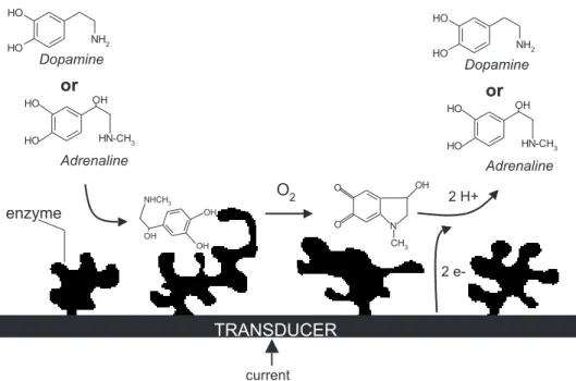
Figure 1 shows a scheme of the enzymatic processes between catecholamine (adrenaline or dopamine) and laccase (PPO) of crude extract incorporated into a carbon paste electrode. Laccase catalyse the oxidation of adrenaline or dopamine to adrenoquinone or dopaminequinone in 1 minute. Then, the quinones produced at the electrode surface are eletrochemically reduced to the corresponding catecholamines at potentials of -174 mV (adrenoquinone) and 238 mV (dopaminequinone) and the resulting cathodic currents correlate directly with the concentration of each catecholamine in the sample solution.
Optimization of the biosensor response
To obtain optimum response conditions for the biosensor based on carbon paste modified with a crude extract of Fungi Laccase, the effect of paste composition, initial stirring time, scan rate, potential pulse amplitude and pH (using hydroquinone, adrenaline and dopamine as substrates) was studied. Table 1 summarizes the range over which each variable was investigated and the optimal values found in those studies.
The effect of enzyme concentration from 0.29 to 1.8 units laccase/mg of carbon paste was investigated. The analytical signals (cathodic peak currents for 2.8 x 10⁻⁴
mol L-1 hydroquinone in 0.1 mol L-1 phosphate buffer
solution at pH 7.0) increased with increases in enzyme
Table 1. Optimization of biosensor parameters
Biosensor parameter Range studied Optimal value
Enzyme concentration 0.29-1.8 1.2
(U/mg of carbon paste)
Stirring time (s) 30-150 6 0
Scan rate (mV s-1) 10-40 3 0
Potential pulse amplitude (mV) 10-60 5 0
p H 3.0-8.0 7.0a; 7.0b
and 6.0c
concentration, using up to 0.94 units laccase/mg of carbon paste and it was practically constant for higher enzyme concentrations. Thus, a concentration of 1.2 units laccase/ mg of carbon paste was used in all biosensors.
The effect of initial stirring time in an interval from 30 to 150 s on the biosensor response for 2.8 x 10⁻⁴ mol L-1
hydroquinone in 0.1 mol L-1 phosphate buffer solution
(pH 7.0) was investigated to determine the best equilibrium time for the reaction. It was observed that 60 s was the best initial stirring to determining these substrates.
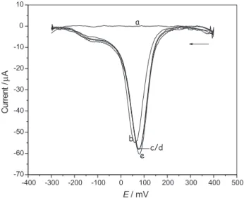
The effects of the pulse amplitudes (Figure 2) and potential scan rate (Figure 3) on the biosensor response for 2.8 x 10⁻⁴ mol L-1 hydroquinone in 0.1 mol L-1 phosphate
buffer solution (pH 7.0) were also investigated. A pulse amplitude of 50 mV and a potential scan rate of 30 mV s-1
were selected, since with these experimental conditions, the highest analytical signal and very good differential pulse voltammogram profiles were obtained.
The effect of pH (Figure 4) from 3.0 to 8.0 for 2.8 x 10⁻⁴
mol L-1 hydroquinone, 2.4 x 10⁻⁴ mol L-1 adrenaline and
3.9 x 10⁻⁴ mol L-1 dopamine solutions was investigated.
The maximum current resulting from the enzyme catalysed reaction was observed at pH 7.0 for hydroquinone and adrenaline and pH 6.0 for the substrate dopamine. Therefore, a pH of 7.0 (hydroquinone and adrenaline) and 6.0 (dopamine) was used in further experiments.
Reproducibility and lifetime studies
The relative standard deviation was smaller than 1.8%
for 2.8 x 10⁻⁴ mol L-1 hydroquinone solution at pH 7.0
(n=10). The reproducibility of three biosensors prepared with the same composition (375 mg of grafite powder (73.8%m/m), 23 mg of crude laccase extract (1.2 units laccase/mg of carbon paste) (4.5%m/m) and 110 mg of Nujol (21.7%m/m)) showed only a slight variation (9.3%) of the analytical curve slope.
After 14 days (over 240 determinations) the response of the biosensor was 75% of the initial response,
Figure 1. Schematic representation of the enzymatic process between catecholamines and laccase at the biosensor surface.
confirming, as expected, the high stability of the crude extract of the fungi laccase.
Biosensor applications
Using the optimized experimental conditions (Table 1), DPV measurements with the proposed biosensor in a recovery study and also in the determination of adrenaline and dopamine in pharmaceutical formulations were carried out.
The effect of excipient substances frequently found with adrenaline and dopamine in pharmaceutical formulations, such as lactose, glucose, sodium chloride, sodium sulfite and phosphate, was evaluated using the proposed procedure. The ratios of the concentrations of adrenaline and dopamine to the excipient substances were fixed at 0.1, 1.0 and 10.0. Of the compounds investigated, only sodium sulfite causes significant interference on biosensor response. In order to eliminate this interference, prior sample treatment with a formaldehyde solution15 in the [formaldehyde]/[sulfite] ratio ≥ 2 was carried out. Recoveries varying from 97.3 to 101% of adrenaline and 95.8 to 102% of dopamine from a pharmaceutical product were obtained using the modified carbon paste electrode (Table 2). In this study, 33.0, 65.0, and 107.0 mg L-1 of adrenaline solutions and 25.0, 49.0,
and 62.0 mg L-1 of dopamine solutions were added to the
samples and the cathodic current peak was obtained. The recovery results obtained suggest the absence of a matrix effect in those determinations.
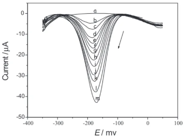
Figures 5 and 6 present the differential-pulse voltammograms using the proposed biosensor in the determination of adrenaline and dopamine in pharma-ceutical formulations. The analytical curve obtained was linear from 6.0 x 10⁻⁵ to 7.0 x 10⁻⁴ mol L-1 of adrenaline
(Icp = 0.17 + 42277[adrenaline] mol L-1); r=0.9989, and
from 7.0 x 10⁻⁵ to 4.0 x 10⁻⁴ mol L-1 of dopamine (Icp = 0.96
+ 63791[dopamine] mol L-1); r=0.9978, with a detection
limit (three times the blank standard deviation/slope) of 7.9 x 10⁻⁶ mol L-1 for adrenaline and 9.8 x 10⁻⁶ mol L-1 for
dopamine. Table 3 presents the results obtained for two commercial samples using the pharmacopeial spectro-photometric procedure37 and the proposed biosensor.
Applying a paired t-test to the results obtained by the two procedures, it was found that all results are in agreement at the 95% confidence level and within an acceptable range of error.
Figure 4. Effect of pH on the biosensor response. : 2.8 x 10⁻⁴ mol L-1 hydroquinone solution; : 2.4 x 10⁻⁴ mol L-1 adrenaline solution and : 3.9 x 10⁻⁴ mol L-1 dopamine solution at 25 ºC.
Figure 3. Differential pulse voltammograms at pulse amplitude of 50 mV and scan rate of 30 mV s-1 for (a) 0.1 mol L-1 phosphate buffer solution (pH 7.0) (blank) and 2.8 x 10⁻⁴ hydroquinone in 0.1 mol L1 phosphate buffer solution (pH 7.0) at pulse amplitude of 50 mV and the following scan rates: (b) 10 mV s-1; (c) 20 mV s-1; (d) 30 mV s-1;(e) 35 mV s-1 and 40 mV s-1 at pulse amplitude of 50 mV.
Table 2. Results of the addition-recovery experiment using adrena-line or dopamine with three different standard concentrations
Adrenaline or dopamine
Sample (mg L-1)a
Added Found Recovery (%)
Adrenaline (Ariston) 33.0 32.1 ±0.8 97.3
65.0 64.5 ±0.9 99.2
107.0 108.1 ±0.6 101
Dopamine (Ariston) 25.0 23.9 ±0.9 95.8
49.0 50.0 ±0.6 102
62.0 61.7 ±0.3 99.5
Conclusions
A biosensor based on carbon paste modified with a crude extract of laccase (obtained from the fungi Pleurotus
ostreatus) is reliable, simple, rapid to prepare, of low cost,
sensitive, precise and accurate. These characteristics make this biosensor an attractive alternative to the procedures presently for pharmaceutical and clinical applications.
Acknowledgments
The authors would like to thank the Fundação de Amparo à Pesquisa do Estado de São Paulo (FAPESP), Coordenacão de Pessoal de Nível Superior (CAPES) and CNPq (Conselho Nacional de Desenvolvimento Científico e Tecnológico), for financial support and the 11º ENQA Organizing Committee.
References
1. Szeponik, J.; Möller, B.; Pfeiffer, D.; Lisdat F.; Wollenberger, U.; Makower, A.; Scheller, F.W.; Biosensors Bioeletronics1997,
12, 947.
2. Hoffman, B.B.; Lefkowitz, R.J. In The Pharmacological
Ba-sis of Therapeutics; Gilman A. G. ed. , 9th ed., McGraw-Hill:
New York, 1996, pp. 211-219.
3. Pesce, A.J.; Kaplan L. A. In Methods in Clinical Chemistry; Bircher, S. ed., The C.V. Mosboy Company: St Louis, MO, 1987, pp. 944-963.
4. Fatibello-Filho, O.; Vieira, I.C.; Analyst1997, 122, 345. 5. Vieira, I. C.; Fatibello-Filho, O.; Talanta1998, 46, 559. 6. Adams, R.N.; Anal. Chem.1976, 48, 1128A.
7. Matysik, F.M.; Nagy, G.; Purgor, E.; Anal. Chim. Acta1992,
264, 177.
8. Brun, A.; Rosset, R.; J.Electroanal. Chem. 1974, 49, 287. 9. Hafizi, S.; Kauk, Z. L.; Stanford, J. A.; J. Electroanal Chem.
1990, 283, 125.
10. Hall, G.F.; Best, D. J.; Turner, A.P.F.; Anal. Chim. Acta 1988,
213, 113.
11. Cosnier, S.; Innocent, C. J.; Electroanal. Chem. 1992, 328, 361. 12. Kulys, J.; Schimid, R.O.; Anal. Lett. 1990, 23, 589. 13. Wang, J.; Ciszewski, A.; Naser, N.; Electroanalysis1992, 4,
777.
14. Rechnitz, G. A.; Science1981, 214, 287.
15. Vieira, I. C.; Fatibello-Filho, O.; Talanta 2000,52, 681. 16. Fatibello-Filho, O.; Vieira, I. C.; Fresenius’ J. Anal. Chem.
2000,368, 338.
17. Fatibello-Filho, O.; Vieira, I C.; J. Braz. Chem. Soc. 2000, 11, 412.
19. Kalcher, K.; Kauffmann, J. M.; Wang, J.; Svancara, I.; Vytras, K.; Neuhold, C.; Yang, Z.; Electroanalysis1995, 7, 5.
Figure 5. Differential pulse voltammograms (adrenaline analytical curve) (a) 0.1 mol L-1 phosphate buffer solution (pH 7.0) (blank) and adrenaline solutions at the following concentrations: (b) 0.6 x 10⁻⁴; (c) 1.2 x 10⁻⁴; (d) 1.8 x 10⁻⁴; (e) 2.4 x 10⁻⁴; (f) 3.0 x 10⁻⁴; (g) 3.6 x 10⁻⁴; (h) 4.7 x 10⁻⁴;(i) 5.8 x 10⁻⁴; (j) 6.9 x 10⁻⁴; (k) 8.0 x 10⁻⁴; (l) 9.1 x 10⁻⁴ and (m) 10.1 x 10⁻⁴ mol L-1 at scan rate of 30 mV s-1, pulse amplitude of 50 mV and 25 oC.
Figure 6. Differential pulse voltammograms (dopamine analytical curve) (a) 0.1 mol L-1 phosphate buffer solution (pH 6.0) and dopam-ine solutions at the following concentrations: (b) 0.7 x 10⁻⁴; (c) 1.3 x 10⁻⁴; (d) 2.0 x 10⁻⁴; (e) 2.6 x 10⁻⁴; (f) 3.3 x 10⁻⁴; (g) 3.9 x 10⁻⁴;(h) 4.5 x 10⁻⁴; (i) 5.1 x 10⁻⁴ and (j) 5.8 x 10⁻⁴ mol L-1, at scan rate of 30 mV s-1, pulse amplitude of 50 mV and 25 oC.
Table 3. Determination of adrenaline and dopamine in pharmaceu-tical formulations using the pharmacopeia37 and the biosensor pro-cedures
Sample Pharmacopeia Biosensor Relative
(mg mL-1) error (%)
Adrenaline
A 1.08 ± 0.04 1.04 ± 0.03 - 3.7
B 0.99 ± 0.03 1.01 ± 0.02 + 2.0
Dopamine
A 4.96 ± 0.06 4.82 ± 0.04 -2.8
B 5.02 ± 0.03 4.97 ± 0.02 -1.0
20. Kissinger, P. T.; Heineman, W. R.; Laboratory Techniques in
Electroanalytical Chemistry, 2nd ed., Marcel Dekker: New York,
1996.
21. Yaropolov, A. I.; Skorobolat’ko, O. V.; Vartanov, S. S.; Varfolomery, S. O.; Appl. Biochem. Biotechnol. 1994, 49.257. 22. Cullen, D.; J. Biotechnol. 1997, 53, 273.
23. Ong, E.; Pollock, W. B. R.; Smith, M.; Gene1997, 196, 113. 24. Collis, P. J.; Dowson, A. D. W.; Appl. Environm. Microbiol.
1997, 63, 3444.
25. Hatamoto, O.; Sekine, H.; Nakano, E.; Abe, K.; Biosci.
Biotechnol. Biochem. 1999, 63, 58.
26. Freire R. S.; Thongngamdee, S.; Duran N.; Wang J.; Kubota, L. T.; Analyst2002, 127, 258.
27. Zouari, N.; Romette, J. T.; Thomas, D.; Biotechnology
Tech-niques1994, 8, 503.
28. Bier, F. F.; EhrentreichForster, E.; Makower, A., Scheller F.
W.; Anal. Chim. Acta 1996, 328, 27.
29. Ghindilis, A. L.; Makower, A.; Bauer, C. G.; Bier, F. F.; Scheller, F. W.; Anal. Chim. Acta 1995, 304, 25.
30. Bier, F. F.; EhrentreichForster, E.; Bauer, C. G.; Scheller F. W.;
Fresenius’ J. Anal. Chem.1996, 354, 861
31. Barton, S. C.; Pickard, M.; Vazquez-Duhalt, R.; Heller, A.;
Biosensors Bioeletronics2002, 17, 1071.
32. Leite, O. D.; Endo, A. S.; Felisbino, M.P.; Obara-Doi, S.M.; Fonseca, R.C.; Barbosa, A. M.; Third Latin American
Biodeg-radation & Biodeterioration Symposium, Book of Abstracts,
Florianópolis, Brazil, September 1998. 33. Vogel, H.; J. Genet. Bull. 1956, 13,42. 34. Hartree, E. S.; Anal. Biochem. 1972, 48, 422.
35. Tienzyme Company, http://TIENZYME.com, accessed in July 2002.
36. Gorton, L.; Electroanalysis1995, 7, 23.
37. United States Pharmacopeia National Formulary XXXIII, US
Pharmacopeial Convention, Rockville, MD, 1995.
Received: October 19, 2002 Published on the web: March 28, 2003