*e-mail: [email protected]
Trabalho apresentado no I Simpósio Mineiro de Ciências dos Materiais, Ouro Preto, Novembro de 2001.
Preparation and Characterization of
Nickel-and Cobalt-doped Magnetites
Maria de Fátima Fontes Lelisa*, José Domingos Fabrisa, Wagner da Nova Mussela,
Armando Yoshihaki Takeuchib
a
Departamento de Química, ICEx, Universidade Federal de Minas Gerais Campus - Pampulha, 31270-901 Belo Horizonte - MG, Brazil
b
Centro Brasileiro de Pesquisas Físicas,
Rua Dr. Xavier Sigaud, 150, 22290-180 Rio de Janeiro - RJ, Brazil
Received: November 11, 2001; Revised: March 3, 2003
Nickel- and cobalt-doped magnetites were prepared by a co-precipitation method and studied in some detail, in an effort to identify some effects of the doping cations on the magnetic, crystallographic and morphological properties of the resulting spinel. The synthetic samples were characterized by conventional chemical analysis, powder X-ray diffractometry, Mössbauer spectroscopy, saturation magnetization and scanning electron microscopy. From chemical analy-sis, the continuous increase of Ni2+ or Co2+ is accompanied by a simultaneous decrease of the Fe2+
contents, in the spinel structure. The magnetization values also decrease continuously with in-creasing doping cation contents. Mössbauer parameters are characteristic of substituted magnetites and indicate the presence of a single phase only. Based on the inverted intensities of the lines 1 (leftmost, on the negative Doppler velocity scale) and 2 of Mössbauer spectra of doped samples, relatively to the pure magnetite, it was assumed that the isomorphical substitution occurs prefer-entially on octahedral coordination sites of the spinel structure. The coercive field of these ferrites decrease steadily with Ni2+ but increases with Co2+ contents, reaching a maximum at x = 0.38, in
the general formula CoxFe3-xO4.
Keywords:Mössbauer, ferrite, magnetization
of the doped-magnetite may vary, depending on the synthe-sis conditions6,7.
The replacement of Fe2+ by Co2+ or Ni2+ does not change essentially the nature of crystallographic structure but its unit cell dimension. The cation distribution in spinels has long been a topic of interest as it affects their magnetic, electric and thermodynamic propertiesb8-10. In addition, it has been found that ferrite particles of similar composition differ on their magnetic properties depending on the prepa-ration method. One reason for such a behavior is believed to be differences in particle size. Decreasing the particle sizes leads to an increase of non-magnetic species on the particle surface6. Various preparation procedures, includ-ing hydrothermal, co-precipition, sol-gel methods and me-chanical alloying have been reportedly used to produce ferrites11. Following the chemical via, some coarse parti-cles may be formed due to agglomeration during the dehy-dration step.
1. Introduction
In the present paper we report the preparation of nickel-and cobalt-doped magnetites by a co-precipitation method and some analysis of their cation distribution, by means of X-ray diffractometry (XRD), Mössbauer spectroscopy, satu-ration magnetization and scanning electron microscopy (SEM).
2. Experimental methods
CoxFe3-xO4 (0 ≤ x ≤ 0.75) and Ni
xFe3-xO4 (0 ≤ x ≤ 0.54) were prepared by co-precipitation. The syntheses were car-ried out by precipitation in aqueous solutions of Co2+, Ni2+ and Fe3+ chlorides (FeCl
3.6H2O, CoCl2.6H2O or NiCl2.6H2O), at room temperature, by adding ammonium hydroxide. The precipitate was washed out with ammonium acetate, dried and decomposed in a N2 atmosphere at 420 °C for 2 h 12. The prepared materials were first characterized by chemical analysis. Nickel, cobalt and iron were deter-mined by dissolving samples with hydrochloric acid 1:1. Nickel and cobalt were analyzed by atomic absorption (Carls Zeiss Jena AAS). Higher contents of iron were determined by volumetric method titration with K2Cr2O713. Fe2+ was determined by dissolving samples in concentrated HCl
un-der a CO2 atmosphere, followed by the volumetric determi-nation with K2Cr2O7. The morphology of the produced grains was determined by scanning electron microscopy (Jeol JSM-840 A). The X-ray diffraction patterns were obtained with a Rigaku Geigerflex diffractometer using CuKa radia-tion. Results confirm the existence of the spinel phase only. NaCl was used as internal standard. The Mössbauer spectra were obtained with a conventional constant acceleration transmission setup and a Co57/Rh source. The isomer shifts are quoted relatively to α-Fe. The saturation magnetization
measurements were performed with a portable magnetometer14. Magnetization curves as a function of the applied magnetic field were performed in a conventional vibrating sample magnetometer, at room temperature.
3. Results and discussion
The proposed chemical formulae and measured mag-netization of all samples are listed in Table 1. The combina-tion of these chemical results and Mössbauer parameters suggests that the amounts of Fe3+ in samples remain nearly unaffected and those of Fe2+ continually decrease with in-creasing amounts of cobalt or nickel. Fitted Mössbauer
pa-Table 1. Chemical formula, saturation magnetization and room-temperature Mössbauer parameters of samples. δ = isomer shift relative to α−Fe; 2ε
Q = quadrupole shift; Bhf = hyperfine field; RA = relative spectral area, and [Fe
3+] and {Fe3+/2+} = tetrahedral and octahedral sites
respectively.
Spinel formula s/J T-1 kg-1 Site d/mm s-1 2ε Q/mm s
-1 B
hf/T RA/% {Fe
3+/2+}
[Fe3+]
Fe3O4 92.5 [Fe3+] 0.28 0.00 49.2 36 1.89
{Fe3+/2+} 0.66 0.01 46.2 64
Fe2.93Co0.07O4 80.9 [Fe3+] 0.25 -0.03 49.4 43 1.41
{Fe3+/2+} 0.69 0.02 46.5 57
Fe2.81Co0.19O4 66.6 [Fe3+] 0.24 -0.04 49.3 45 1.28
{Fe3+/2+} 0.68 0.01 47.1 54
Fe2.62Co0.38O4 50.0 [Fe3+] 0.26 -0.03 49.3 50 1.06
{Fe3+/2+} 0.70 0.02 47.2 50
Fe2.37Co0.63O4 46.3 [Fe3+] 0.26 -0.03 49.3 55 0.87
{Fe3+/2+} 0.68 0.02 47.4 45
Fe2.25Co0.75O4 39.9 [Fe3+] 0.27 -0.02 49.1 53 0.94
{Fe3+/2+} 0.69 0.03 47.4 47
Fe2.96Ni0.04O4 80.6 [Fe3+] 0.26 -0.03 49.1 44 1.35
{Fe3+/2+} 0.70 0.02 46.4 56
Fe2.90Ni 0.10O4 76.6 [Fe3+] 0.26 -0.03 49.2 47 1.20
{Fe3+/2+} 0.69 0.01 46.5 53
Fe2.86Ni 0.14O4 73.4 [Fe3+] 0.26 -0.03 49.4 49 0.92
{Fe3+/2+} 0.70 0.01 46.8 51
Fe2.72Ni 0.28O4 70.1 [Fe3+] 0.28 -0.02 49.3 52 0.98
{Fe3+/2+} 0.70 0.01 47.1 48
Fe2.46Ni 0.54O4 66.3 [Fe3+] 0.28 -0.02 49.3 53 0.94
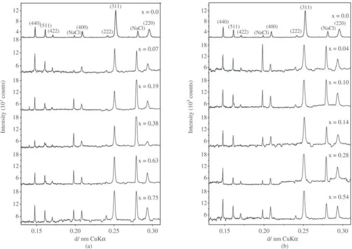
Figure 2. Powder X-ray patterns for the (a) Fe3-xCoxO4 (0 ≤x≤ 0.75); (b) Fe3-xNixO4 (0 ≤x≤ 0.54) samples. NaCl is the internal standard. Figure 1. Scanning electron micrograph of the (a) Fe3O4; (b) Fe2.46Ni0.54O4; (c) Fe2.25Co0.75O4.
rameters indicate a progressive decreasing of the relative subspectral areas corresponding to the octahedral iron of the spinel structure. The decrease in magnetization in doped-samples also suggests that cobalt or nickel is actually re-placing iron in the lattice as Fe2+ in octahedral coordination
has higher magnetic moment than Ni2+15.
ag-glomerates of very small particles. Differently, Co-doped magnetites grow particles of octahedral habit.
XRD patterns show that all samples are well crystal-lized (Fig. 2) with a cubic structure16, there is no appreci-able line broadening or detectappreci-able sign of any other crystal-line or amorphous phase.
The room temperature Mössbauer spectra show a typi-cal hyperfine pattern consisting of two overlapping six-line magnetic splitting structure, in all samples (Fig. 3), assign-able to 57Fe in octahedral and in tetrahedral sites of the spinel lattice. The recoilless fraction of the octahedral iron (f{Fe3+/2+}) is assumed to be 6 % lower than that of the tetra-hedral site (f[Fe3+]), at room temperature6. The relative occu-pancy of iron ions in octahedral {Fe3+/2+} and tetrahedral [Fe3+] coordination sites can be then estimated from the rela-tive subspectral areas (RA; Table 1) of the corresponding Mössbauer spectrum:
] [Fe
} {Fe 3
/2 3
3 /2 3
RA x 0.94
RA
] [Fe
} {Fe
+ + +
= +
+ +
(1)
The so deduced values are presented in Table 1. The
ratio decreases with increasing cobalt or nickel
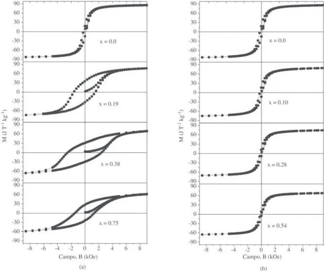
contents. Also, the hyperfine magnetic field of the tetrahe-dral site remains essentially unaltered whereas that of the octahedral iron increases with increasing cobalt contents in the spinel structure. This may indicate that the foreign ions tend to replace referentially iron in octahedral coordination. The saturation magnetization and the coercive field at room temperature depend on the concentration of Ni2+ or Co2+. The hysteresis loop of the pure magnetite is compared with those of the doped-samplles in Fig. 4. It can be ob-served that the coercive field decreases steadily with Ni2+ content but increases with Co2+, reaching a maximum at
x = 0.38, in the general formula, CoxFe3-xO4. One can also see that the saturation magnetization limit is higher and that the coercive field strongly depends on the cobalt propor-tion. It is well known that the saturation magnetization of a spinel ferrite largely depends on its composition and parti-cle size, while the coercive field depends on composition,
particle size and shape. Likewise, the coercive field of these ferrites decreases with the degree of Ni2+ substitution. This can be interpreted as being due to the fact that the magneto-crystalline anisotropy constant of Fe3O4 is higher than that of NiFe2O4.
4. Conclusions
The present study showed that for CoxFe3-xO4 (0 ≤ x ≤ 0.75) and Ni
xFe3-xO4 (0 ≤ x ≤ 0.54) samples ob-tained by a co-precipitation synthesis, room temperature Mössbauer spectra evidence an increase of the hyperfine magnetic field due to 57Fe in octahedral coordination sites, according to the doping degree of the resulting spinel. In both cases, Ni2+ and Co2+, doping-cations tend to replace iron preferentially in octahedral coordination sites. The more the magnetite is doped with nickel the more it tends to form
uniform agglomerates of very small particles, whilst Co-doped magnetites tend to grow particles of octahedral habit. The room temperature magnetic hysteresis loops of these doped-magnetites depend on their composition and parti-cle size. The coercive field decreases with Ni2+ and increases with the Co2+ substitution.
Acknowledgements
This work was financially supported by FAPEMIG, CAPES, CNPq and FINEP (Brazil).
References
1. Adam, J.D., Krishnaswamy, S.V., Talisa S. H. & Yoo, K.C., J. Magn. Mag. Mater, v. 83, p. 419-424, 1990. 2. Lee, J.G., Park, J.Y.; Oh, Y-J & Kim, C.S., Journal of
Applied Physics, v. 84, n. 5, p. 2801-2804, 1998.
3. Petrosius, S.C.; Drago, R.S.; Young, V. & Grunewald, G.C. J. Am. Chem. Soc., v. 115, p. 6131-6137, 1993. 4. Verwey, E.J.W.; Haayman, P.W. Physica 8 , p. 979-987,
1941.
5. Ferguson Jr, G.A.; Haas, M. Physical Review, v. 112 , p. 1130-1139, 1958.
6. Sawatzky, G.A; Van Der Woude, F.; Morrish, A.H. Physi-cal Review. 187:747-757, 1969.
7. Haneda, K.; Morrish, A. H. J.Appl. Phys, v. 63, n. 8, p. 765-771, 1988.
8. Goss, C.J. Phys Chem Minerals, v. 16, p. 164-171, 1988. 9. Lenglet, M.; Lefez, B. Solid State Communications, v.
98, n. 8, p. 689-694, 1996.
10. Mendelovici, E., Villalba, R.; Sagarzazu, A.
Thermochimica Acta, v. 318, p. 51-56, 1998.
11. Deshpande, C.E.; Date, S.K. Indian Journal of Chemis-try, v. 35A, p. 353-365, 1996.
12. De Abreu Filho, P.P.; Pinheiro, E.A. ; Galembeck, F.; Labaki, L.C. Reactivity of solids, v. 3, p. 241-250, 1987. 13. Jeffery, P.G.; Hutchison, D. Chemical Methods of Rock Analysis, 3.ed. Oxford, Pergamon Press, p. 379, 1981. 14. Coey, J.M.D.; Cugat, O.; Mac Cauley, J.; Fabris, J.D.
Revista de Física Aplicada e Instrumentação. São Paulo, v. 7, p 25-30, 1992.
15. Huang, C.; Matijevic, E. Solid State Ionics, v. 84, p. 249-258, 1996.