Cardiovascular risk factors and
cognitive performance in aging
Juliana Rumy Tsuchihashi Takeda1, Tatiane Martins Matos2, Juliana Nery de Souza-Talarico3
ABSTRACT. Background. Atherosclerosis in cerebral blood vessels, especially those which compose the Circle of Willis, can lead to reduced supply of oxygen and nutrients to different cortical structures, affecting cognitive function. Objective: To analyze whether cardiovascular risk factors negatively influence cognitive performance in adults and elderly. Methods: One hundred twenty-nine participants of both sexes, aged over 50 years, without cognitive or functional impairment were included. Body mass index (BMI), hypertension (HTN), diabetes mellitus (DM), smoking history, plasma levels of total cholesterol, low density lipoproteins (LDL), high density lipoproteins (HDL) and very low density lipoproteins (VLDL) cholesterol, triglycerides, and glucose were the cardiovascular risk factors analyzed. Cognitive assessment was performed using tests of attention, working memory, category fluency and declarative memory. Results: Controlling for age and education, multivariate linear regression models revealed that higher concentrations of triglycerides, as well as total, LDL and VLDL cholesterol, were associated with poorer performance on the digit span and category fluency tests. Higher HDL concentrations were associated with higher scores on category fluency tasks. Furthermore, higher BMI was associated with poorer delayed recall performance. Conclusion: The findings revealed that cardiovascular risk factors may negatively impact cognitive performance in aging.
Key words: memory, cognitive performance, cardiovascular risk factor, preventive health.
FATORES DE RISCO CARDIOVASCULAR E DESEMPENHO COGNITIVO NO ENVELHECIMENTO
RESUMO. Introdução: A aterosclerose nos vasos sanguíneos cerebrais, especialmente naqueles que compõem o Círculo de Willis, pode levar à redução da oferta de oxigênio e nutrientes para diferentes estruturas corticais, afetando a função cognitiva. Objetivo: Analisar se fatores de risco cardiovascular influenciam negativamente o desempenho cognitivo em adultos e idosos. Métodos: Foram incluídos cento e vinte e nove participantes de ambos os sexos, com idade superior a 50 anos, sem comprometimento cognitivo ou funcional. Índice de massa corporal (IMC), hipertensão arterial (HAS), diabetes mellitus (DM), história de tabagismo, níveis plasmáticos de colesterol total e lipoproteínas de baixa densidade (LDL), lipoproteínas de alta densidade (HDL) e lipoproteínas de baixa densidade (VLDL), triglicerídeos e glicemia foram os fatores de risco cardiovascular analisados. A avaliação cognitiva foi realizada utilizando testes de atenção, memória operacional, fluência verbal e memória declarativa. Resultados: Controlando para idade e escolaridade, os modelos de regressão linear multivariada revelaram que concentrações mais elevadas de triglicerídeos, bem como colesterol total, LDL e VLDL, estão associadas com pior desempenho nos testes de extensão de dígitos e fluência verbal. Além disso, concentrações maiores de HDL se correlacionaram com pontuações maiores nas tarefas de fluência verbal. IMC maior se associou com menor desempenho na evocação tardia. Conclusão: Os achados revelaram que fatores de risco cardiovascular podem afetar negativamente o desempenho cognitivo durante o envelhecimento.
Palavras-chave: memória, desempenho cognitivo, fatores de risco cardiovascular, saúde preventiva.
INTRODUCTION
C
omplaints of memory decline duringaging have been the subject of many studies, in an attempt to identify factors that might explain the cognitive variability seen
among adults and the elderly. Many elders have cognitive performance that is equal to or even better than young adults; although com-mon, memory decline cannot be considered part of natural aging.1-6
This study was conducted at the Heart Institute (InCor), Faculty of Medicine, University of São Paulo, SP, Brazil.
1RN, Heart Institute (InCor), Faculty of Medicine, University of São Paulo, SP, Brazil. 2Master degree, Graduate Program in Adult Healthcare Nursing, School of Nursing,
University of São Paulo, SP, Brazil.3PhD, Department of Medical-Surgical Nursing, School of Nursing, University of São Paulo, SP, Brazil.
Juliana Nery de Souza-Talarico. Department of Medical-Surgical Nursing / School of Nursing / Universidade de São Paulo – Av. Dr Enéas de Carvalho Aguiar, 419 – 05403-000 São Paulo SP – Brazil. E-mail address: [email protected]
Disclosure: The authors report no conflits of interest.
Received October 22, 2017. Accepted in final form November 16, 2017.
Genetic, epigenetic and environmental factors have been considered in research investigating how healthy habits and lifestyle can inluence brain function and
human behavior throughout life.4
Most available evidence describes the efect of car-diovascular risk on cognitive performance in individu-als who already have some degree of cognitive impair-ment.7 However, it is still unclear whether these factors also negatively afect cognitive performance in healthy adults and elderly. his is particularly relevant, since actions for prevention and control of cardiovascular risk factors can reduce not only the risk for ischemic cardiomyopathies, but can also promote wellbeing and quality of life during aging.
Anatomical and functional age-related changes allied with exposure to cardiovascular risk factors may com-promise cerebral blood low which may in turn nega-tively inluence cognitive performance.3,5
Changes in cardiac output, increase in cholesterol levels and peripheral vascular resistance, systemic blood pressure, body mass index (BMI), and body fat number among the cardiovascular risk factors observed during
aging which may inluence memory decline.6
Atherosclerosis in cerebral blood vessels, especially those that comprise the Circle of Willis, can lead to a reduced supply of oxygen and nutrients to diferent cor-tical structures, afecting cognitive function in patients with cardiovascular diseases.8 he very high blood pres-sure can interfere with microcirculation and cause cere-bral ischemia.2,8
he control of cardiovascular risk factors, such as hypertension, diabetes and dyslipidemia, is associ-ated with better cognitive performance.2,8 Additionally, post-mortem analysis has revealed a positive associa-tion between high cholesterol and amyloid deposits, one of the most important biomarkers of Alzheimer’s disease (AD).9 Similarly, previous studies have showed that cardiovascular risk factors are associated with poor memory performance and executive function in midlife adults from developed countries.10,11 However, this is poorly understood among individuals from low- and middle-income countries, where socioeconomic inequal-ity may limit access to qualinequal-ity health services and thus, the control of cardiovascular risk factors.
his study aimed to examine whether modiiable car-diovascular risk factors negatively inluence cognitive performance in Brazilian adults and older adults.
METHODS
Ethical procedures. All participants were volunteers and signed the consent form for the study, approved by the
Research Ethics Committee of the Federal University of São Paulo - UNIFESP (n. 0823/09).
Participants. One hundred twenty-nine elderly indi-viduals were included, with preserved, intact cognitive function; 83.6% (107 individuals) were female, with mean age of 65.5 (SD ± 8.0) years, and mean education of 9.78 (SD ± 4.45) years of study.
Eligibility criteria for the study participants were: aged over 50 years, both sexes, literate, with education ≥ four years; intact cognitive and functional features preserved (according to performance evaluation on the Mini-Mental State Examination (MMSE) and the Infor-mant Questionnaire on Cognitive Decline in the Elderly (IQCODE) for dementia detection). Individuals were excluded according to the following criteria: diagnosis of neurological, neurodegenerative and/or psychiatric disease; use of psychoactive drugs; a history of alcohol or drug abuse in the past year, or previously for a long period; smokers or ex-smokers for less than ten years.
he risk factors established by the American Heart Association12 include BMI, hypertension (HTN), diabe-tes mellitus (DM), smoking history, blood glucose, tri-glycerides, total cholesterol and LDL, HDL and VLDL
cholesterol. Blood samples were analyzed by the
Asso-ciação Fundo de Incentivo à Pesquisa (AFIP).
Cognitive assessment. he cognitive abilities evaluated were: attention, working memory, category luency, and short and long-term declarative memory.
Attention and working memory performance were evaluated using the Forward Digit Span (FDS) and For-ward Corsi Blocks (FCB) tests which evaluate attention, and the Backward Digit Span (BDS) and Backward Corsi Blocks (BCB) tests that evaluate working memory. In the FDS, a sequence of digits was read out at a rate of one number per second. After reading, the individual is asked to repeat the numbers in the order they were read out. If errors occur, the individual can retake the test with a sequence of the same number of digits. In the BDS, the individual must remember the sequence backwards. he test assesses the storage capacity and reverberation in immediate verbal memory (phonologi-cal loop), and the ability to maintain and manipulate information (central executive). he scores for both tests were the maximum number of digits the individu-als repeated in sequence correctly, which ranged from three to six digits.
two blocks changed colors sequentially at one-second interval, and the individual had to indicate that block change sequence. When answering correctly, the length of the sequence was increased until the participant committed two errors in the same sequence. For the indirect order (BCB), the individual had to indicate the sequence backwards. he scores for both tests were the maximum number of digits or blocks correctly repeated in the sequence, which ranged from three to six digits or blocks.
For assessment of category luency, the FAS version was used, in which the participant had to state as many words as possible beginning with the letters F, A, and S, with one minute for each letter. he objective of this test is to evaluate spontaneity in generating words. It consti-tutes an executive function test, because the participant must apply a set of rules with which he/she must comply during the generation of words. he score corresponds to the number of words stated for each letter, and mis-takes (for example, repeated words or those that did not begin with the proposed letter). Category luency is also evaluated, using animals, fruits and musical instru-ments stated in one minute.
Declarative memory was assessed using the Cali-fornia Verbal Learning Test (CVLT). he test measures the verbal learning process, and the amount of material acquired and retained using a verbal memory task. A list containing 16 items was presented, classiied into categories of clothing, vegetables, tools, and fruit. he individual had ive attempts to recall the items and remember the list immediately after its presentation. hirty minutes after the immediate recall list (learning), the participant had to recall the items again, without being reread the list (delayed recall). he score is the number of items recalled during the ive attempts, that is, a point is given for each correct item.
Data collection procedures. Initially the study was publi-cized in the media, through the radio, newspaper and internet, spontaneously attracting volunteers inter-ested in participating in the study. hese individuals contacted the telephone survey team to express their interest in participating. During this telephone contact, the study candidate answered questions relevant to inclusion and exclusion criteria. hose who were included in the study, in accordance with the criteria, were scheduled for individual interviews at the Depart-ment of Psychobiology of UNIFESP. In this interview, participants were assessed for functional and cogni-tive performance, and those with incompatible perfor-mance for age and level of education, and with
pre-established scores on the MMSE and IQCODE, were excluded.13,14
Informed consent was obtained from eligible partici-pants. Blood samples were collected for laboratory anal-ysis, and then sociodemographic and cognitive assess-ment questionnaires were immediately administered. Anthropometric measurements (weight and height) were obtained using properly calibrated manual scales.
Statistical analyses. Data were analyzed descriptively using means, standard deviation (SD) and frequency (absolute and relative). For analyses of the learning curve in the CVLT test, the analysis of variance (ANOVA) for repeated measures was used with the Greenhouse correction method in the absence of sphe-ricity. To investigate the association between cognitive assessment test scores and cardiovascular risk factors, multivariate linear regression analysis was performed with age and education as potential confounders. he Statistical Package for the Social Sciences (SPSS), version 14.0 was used. he signiicance level was set at p < 0.05.
RESULTS
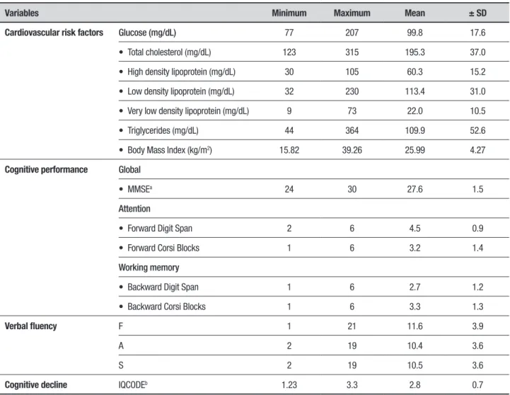
Cardiovascular risk factor profiles and cognitive assess-ment. More than half the sample (58.6%) had HTN, 23.8% had stopped smoking for at least 10 years, and 8.7% had type 2 DM. About 40% of the sample was overweight or obese, according to the BMI cutof value > 25 for individuals up to 59 years, and BMI > 27 for elderly aged 60 or older.15 Regarding medication, 59.7% (n = 77) used antihypertensive drugs, statins, oral hypoglycemic agents and antidiuretics. he lipid proile and mean glucose concentrations are shown in Table 1. he participants had cognitive and functional perfor-mance considered to be within normal range.13,14 Table 1 shows the cognitive performance of participants.
Regarding the CVLT test, an efect of time on imme-diate short- and long-term recall of the list items was observed (F (762.685) = 172.9, p < 0.001; Figure 1), evi-dencing a learning efect after ive attempts to memo-rize the list.
Table 1. Sample characteristics according to cognitive and functional performance and cardiovascular risk factors.
Variables Minimum Maximum Mean ± SD
Cardiovascular risk factors Glucose (mg/dL) 77 207 99.8 17.6
• Total cholesterol (mg/dL) 123 315 195.3 37.0
• High density lipoprotein (mg/dL) 30 105 60.3 15.2
• Low density lipoprotein (mg/dL) 32 230 113.4 31.0
• Very low density lipoprotein (mg/dL) 9 73 22.0 10.5
• Triglycerides (mg/dL) 44 364 109.9 52.6
• Body Mass Index (kg/m2) 15.82 39.26 25.99 4.27
Cognitive performance Global
• MMSEa 24 30 27.6 1.5
Attention
• Forward Digit Span 2 6 4.5 0.9
• Forward Corsi Blocks 1 6 3.2 1.4
Working memory
• Backward Digit Span 1 6 2.7 1.2
• Backward Corsi Blocks 1 6 3.3 1.3
Verbal fluency F 1 21 11.6 3.9
A 2 19 10.4 3.6
S 2 19 10.5 3.6
Cognitive decline IQCODEb 1.23 3.3 2.8 0.7
aMini-Mental State Exam; bInformant Questionnaire on Cognitive Decline in the Elderly.
Figure 1. Means of immediate short-term and delayed recall of California Verbal Learning Test.
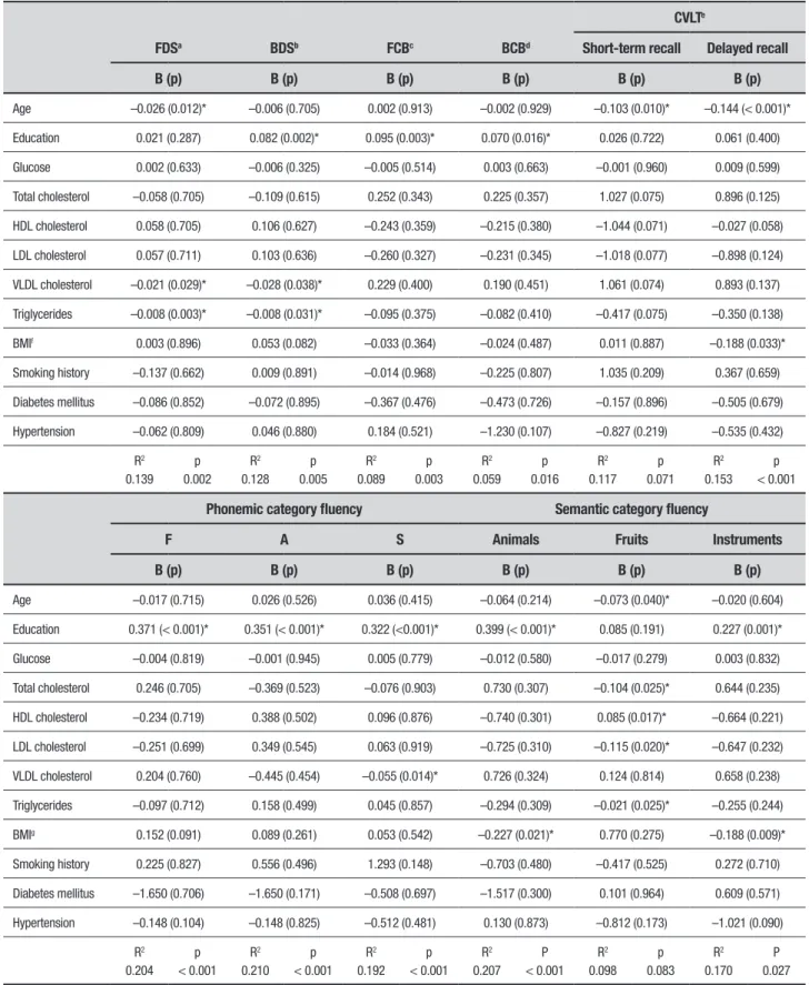
Table 2. Multivariate linear regression coefficients between cardiovascular risk factors and cognitive test scores.
CVLTe
FDSa BDSb FCBc BCBd Short-term recall Delayed recall
B (p) B (p) B (p) B (p) B (p) B (p)
Age –0.026 (0.012)* –0.006 (0.705) 0.002 (0.913) –0.002 (0.929) –0.103 (0.010)* –0.144 (< 0.001)*
Education 0.021 (0.287) 0.082 (0.002)* 0.095 (0.003)* 0.070 (0.016)* 0.026 (0.722) 0.061 (0.400)
Glucose 0.002 (0.633) –0.006 (0.325) –0.005 (0.514) 0.003 (0.663) –0.001 (0.960) 0.009 (0.599)
Total cholesterol –0.058 (0.705) –0.109 (0.615) 0.252 (0.343) 0.225 (0.357) 1.027 (0.075) 0.896 (0.125)
HDL cholesterol 0.058 (0.705) 0.106 (0.627) –0.243 (0.359) –0.215 (0.380) –1.044 (0.071) –0.027 (0.058)
LDL cholesterol 0.057 (0.711) 0.103 (0.636) –0.260 (0.327) –0.231 (0.345) –1.018 (0.077) –0.898 (0.124)
VLDL cholesterol –0.021 (0.029)* –0.028 (0.038)* 0.229 (0.400) 0.190 (0.451) 1.061 (0.074) 0.893 (0.137)
Triglycerides –0.008 (0.003)* –0.008 (0.031)* –0.095 (0.375) –0.082 (0.410) –0.417 (0.075) –0.350 (0.138)
BMIf 0.003 (0.896) 0.053 (0.082) –0.033 (0.364) –0.024 (0.487) 0.011 (0.887) –0.188 (0.033)*
Smoking history –0.137 (0.662) 0.009 (0.891) –0.014 (0.968) –0.225 (0.807) 1.035 (0.209) 0.367 (0.659)
Diabetes mellitus –0.086 (0.852) –0.072 (0.895) –0.367 (0.476) –0.473 (0.726) –0.157 (0.896) –0.505 (0.679)
Hypertension –0.062 (0.809) 0.046 (0.880) 0.184 (0.521) –1.230 (0.107) –0.827 (0.219) –0.535 (0.432)
R2 0.139
p 0.002
R2 0.128
p 0.005
R2 0.089
p 0.003
R2 0.059
p 0.016
R2 0.117
p 0.071
R2 0.153
p < 0.001
Phonemic category fluency Semantic category fluency
F A S Animals Fruits Instruments
B (p) B (p) B (p) B (p) B (p) B (p)
Age –0.017 (0.715) 0.026 (0.526) 0.036 (0.415) –0.064 (0.214) –0.073 (0.040)* –0.020 (0.604)
Education 0.371 (< 0.001)* 0.351 (< 0.001)* 0.322 (<0.001)* 0.399 (< 0.001)* 0.085 (0.191) 0.227 (0.001)*
Glucose –0.004 (0.819) –0.001 (0.945) 0.005 (0.779) –0.012 (0.580) –0.017 (0.279) 0.003 (0.832)
Total cholesterol 0.246 (0.705) –0.369 (0.523) –0.076 (0.903) 0.730 (0.307) –0.104 (0.025)* 0.644 (0.235)
HDL cholesterol –0.234 (0.719) 0.388 (0.502) 0.096 (0.876) –0.740 (0.301) 0.085 (0.017)* –0.664 (0.221)
LDL cholesterol –0.251 (0.699) 0.349 (0.545) 0.063 (0.919) –0.725 (0.310) –0.115 (0.020)* –0.647 (0.232)
VLDL cholesterol 0.204 (0.760) –0.445 (0.454) –0.055 (0.014)* 0.726 (0.324) 0.124 (0.814) 0.658 (0.238)
Triglycerides –0.097 (0.712) 0.158 (0.499) 0.045 (0.857) –0.294 (0.309) –0.021 (0.025)* –0.255 (0.244)
BMIg 0.152 (0.091) 0.089 (0.261) 0.053 (0.542) –0.227 (0.021)* 0.770 (0.275) –0.188 (0.009)*
Smoking history 0.225 (0.827) 0.556 (0.496) 1.293 (0.148) –0.703 (0.480) –0.417 (0.525) 0.272 (0.710)
Diabetes mellitus –1.650 (0.706) –1.650 (0.171) –0.508 (0.697) –1.517 (0.300) 0.101 (0.964) 0.609 (0.571)
Hypertension –0.148 (0.104) –0.148 (0.825) –0.512 (0.481) 0.130 (0.873) –0.812 (0.173) –1.021 (0.090)
R2 0.204
p < 0.001
R2 0.210
p < 0.001
R2 0.192
p < 0.001
R2 0.207
P < 0.001
R2 0.098
p 0.083
R2 0.170
P 0.027
luency tasks. Furthermore, higher BMI was associ-ated with poorer delayed recall performance. No asso-ciation was observed between cognitive performance and glucose, HTN, DM or previous smoking history (Table 2).
DISCUSSION
his study allowed us to ascertain that cardiovascular factors are associated with low cognitive performance. Individuals with higher levels of LDL, VLDL, triglycer-ides and BMI showed poorer performance in attention, working memory, category luency and delayed recall, suggesting a negative inluence of lipids on memory during aging.
Corroborating our indings, previous studies show increased LDL in patients with dementia or in those
who have sufered a stroke.7 Furthermore, changes in
serum lipids and lipoproteins are associated with the development of cerebrovascular disease.7 Accordingly, adults with high HDL had better performance on lan-guage tests,16 immediate recall and the MMSE,17 rein-forcing that the bad cholesterol fraction can negatively afect memory performance in older adults. However, indings obtained in cognitively healthy, nonagenarian and centenarian elderly revealed no signiicant asso-ciation between dyslipidemia and cognitive impair-ment.18-20 Also, treatment with lipid-lowering agents did not result in improvement in memory performance or language.18
In addition, we found that the greater the BMI, the poorer the category luency and delayed recall perfor-mance. Given the category luency test evaluates not only executive function, but also the ability to generate words, it is reasonable to assume that the negative corre-lation between BMI and category luency may be related to a slowing in information processing speed. Support-ing this interpretation, some authors found a signii-cant association between high BMI and worse overall cognitive performance,21 memory decline,22 poor
atten-tion and processing speed.23 Although these authors
attributed age to slower processing speed, in the current study, BMI was negatively associated with category lu-ency even after controlling for age and education level.
Although the mechanisms by which cardiovascular risk factors may compromise cognitive performance during aging are unclear, some hypotheses have been discussed. High concentration of LDL, as an indepen-dent predictor of coronary artery disease and carotid artery atherosclerosis,2,7 can lead to embolism and cere-bral hypoperfusion and subsequent cognitive decline. Hypertension may induce atherosclerosis and cause
capillary damage, leading to hypoxia, cerebral ischemia, and clinical and subclinical brain damage.24 Obesity can reduce cardiovascular capacity, promote inlammation, and lead to endocrine disruption, thereby inluencing
cognitive performance.21 Furthermore, a high BMI is
associated with decreased white matter in the brain when weight loss is induced by dieting.23
It is noteworthy that the associations observed in this study were obtained with mean cholesterol and triglyceride levels within the normal range, and only a few individuals actually had dyslipidemia. his suggests that lipid indicators such as cholesterol and triglycerides do not necessarily need to be overly high to negatively inluence cognitive performance. hus, it is possible that cognitive performance is modulated by lipid indicators in an inverted U-shape function, in which optimal lipid concentrations are associated with improved memory performance, whereas overly high or low concentrations can produce cognitive decline. Supporting this interpre-tation, some studies have found a positive correlation
between cholesterol, BMI and cognitive performance.25
It should be noted that these lipids are natural compo-nents of the body and necessary for the formation of neuronal myelin sheath, and hence for the conduction of nerve impulses.
It is important to consider that the sample analyzed in this study was composed of predominantly female individuals over 60 years of age, which raises the pos-sibility of selection bias in the results. However, we emphasize that the variable “age” was included as a covariate in the statistical analysis. In addition, non-random selection of participants can compromise the external validity of the results, since it may not relect the actual health scenario of the adult and elderly populations. In this sense, the results may have been underestimated and the observed correlations might be stronger, if we consider that many older people do not spontaneously seek health services to assess their metabolic status and cognitive performance.
sug-gest that such strategies may have a positive impact on cognitive performance during aging.
Author contribution. JNST coordinated the study design, collected data, performed the statistical analysis, and contributed to the inal version of the manuscript. JRTT contributed to the statistical analysis, searched
the literature and prepared the irst draft of the manu-script. TMM searched the literature and prepared the irst draft of the manuscript. All authors read and approved the inal manuscript.
Funding. São Paulo Research Foundation (FAPESP; Grant # 2009/13911-6)
REFERENCES
1. Alosco ML, Gunstad J, Jerskey BA, Xu X, Clark US, Hassenstab J, et al. The adverse effects of reduced cerebral perfusion on cognition and brain structure in older adults with cardiovascular disease. Brain Behav. 2013;3:626-36.
2. Alves TCTF, Wajngarten M, Busatto Filho G. Cognitive decline, cardio-vascular risk factors, and neuroimaging abnormalities. Rev Psiq Clin. 2005;32:160-9.
3. Chen JJ, Rosas HD, Salat DH. Age-associated reductions in cerebral blood flow are independent from regional atrophy. Neuroimage. 2011; 55:468-78.
4. Lupien SJ, Wan N. Successful ageing: from cell to self. Philos Trans R Soc Lond B Biol Sci. 2004;359:1413-26.
5. Matsudo SM, Matsudo VKM, Barros Neto TL. The impact of aging on anthropometric, neuromotor, and metabolic variables of physical fitness. Rev Bras Cienc Mov. 2000;8:21-32.
6. Glisky EL. Changes in Cognitive Function in Human Aging. In: Riddle DR, editor. Brain Aging: Models, Methods, and Mechanisms. Boca Raton, FL: CRC Press, 2007: 3-20.
7. Moroney JT, Tang MX, Berglund L, Small S, Merchant C, Bell K, Stern Y, Mayeux R. Low-density lipoprotein cholesterol and the risk of dementia with stroke. JAMA. 1999;281:254-60.
8. Leritz EC, McGlinchey RE, Kellison I, Rudolph JL, Milberg WP. Cardio-vascular disease risk factors and cognition in the elderly. Curr Cardiovasc Risk Rep. 2011; 5:407-12.
9. Pappolla MA, Bryant-Thomas TK, Herver D, Pacheco J, Fabra GM. Mild hypercholesterolemia is an early risk factor for the development of Alzheimer amyloid pathology. Neurology. 2003;61:199-205.
10. Gupta A, Preis SR, Beiser A, Devine S, Hankee L, Seshadri S, Au R. Midlife cardiovascular risk impacts memory function: The Framingham Offspring Study. Alzheimer Dis Assoc Disor. 2015;29:117-23. 11. Nishtala A, Preis SR, Beiser A, Devine S, Hankee L, Seshadri S, et al.
Midlife cardiovascular risk impacts executive function: Framingham offspring study. Alzheimer Dis Assoc Disor. 2014;28:16-22.
12. American Heart Association. Guidelines for primary prevention of cardio-vascular disease and stroke: update consensus panel guide to compre-hensive risk reduction for adult patients without coronary or other athero-sclerotic vascular diseases. Circulation. 2002;106:388-91.
13. Brucki SMD, Nitrini R, Caramelli P. Suggestions for utilization of the
mini-mental state examination in Brazil. Arq Neuropsiquiatr. 2003;61: 777-81.
14. Bustamante SEZ, Bottino CMC, Lopes MA, Azevedo D, Hototian SR, Litvoc J. Combined instruments on the evaluation of dementia in the elderly: preliminary results. Arq Neuropsiquiatr. 2003;61:601-6. 15. Lipschitz DA. Screening for nutritional status in the elderly. Prim Care.
1994;21:55-67.
16. Reynolds CA, Gatz M, Prince JA, Berg S, Pedersen NL. Serum lipid levels and cognitive change in late life. JAGS. 2010;58:501-9. 17. Panza F, D’Introno A, Colacicco AM, Capurso C, Pichichero G, Capurso
SA, Stern Y, Mayeux R. Lipid metabolism in cognitive decline and dementia. Brain Res Rev. 2006; 51:275-92.
18. Reitz C, Luchsinger J, Tang MX, Mayeux R. Impact of plasma lipids and time on memory performance in healthy elderly without dementia. Neurology. 2005;64:1378-83.
19. Huang CQ, Dong BR, Wu HM, Zhang YL, Wu JH, Lu ZC, Flaherty JH. Association of cognitive impairment with serum lipid / lipoprotein among chinese nonagerian and centenarian. Dement Geriatr Cogn Disord. 2009;27:111-6.
20. Okusaga O, Stewart MCW, Butcher I, Deary I, Fowkes GR, Price JF. Smoking, hypercholesterolaemia and hypertension as risk factors for cognitive impairment in older adults. Age Ageing. 2013;42:306-11. 21. Gunstad J, Lhotsky A, Wendell CR, Ferrucci L, Zonderman AB.
Longitu-dinal examination of obesity and cognitive function: results from the Balti-more longitudinal study of aging. Neuroepidemiology. 2010;34:222-9. 22. Dregan A, Stewart R, Gulliford MC. Cardiovascular risk factors and
cognitive decline in adults aged 50 and over: a population-based cohort study. Age Ageing. 2013; 42:338-45.
23. Stanek KM, Strain G, Devlin M, Cohen R, Paul R, Crosby RD, et al. Body mass index and neurocognitive functioning across the adult lifespan. Neuropsychology. 2013;27:141-51.