Cephalometric changes in Class II division 1 patients
treated with two maxillary premolars extraction
Marisana Piano Seben1, Fabricio Pinelli Valarelli2, Karina Maria Salvatore de Freitas3, Rodrigo Hermont Cançado3, Aristeu Correa Bittencourt Neto1
Objective:The purpose of this study was to evaluate the cephalometric alterations in patients with Angle Class II divi-sion 1 maloccludivi-sion, orthodontically treated with extraction of two maxillary premolars. Methods: The sample com-prised 68 initial and inal lateral cephalograms of 34 patients of both gender (mean initial age of 14.03 years and mean inal age of 17.25 years), treated with full ixed appliances and extraction of the irst maxillary premolars. In order to evaluate the alterations due the treatment between initial and inal phases, the dependent t test was applied to the studied cephalometric variables. Results: The dentoskeletal alterations due to extraction of two maxillary premolars in the Class II division 1 malocclusion were: maxillary retrusion, improvement of the maxillomandibular relation, increase of lower anterior face height, retrusion of the maxillary incisors, buccal inclination, protrusion and extrusion of the mandibular incisors, besides the reduction of overjet and overbite. The tissue alterations showed decrease of the facial convexity and retrusion of the upper lip. Conclusions: The extraction of two maxillary premolars in Class II division 1 malocclusion promotes dentoskeletal and tissue alterations that contribute to an improvement of the relation between the bone bases and the sot tissue proile.
Keywords:Corrective Orthodontics. Cephalometry. Retrospective studies. Tooth extraction.
How to cite this article: Seben MP, Valarelli FP, Freitas KMS, Cançado RH, Bittencourt Neto AC. Cephalometric changes in Class II division 1 patients treated with two maxillary premolars extraction. Dental Press J Orthod. 2013 July-Aug;18(4):61-9.
Submitted: August 25, 2010 - Revised and accepted: May 03, 2011
Contact address: Fabrício Pinelli Valarelli
Rua Manoel Pereira Rolla, 12-75 – Apto 503 – Bauru/SP – Brazil CEP: 17012-190 – E-mail: [email protected] 1 MSc in Orthodontics, Inga Dental School, Maringá/PR, Brazil.
2 Assistant Professor, MSc Program at Inga Dental School, Maringá/PR, Brazil. 3 Post-doc in Orthodontics, University of Toronto. Assistant Professor, Inga
Dental School, Maringá/PR, Brazil.
» The authors report no commercial, proprietary or inancial interest in the prod-ucts or companies described in this article.
Objetivo:avaliar as alterações cefalométricas em pacientes com má oclusão Classe II, divisão 1, de Angle, tratados or-todonticamente com extrações de dois pré-molares superiores. Métodos: a amostra consistiu de 68 telerradiograias iniciais e inais de 34 pacientes de ambos os sexos (idade inicial média de 14,03 anos e idade inal média de 17,25 anos), tratados com aparelho ixo completo e extrações de primeiros pré-molares superiores. Para avaliar as alterações decor-rentes do tratamento entre as fases inicial e inal, foi realizado o teste t dependente aplicado às variáveis cefalométricas estudadas. Resultados: as alterações dentoesqueléticas decorrentes da extração de dois pré-molares superiores na má oclusão de Classe II, divisão 1, foram retrusão da maxila, melhora da relação maxilomandibular, aumento da altura facial anteroinferior, retrusão dos incisivos superiores, vestibularização, protrusão e extrusão dos incisivos inferiores, além da diminuição dos trespasses horizontal e vertical. As alterações tegumentares mostraram diminuição da conve-xidade facial e retrusão do lábio superior. Conclusões: a extração de dois pré-molares superiores na má oclusão de Classe II, divisão 1, propicia alterações dentoesqueléticas e tegumentares que contribuem para uma melhora da relação entre as bases ósseas e do peril mole.
INTRODUCTION
Nowadays, the protocol for Class II treatment with extraction of two maxillary premolars is the sec-ond most used protocol of extraction in orthodontic treatments (20.2%), being only inferior to the proto-col of extraction of the four first premolars (42.9%). It is especially recommended when there is no ceph-alometric discrepancy and severe crowding on the lower arch. This treatment protocol favors the patient regarding to collaboration on the use of anchorage re-inforcement, once it will be required a shorter period
of use of such appliances.12
Some authors speculate that dental extractions may cause some problems to the patient such as:
Tem-poromandibular disorder,2,6 lack of treatment
stabil-ity,10,17 and unwanted profile flattening, which would
compromise the patient’s esthetics by the end of the
treatment.22,23 However, other authors point the
nu-merous favorable results obtained on treatment with extraction of two upper premolars with good occlusal
stability in the long term13,14 and without direct
influ-ence on the flattening of the patient’s profile.11,12,15
The cephalometric alterations promoted by this treatment protocol and oten mentioned in literature are: Increase of nasolabial angle, retraction of upper lip, reduction of proile convexity and retraction with
ver-ticalization of upper incisors,24-27 i.e., the orthodontic
treatment with extractions of maxillary premolars has little inluence in relation to skeletal changes and pro-vides greater dental and proile alterations.
Nevertheless, some doubts and questioning still persist about the real impact of extraction of upper premolars on skeletal, dentoalveolar and tissue com-ponents of patients with Class II malocclusion. Before that, this work aims to assess the cephalometric, den-toalveolar and tissue alterations in patients with An-gle Class II malocclusion, division 1, orthodontically treated with extraction of two maxillary premolars.
MATERIAL AND METHODS
Material
The sample used in this retrospective study consist-ed of 68 initial and final teleradiographs of 34 patients (15 females, 19 males, mean age of 14.03 years ± 2.65, with amplitude of 10.83 to 25.83) treated on the course of specialization in Orthodontics at Uningá, Bauru, for a mean period of 3.21 ± 1.43 years, with
amplitude of 1.25 to 7.83 and finished the orthodontic treatment with a final mean age of 17.25 ± 2.59 years, with amplitude of 13.49 to 28.24.
The criteria for inclusion of patients in the se-lected sample was based on presence of the following characteristics: Angle Class II malocclusion division 1 with molar relation of at least ½ Class II (cusp-to-cusp relation), absence of crowding or with mild crowding, presence of all permanent teeth erupted
until the first premolars, overjet of at least 5 mm4,25
and orthodontically treated with extractions of upper first premolars. Patients with Class II malocclusion subdivision were excluded from the sample.
Patients in the sample were treated with Edgewise technique braces, slot 0.022 x 0.028-in. The most used sequence of alignment and leveling was 0.015-in twist-flex or 0.014-in NiTi at the beginning of treat-ment, followed by arches 0.016, 0.018 and 0.020-in of stainless steel. For the anterior superior retraction phase it was used the arch 0.019 x 0.025-in of stain-less steel and some patients used during this phase intermaxillary elastics of Class II and/or headgear for anchorage reinforcement. By the end of active treat-ment, the patients used a Hawley plate on the upper arch and a retainer 3 x 3 attached on the lower arch.
Methods
Lateral teleradiographs were obtained from all
pa-tients at the beginning (T1) and end (T2) of the
orth-odontic treatment. These teleradiographies were ob-tained in 4 different radiographic units that presented magnification factors ranging from 6 to 9.8%.
The teleradiographies were scanned with flatbed
scanner Microtek ScanMaker i800 (9600 x 4800 dpi,
from Microtek International, Inc., Carson, CA, USA)
and attached to a microcomputer Pentium. The
im-ages were transferred to Dolphin Imaging Premium 10.5
(Dolphin Imaging & Management Solutions, Chatsworth,
CA, USA) through which it were unmarked the
points by the same examiner and it were performed the measurements of skeletal, dental and tissue mea-sures (Figs 1, 2, and 3).
Skeletal cephalometric measures
» SNA (°): Angle formed by lines SN and NA.
» Co-A (mm): Distance between the points Con-dyle and A. It represents the effective length of the mean face (maxilla).
» A-Nperp (mm): Distance between the point A
and the line N perpendicular to Frankfurt’s plane. It de-ines the sagittal position of the maxilla.
» SNB (°): Angle formed by lines SN and NB.
It indicates the sagittal relation of the mandible in re-lation to the skull base.
» Co-Gn (mm): Distance between the points
Con-dyle and Gnathion. It deines the efective mandibular length.
» ANB (°): Angle between the lines NA and NB.
It represents the degree of sagittal discrepancy be-tween maxilla and mandible.
» SN.GoGN (°): Defines the orientation of facial
growth pattern.
» FMA (°): Angle formed by Frankfurt’s and
man-dibular planes.
» OP.SN (°): Angle formed between the line SN
and the occlusal plane. It relates the occlusal plane inclination to the skull base.
» AIFH (mm): Distance between the points
ante-rior nasal spine and mentalis. It indicates the height of the lower third of the face.
Dental cephalometric measures
» 1-Aperp (mm): Distance from the vestibular
por-tion of the upper central incisor to the line A-perp.
» 1-PP (mm): Distance between the incisal edge
of the upper central incisor and the palatal plane per-pendicularly measured. It relates the vertical posi-tioning of the upper incisor to the maxilla.
» 1-NA (mm): Distance between the anterior
point of the crown of upper central incisor and the line NA. It relates the sagittal position of upper incisor in relation to maxilla and to Nasion.
Figure 3 - Tissue cephalometric measures: 23) G’.Sn.Pog’ (°); 24) A-NPog (mm); 25) Sn-H (mm); 26) ANL (°); 27) UL-SnPog (mm); 28) LL-SnPog (mm); 29) UL-E (mm); 30) LL-E (mm).
14
12
13 11 15
22
23
26 25
29
30 28
27 24
21 18
19 20
16 17
6 4
2
5 9
8
10
1
3
7
Figure 2 - Dental cephalometric measures: 11) 1-Aperp (mm); 12) 1-PP (mm); 13) 1-NA (mm); 14) 1.NA (°); 15) 1-AP (mm); 16) IMPA (°); 17) 1-GoGn (mm); 18) 1-NB (mm); 19) 1.NB (°); 20) Molar relation (mm); 21) Overbite (mm); 22) Overjet (mm).
For evaluation of intraexaminer error, 20 teleradio-graphs randomly selected were scanned and measured again ater a minimum interval of 4 weeks. For evalu-ation of systematic error, it was applied the dependent t test and the magnitude of random error was calcu-lated by Dahlberg’s formula.
It was performed the evaluation of data normal-ity through Kolmogorov-Smirnov test. The results showed that all variables presented normal distribu-tion. This way, it was applied the dependent t test on the studied cephalometric variables to verify alterations
due treatment between initial (T1) and inal (T2) phases.
The statistical analysis was performed with Statistica for Windows (Statistica for Windows - Release 7.0 - Copy-right Statsot, Inc. 2005). It were considered statistically signiicant the results with p value of p < 0.05.
RESULTS
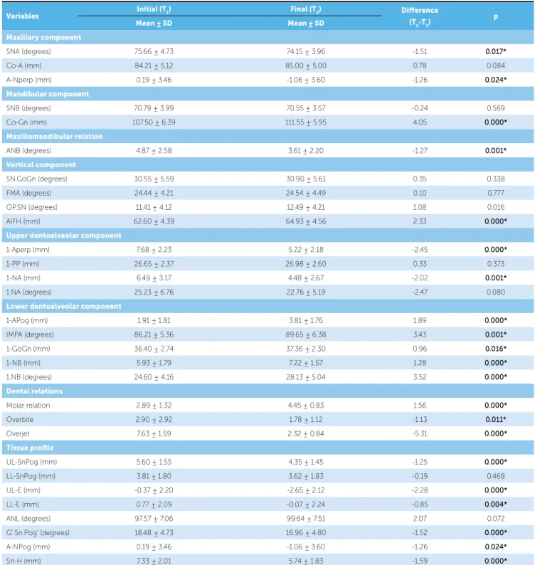
Among the 30 studied cephalometric variables, only 3 presented significant systematic error (FMA, 1-PP and IMPA). The magnitude of random errors ranged from 0.22 (overjet) to 2.12 (IMPA). The re-sults are displayed on Table 1.
Patients with Class II malocclusion, division 1, treated with extraction of two upper premolars pre-sented the following cephalometric alterations: max-illary retrusion in relation to the skull base, increase on the mandibular length and anterior inferior facial height (AIFH), improvement on the maxillomandib-ular relation, upper incisors retrusion, vestibmaxillomandib-ulariza- vestibulariza-tion and protrusion of the lower dentoalveolar com-ponent, improvement on molar relation, reduction of overbite and overjet, reduction of facial convexity and upper lip retrusion.
DISCUSSION
Evaluating the results found in this work it is possible to establish and assess the alterations caused on dento-skeletal and tissue components due the extraction of up-per irst premolars, in Class II division 1 patients.
Regarding the maxillary component, the maxilla experienced a signiicant retrusion noticed by the sta-tistically signiicant reduction of variables SNA and A-NPerp (Table 1). The reduction of these variables oc-curred due the necessity of correction of Class II relation of canines and overjet normalization, due the use of inter-maxillary elastic and EOA as anchorage reinforcement.
» 1.NA (°): Angle between the long axis of upper
cen-tral incisor and line NA. It deines the degree of inclination of the central incisor in relation to maxilla and Nasion.
» 1-APog (mm): Distance from the incisal edge of lower incisor to line Apog.
» IMPA (°): Angle between the long axis of lower central incisor and the mandibular plane GoMe. It indicates the inclination of this tooth in relation to the mandible.
» 1-GoGn (mm): Distance from incisal edge of lower incisor to mandibular plane GoGn, measured perpendicularly to this plane.
» 1-NB (mm): Distance between the anterior point of the crown of lower central incisor and line NB. It relates the sagittal position of lower incisor in relation to mandible and Nasion.
» 1.NB (°): Angle between the long axis of lower incisor and line NB. It relates the inclination of this tooth to the mandible and Nasion.
» Molar relation (mm): Distance between the me-sial cusp of upper and lower first premolars perpen-dicularly projected on the occlusal plane.
» Overbite (mm): Distance between the incisal edges of upper and lower central incisors measured perpendicularly to the occlusal plane.
» Overjet (mm): Distance between the incisal edges of upper and lower central incisors perpendicu-larly projected to the occlusal plane.
Tissue profile
» UL-SnPog (mm): Distance from the upper lip to
line Subnasal Pogonion.
» LL-SnPog (mm): Distance from the lower lip to
line Subnasal Pogonion.
» UL-E (mm): Distance from the upper lip to line
Pronasal Pogonion.
» LL-E (mm): Distance from the lower lip to line
Pronasal Pogonion.
» ANL - nasolabial angle: Formed by lines columella
to Subnasal and from Subnasal to upper lip.
» G’.Sn.Pog’ (°): Facial convexity angle. It’s formed
by lines tissue glabella to Subnasal and from Subnasal to tissue Pogonion.
» A-NPog (mm): Distance from point A to line
Nasion to the Pogonion.
» Sn-H (mm): Shortest distance from point Subnasal
alterations on point A. But in this study the variable Co-A did not present statistically signiicant altera-tion. It is speculated that there was no alteration due the fact that most patients were in growth stage, which disguised the retraction experienced by point A.
On the mandibular component, it was evidenced a sig-niicant increase of the mandible efective length (Co-Gn) It is notable that the antero-superior teeth retraction
may affect the positioning of point A in relation to
the skull base.4,25
Scott Conley and Jernigan25 also observed a
statisti-cally signiicant reduction of the variable A-Nperp in cases treated with extraction of two upper premolars.
However, Rains and Nanda24 did not ind signiicant
Variables Initial (T1) Final (T2) Diference
(T2-T1) p
Mean ± SD Mean ± SD
Maxillary component
SNA (degrees) 75.66 ± 4.73 74.15 ± 3.96 -1.51 0.017*
Co-A (mm) 84.21 ± 5.12 85.00 ± 5.00 0.78 0.084
A-Nperp (mm) 0.19 ± 3.46 -1.06 ± 3.60 -1.26 0.024*
Mandibular component
SNB (degrees) 70.79 ± 3.99 70.55 ± 3.57 -0.24 0.569
Co-Gn (mm) 107.50 ± 6.39 111.55 ± 5.95 4.05 0.000*
Maxillomandibular relation
ANB (degrees) 4.87 ± 2.58 3.61 ± 2.20 -1.27 0.001*
Vertical component
SN.GoGn (degrees) 30.55 ± 5.59 30.90 ± 5.61 0.35 0.338
FMA (degrees) 24.44 ± 4.21 24.54 ± 4.49 0.10 0.777
OP.SN (degrees) 11.41 ± 4.12 12.49 ± 4.21 1.08 0.016
AIFH (mm) 62.60 ± 4.39 64.93 ± 4.56 2.33 0.000*
Upper dentoalveolar component
1-Aperp (mm) 7.68 ± 2.23 5.22 ± 2.18 -2.45 0.000*
1-PP (mm) 26.65 ± 2.37 26.98 ± 2.60 0.33 0.373
1-NA (mm) 6.49 ± 3.17 4.48 ± 2.67 -2.02 0.001*
1.NA (degrees) 25.23 ± 6.76 22.76 ± 5.19 -2.47 0.080
Lower dentoalveolar component
1-APog (mm) 1.91 ± 1.81 3.81 ± 1.76 1.89 0.000*
IMPA (degrees) 86.21 ± 5.36 89.65 ± 6.38 3.43 0.001*
1-GoGn (mm) 36.40 ± 2.74 37.36 ± 2.30 0.96 0.016*
1-NB (mm) 5.93 ± 1.79 7.22 ± 1.57 1.28 0.000*
1.NB (degrees) 24.60 ± 4.16 28.13 ± 5.04 3.52 0.000*
Dental relations
Molar relation 2.89 ± 1.32 4.45 ± 0.83 1.56 0.000*
Overbite 2.90 ± 2.92 1.78 ± 1.12 -1.13 0.011*
Overjet 7.63 ± 1.59 2.32 ± 0.84 -5.31 0.000*
Tissue proile
UL-SnPog (mm) 5.60 ± 1.55 4.35 ± 1.45 -1.25 0.000*
LL-SnPog (mm) 3.81 ± 1.80 3.62 ± 1.83 -0.19 0.468
UL-E (mm) -0.37 ± 2.20 -2.65 ± 2.12 -2.28 0.000*
LL-E (mm) 0.77 ± 2.09 -0.07 ± 2.24 -0.85 0.004*
ANL (degrees) 97.57 ± 7.06 99.64 ± 7.51 2.07 0.072
G’.Sn.Pog’ (degrees) 18.48 ± 4.73 16.96 ± 4.80 -1.52 0.000*
A-NPog (mm) 0.19 ± 3.46 -1.06 ± 3.60 -1.26 0.024*
Sn-H (mm) 7.33 ± 2.01 5.74 ± 1.83 -1.59 0.000*
Table 1 - Results of dependent t test comparing initial phase (T1) and inal phase (T2) of treatment.
(Table 1). This was expected since the evaluated pa-tients in this research were in growth stage, as explained above. In this study, this increase was greater than the expected being corroborated by some authors that
re-ported similar results in literature. 4,7,23
On the maxillomandibular relation, there was a sig-niicant alteration of the anteroposterior skeletal discrep-ancy, shown by the reduction of angle ANB (Table 1). This alteration was expected because the orthodontic treatment aimed the correction of Class II malocclusion and of the overjet initially increased by upper incisors retraction. Besides, the potential of mandibular growth
on this age group helps the reduction of angle ANB. 25,26
Tadic and Woods26 observed that the greater the overjet
at the beginning of treatment, greater the probability of reduction of angle ANB, in cases treated with extraction
of two upper premolars. Oliveira et al20 observed that
there was improvement on the anteroposterior maxil-lomandibular relation, shown by the reduction of ANB.
Scott Conley and Jernigan25 also observed a statistically
signiicant reduction of the angle ANB contributing for an improvement of the existent relation between dental arches, and a signiicant increase of AIFH as occurred in the present study.
In the work by Chua, Lim and Lubit5 it was found
an increase of AIFH associated to a clockwise rotation of the mandible only in cases treated without dental extractions, while in treatments with extractions it was not observed any signiicant alteration on AIFH. Dif-ferently from the indings of these authors, most works found in literature are in agreement with the present study, in which AIFH presented a statistically signii-cant increase during treatment. It is believed that the responsible for this alteration was the potential growth still present in patients and the orthodontic mechanics that used intermaxillary elastics during closing of spac-es ater extractions. Some authors observed that the in-crease of the lower third of the face is related to age and growth potential of the patients, besides the use of
in-termaxillary elastics.19 On the work by Oliveira et al20
the AIFH increased due the compensatory extrusion of molars during anterior retraction phase. Now
Mer-riield and Cross19 emphasized that any mechanics that
promote dental extrusion causes the increase of AIFH. On the variables related to growth pattern, both SN.GoGn and FMA did not present statistically sig-niicant alterations between initial and inal phases of
treatment (Table 1). Scott Conley and Jernigan25 also
did not ind signiicant alterations in relation to the change on growth pattern in cases treated with extrac-tion of two premolars. The variable OP.SN presented a statistically signiicant increase during treatment. This increase does not mean in alteration of growth pattern impressing a vertical characteristic for the pa-tients, because the other variables of this component did not present signiicant alteration. It is believed that the alterations observed on the variable OP.SN are re-lated to dentoalveolar changes, for instance, the curve of Spee lattening, occurred during the treatment of patients. At the beginning of treatment, it is frequently observed an accented extrusion of lower incisors due
to increased overjet.18,28 During treatment there was
lattening of curve of Spee, which led to a clockwise rotation of the occlusal plane. This efect was enough to promote statistically signiicant change of variable OP.SN being only a relex of dental alterations and not skeletal since the other variables, SN.GoGn and FMA, that characterize the component, did not present sta-tistically signiicant changes.
The study by Oliveira et al20 showed
compensa-tory extrusion of molars during retraction phase in cases with extraction of four premolars, and also did not report statistically significant differences on vari-ables SN.GoGn and SN.GoMe that characterized the growth pattern.
On the upper alveolar component, the upper in-cisors presented a statistically significant retrusion (1-Aperp, 1-NA). There was no significant altera-tion of upper incisors on the vertical direcaltera-tion or in relation to inclination (1-PP and 1.NA, respectively) (Table 1). According to several authors, the skeletal effects of the treatment were more evident on the maxilla and on the superior teeth than in the man-dible or lower teeth, in cases treated with extraction
of two upper premolars.25,27
Tadic and Woods26 veriied a signiicant increase on
upper incisors at the beginning and end of treatment, which difers from most works with extraction of two premolars. It can be asserted that there is a tendency of these teeth to obtain a more palatine inclination ater the anterior retraction because the numeric value was reduced, but with no statistical signiicance.
Paiva et al21 also in cases treated with extraction of
4 premolars reported a statistically signiicant reduc-tion of linear values (1-NA), however the reducreduc-tion on angular values (1.NA), was not statistically signiicant, which is in agreement with the indings in this study.
On the lower dentoalveolar component, all vari-ables related to lower incisors experienced statistically significant alteration (Table 1). It was verified a pro-trusion, vestibularization and extrusion of lower inci-sors (1-APog, IMPA, 1-GoGn, 1-NB, 1.NB). This can be explained by the use of intermaxillary elastics of Class II that were necessary in most patients at fi-nal stage. Side effects on the inclination of incisors in patients treated with intermaxillary elastics are widely
reported in literature, confirming this supposition.19
On the other hand, Scott Conley and Jernigan25 did
not find statistically significant alterations on IMPA. By evaluating the dental relations, it was observed a reduction of the value of molar relation, pointing the increase of Class II molar relation, which was expect-ed, for only two upper premolars were extractexpect-ed, and the cases would be inished with a full Class II molar relation in both sides (Table 1). It was observed a sig-niicant improvement on the overbite which was also already expected, for the deep overbite was corrected during treatment with the use of reverse curve wires on the lower arch and accentuated on the upper arch, including during the anterior retraction (Table 1). The overjet reduced signiicantly with the treatment, which was also expected, because it was taken as criteria for inclusion on the sample that the patients presented an overjet of at least 5 mm and inished the treatment with the malocclusion corrected with canines in Class I relation and normalized overjet (Table 1).
Also on the evaluation of tissue proile results, the data presented an upper lip retrusion, veriied on mea-sures UL-SnPog, A-NPog, UL-E, and a lower lip re-trusion on measure E, however the variable LL-SnPog did not present statistically signiicant difer-ence despite presenting a tendency to reduction in its value which shows a tendency to lower lip retrusion,
contradicting the expected lower lip protrusion due to vestibularization and protrusion veriied on lower incisors (Table 1). These results are in agreement with
the works presented in literature.25,27 Scott Conley and
Jernigan25 also found an unexpected result. They
as-signed this reduction on the lower lip projection to the presence of an everted lower lip, because with a deep overbite, an accentuated overjet and a Class II dental relation, the lower lip may be artiicially kept in a more protruded position trapped in the space between lower and upper incisors and ater the upper incisors retrac-tion, returns to its normal position.
Some authors that evaluated cases with extraction of two upper premolars, realized that according to the treatment protocol used, the lip thickness at the be-ginning of treatment, the vertical control and variety of facial patterns, the lips may be affected by dental moves on the anteroposterior direction, however the magnitude of this alterations is of difficult predict-ability.26,27
Several authors did not find significant differ-ences on the alterations of soft tissue between groups treated with extraction of four premolars and without
extractions7,30 while other authors observed a tissue
profile retrusion.1,3,4
Considering that a retraction of upper incisors
necessarily imply in a retrusion of the upper lip,30 it is
known that the muscle-skeletal-functional complex of the upper lip contributes for the variability ob-served on alterations of upper lip with the treatment
protocol with premolars extractions.29
In this work the nasolabial angle did not present statistically signiicant alteration (Table 1). Despite the upper teeth retraction suggests that occurs an increase of the nasolabial angle, this result was not observed.
Some authors assign this result to nasal growth, occurring a down inclination of the columella and the pronasal, reducing the nasolabial angle. Theses authors assert that the nasolabial angle is formed by soft tissue (pronasal) and cartilage (columella), which continues to grow forth as well as the soft tissue of the upper lip and observed that only a small statistically insignificant change occurred of nasal base retraction (subnasal), therefore, if the projection of the upper lip tends to reduce, while the base nasal projection remains the same, the nasolabial angle must become
Kocadereli16 and Uehara et al28 observed mean
val-ues statistically equal of the nasolabial angle both for patients that did not experience dental extraction and those who were submitted to extractions of four
pre-molars. Tadic and Woods26 also did not ind statistically
signiicant alteration of the nasolabial angle in patients treated with extraction of upper irst premolars.
However, Freitas et al8 observed an increase of the
nasolabial angle in cases treated with extraction of four premolars in a proportion of increase of naso-labial angle in 1,49° for each millimeter of retraction on upper teeth which also was confirmed by other
authors.27 Scott Conley and Jernigan25 also found
sta-tistically significant alterations of the nasolabial angle which had an increase of 6.38°.
Erdinç et al7 observed that the nasolabial angle
re-duced significantly in groups treated with extraction of four premolars and in groups without extractions these alterations were insignificant.
Despite the generalized idea that extractions cause an increase of the nasolabial angle, the results from this work are in agreement with latest conclusions of works mentioned above.
CLINICAL CONSIDERATIONS
The obtained results showed that profile altera-tions occur as effect of orthodontic treatment.
How-ever, each patient must be individually analyzed so the professional can plan the treatment and instruct the patient about these aspects. Concomitantly, it al-lows the clinician a greater predictability of possible alterations that the treatment will cause and thus in-crease the percentage of success in this type of treat-ment and the patient’s satisfaction.
The lower lip retrusion is an essential data when planning the treatment and its positioning must be eval-uated in the beginning of treatment and veriied if it is afected by the positioning of upper incisors, for this will lead to lower lip retrusion and the patient must be aware. Generally this detail go unnoticed by the clinician, for he only reports the upper lip retrusion due the chosen treatment does not include extraction on the lower arch.
CONCLUSION
Based on the evaluated sample and the used method-ology, the alterations caused by extraction of two pre-molars on Class II division 1 malocclusion were:
» Maxillary retrusion, improvement of the
maxil-lomandibular relation, increase of the anteroinferior fa-cial height, upper incisors retrusion, vestibularization, protrusion and extrusion of lower incisors, besides the reduction of overbite and overjet.
» The proile alterations were: Reduction of the
1. Basciftci FA, Uysal T, Buyukerkmen A, Demir A. The inluence of extraction treatment on Holdaway soft-tissue measurements. Angle Orthod. 2004;74(2):167-73.
2. Bowbeer GR. The 6th key to facial beauty and TMJ health. Funct Orthod.
1987;4(4):10-11, 13-15.
3. Bowman SJ. More than lip service: facial esthetics in orthodontics. J Am Dent Assoc. 1999;130(8):1173-81.
4. Bravo LA, Canut JA, Pascual A, Bravo B. Comparison of the changes in
facial proile after orthodontic treatment, with and without extractions. Br J Orthod. 1997;24(1):25-34.
5. Chua AL, Lim JY, Lubit EC. The efects of extraction versus nonextraction
orthodontic treatment on the growth of the lower anterior face height. Am J Orthod Dentofacial Orthop. 1993;104(4):361-8.
6. Eirew HL. An orthodontic challenge. Int J Orthod. 1976;14(4):21-5.
7. Erdinc AE, Nanda RS, Dandajena TC. Proile changes of patients treated
with and without premolar extractions. Am J Orthod Dentofacial Orthop. 2007;132(3):324-31.
8. Freitas MR, Henriques JFC, Pinzan A, Janson GRP, Siqueira VCV. Estudo
longitudinal das alterações do ângulo naso-labial, em jovens com Classe II, 1ª divisão, que se submeteram ao tratamento ortodôntico corretivo. Ortodontia. 1999;32(1):8-16.
9. Gottlieb EL, Nelson AH, Vogels DS 3rd. 1990 JCO study of orthodontic
diagnosis and treatment procedures. 1. Results and trends. J Clin Orthod. 1991;25(3):145-56.
10. Graber T, Vanarsdall R Jr. Ortodontia: princípios e técnicas atuais. 3a ed. Rio de Janeiro: Guanabara Koogan; 2000.
11. Janson G, Barros SE, de Freitas MR, Henriques JF, Pinzan A. Class II treatment eiciency in maxillary premolar extraction and nonextraction protocols. Am J Orthod Dentofacial Orthop. 2007;132(4):490-8. 12. Janson G, Brambilla Ada C, Henriques JF, de Freitas MR, Neves LS.
Class II treatment success rate in 2- and 4-premolar extraction protocols. Am J Orthod Dentofacial Orthop. 2004;125(4):472-9.
13. Janson G, Camardella LT, Araki JD, de Freitas MR, Pinzan A. Treatment stability in patients with Class II malocclusion treated with 2 maxillary premolar extractions or without extractions. Am J Orthod Dentofacial Orthop. 2010;138(1):16-22.
14. Janson G, Leon-Salazar V, Leon-Salazar R, Janson M, de Freitas MR. Long-term stability of Class II malocclusion treated with 2- and 4-premolar extraction protocols. Am J Orthod Dentofacial Orthop. 2009;136(2):154.e1-10; discussion 154-5.
REFERENCES
15. Janson G, Maria FR, Barros SE, Freitas MR, Henriques JF. Orthodontic treatment time in 2- and 4-premolar-extraction protocols. Am J Orthod Dentofacial Orthop. 2006;129(5):666-71.
16. Kocadereli I. Changes in soft tissue proile after orthodontic treatment with and without extractions. Am J Orthod Dentofacial Orthop. 2002;122(1):67-72.
17. Mailankody J. Enigma of Class II molar inishing. Am J Orthod Dentofacial Orthop. 2004;126(6):a15-16; author reply a16-17.
18. McNamara JA Jr. Components of class II malocclusion in children 8-10 years of age. Angle Orthod. 1981;51(3):177-202.
19. Merriield LL, Cross JJ. Directional forces. Am J Orthod. 1970;57(5):435-64. 20. Oliveira GF, Almeida MR, Almeida RR, Ramos al Alterações
dentoesqueléticas e do peril facial em pacientes tratados
ortodonticamente com extração de quatro primeiros pré-molares. Rev Dent Press Ortod Ortop Facial. 2008;13(2):105-14.
21. Paiva J, Rino Neto J, Batista K. Análise do lábio superior após o tratamento ortodôntico. Ortodontia. 2004;37(2):8-13.
22. Proit WR. Forty-year review of extraction frequencies at a university orthodontic clinic. Angle Orthod. 1994;64(6):407-14.
23. Proit WR, Phillips C, Tulloch JF, Medland PH. Surgical versus orthodontic correction of skeletal Class II malocclusion in adolescents: efects and indications. Int J Adult Orthodon Orthognath Surg. 1992;7(4):209-20. 24. Rains MD, Nanda R. Soft-tissue changes associated with maxillary incisor
retraction. Am J Orthod. 1982;81(6):481-8.
25. Scott Conley R, Jernigan C. Soft tissue changes after upper premolar extraction in Class II camoulage therapy. Angle Orthod. 2006;76(1):59-65. 26. Tadic N, Woods MG. Incisal and soft tissue efects of maxillary premolar
extraction in Class II treatment. Angle Orthod. 2007;77(5):808-16. 27. Talass MF, Talass L, Baker RC. Soft-tissue proile changes resulting
from retraction of maxillary incisors. Am J Orthod Dentofacial Orthop. 1987;91(5):385-94.
28. Uehara SY, et al Peril facial após tratamento de Classe II-1 com ou sem extrações. RGO: Rev Gaúch Odontol. 2007;55(1):61-8.
29. Waldman BH. Change in lip contour with maxillary incisor retraction. Angle Orthod. 1982;52(2):129-34.