Annals of Oncology0: 1–37, 2016 doi:10.1093/annonc/mdw235
ESMO consensus guidelines for the management
of patients with metastatic colorectal cancer
E. Van Cutsem
1*, A. Cervantes
2, R. Adam
3, A. Sobrero
4, J. H. Van Krieken
5, D. Aderka
6, E. Aranda
Aguilar
7, A. Bardelli
8, A. Benson
9, G. Bodoky
10, F. Ciardiello
11, A. D
’
Hoore
12, E. Diaz-Rubio
13,
J.-Y. Douillard
14, M. Ducreux
15, A. Falcone
16,17, A. Grothey
18, T. Gruenberger
19, K. Haustermans
20,
V. Heinemann
21, P. Hoff
22, C.-H. Köhne
23, R. Labianca
24, P. Laurent-Puig
25, B. Ma
26, T. Maughan
27,
K. Muro
28, N. Normanno
29, P. Österlund
30,31, W. J. G. Oyen
32, D. Papamichael
33,
G. Pentheroudakis
34, P. Pfeiffer
35, T. J. Price
36, C. Punt
37, J. Ricke
38, A. Roth
39, R. Salazar
40,
W. Scheithauer
41, H. J. Schmoll
42, J. Tabernero
43, J. Taïeb
25, S. Tejpar
1, H. Wasan
44,
T. Yoshino
45, A. Zaanan
25& D. Arnold
461Digestive Oncology, University Hospitals Gasthuisberg Leuven and KU Leuven, Leuven, Belgium;2Medical Oncology Department, INCLIVA University of Valencia,
Valencia, Spain;3Hepato-Biliary Centre, Paul Brousse Hospital, Villejuif, France;4Medical Oncology, IRCCS San Martino Hospital, Genova, Italy;5Research Institute for
Oncology, Radboud University Nijmegen Medical Center, Nijmegen, The Netherlands;6Division of Oncology, Sheba Medical Center, Tel Aviv University, Tel Aviv, Israel; 7Medical Oncology Department, University Hospital Reina Sofia, Cordoba, Spain;8School of Medicine, University of Turin, Turin, Italy;9Division of Hematology/Oncology,
Northwestern Medical Group, Chicago, USA;10Department of Oncology, St László Hospital, Budapest, Hungary;11Division of Medical Oncology, Seconda Università di
Napoli, Naples, Italy;12Abdominal Surgery, University Hospitals Gasthuisberg Leuven and KU Leuven, Leuven, Belgium;13Medical Oncology Department, Hospital Clínico
San Carlos, Madrid, Spain;14Medical Oncology, Institut de Cancérologie de l’Ouest (ICO), St Herblain;15Department of Medical Oncology, Gustave Roussy, Université
Paris-Saclay, Villejuif, France;16Department of Medical Oncology, University of Pisa, Pisa, Italy;17Division of Medical Oncology, Department of Oncology, University Hospital
‘S. Chiara’, Istituto Toscano Tumori, Pisa, Italy;18Division of Medical Oncology, Mayo Clinic, Rochester, USA;19Department of Surgery I, Rudolfstiftung Hospital, Vienna,
Austria;20Department of Radiation Oncology, University Hospitals Gasthuisberg and KU Leuven, Leuven, Belgium;21Comprehensive Cancer Center, University Clinic
Munich, Munich, Germany;22Instituto do Câncer do Estado de São Paulo, University of São Paulo, São Paulo, Brazil;23Northwest German Cancer Center, University
Campus Klinikum Oldenburg, Oldenburg, Germany;24Cancer Center, Ospedale Giovanni XXIII, Bergamo, Italy;25Digestive Oncology Department, European Hospital
Georges Pompidou, Paris, France;26Department of Clinical Oncology, Prince of Wales Hospital, State Key Laboratory in Oncology in South China, Chinese University of
Hong Kong, Shatin, Hong Kong;27CRUK/MRC Oxford Institute for Radiation Oncology, Gray Laboratories, University of Oxford, Oxford, UK;28Department of Clinical
Oncology and Outpatient Treatment Center, Aichi Cancer Center Hospital, Nagoya, Japan;29Cell Biology and Biotherapy Unit, I.N.T. Fondazione G. Pascale, Napoli, Italy; 30Helsinki University Central Hospital, Comprehensive Cancer Center, Helsinki, Finland;31Department of Oncology, University of Helsinki, Helsinki, Finland;32The Institute of
Cancer Research and The Royal Marsden Hospital, London, UK;33Department of Medical Oncology, Bank of Cyprus Oncology Centre, Nicosia, Cyprus;34Department of
Medical Oncology, University of Ioannina, Ioannina, Greece;35Department of Oncology, Odense University Hospital, Odense, Denmark;36Haematology and Medical
Oncology Unit, Queen Elizabeth Hospital, Woodville, Australia;37Department of Medical Oncology, Academic Medical Centre, University of Amsterdam, Amsterdam, The
Netherlands;38Department of Radiology and Nuclear Medicine, University Clinic Magdeburg, Magdeburg, Germany;39Digestive Tumors Unit, Geneva University Hospitals
(HUG), Geneva, Switzerland;40Catalan Institute of Oncology (ICO), Barcelona, Spain;41Department of Internal Medicine I and Comprehensive Cancer Center, Medical
University of Vienna, Vienna, Austria;42Department of Internal Medicine IV, University Clinic Halle, Martin-Luther-University Halle-Wittenberg, Halle, Germany;43Medical
Oncology Department, Vall d’Hebron University Hospital, Vall d’Hebron Institute of Oncology (V.H.I.O.), Barcelona, Spain;44Department of Cancer Medicine,
Hammersmith Hospital, Imperial College Healthcare NHS Trust, London, UK;45Department of Gastroenterology and Gastrointestinal Oncology, National Cancer Center
Hospital East, Chiba, Japan;46Instituto CUF de Oncologia (ICO), Lisbon, Portugal
Received 31 March 2016; revised 27 May 2016; accepted 31 May 2016
Colorectal cancer (CRC) is one of the most common malignancies in Western countries. Over the last 20 years, and the last decade in particular, the clinical outcome for patients with metastatic CRC (mCRC) has improved greatly due not only to an increase in the number of patients being referred for and undergoing surgical resection of their localised metastatic disease but also to a more strategic approach to the delivery of systemic therapy and an expansion in the use of ablative
techniques. This reflects the increase in the number of patients that are being managed within a multidisciplinary team
environment and specialist cancer centres, and the emergence over the same time period not only of improved imaging techniques but also prognostic and predictive molecular markers. Treatment decisions for patients with mCRC must be evidence-based. Thus, these ESMO consensus guidelines have been developed based on the current available evidence
*Correspondence to: Prof. Eric Van Cutsem, ESMO Guidelines Committee, ESMO Head Office, Via L. Taddei 4, CH-6962 Viganello-Lugano, Switzerland.
E-mail: [email protected]
special
article
© The Author 2016. Published by Oxford University Press on behalf of the European Society for Medical Oncology. All rights reserved. For permissions, please email: [email protected].
at KU Leuven University Library on July 5, 2016
http://annonc.oxfordjournals.org/
to provide a series of evidence-based recommendations to assist in the treatment and management of patients with mCRC in this rapidly evolving treatment setting.
Key words:colorectal cancer, ESMO, consensus, clinical practice guidelines
introduction
Colorectal cancer (CRC) is the second most commonly diag-nosed cancer in Europe and a leading cause of death both in Europe and worldwide [1,2]. In 2012, there were 447 000 new cases of CRC in Europe with 215 000 deaths and worldwide, there were 1.4 million new cases with 694 000 deaths. Over the last decade in particular, the clinical outcome for patients with metastatic CRC (mCRC) has improved. Today, the median overall survival (OS) for patients with mCRC being treated both in phase III trials and in large observational series or registries is
∼30 months and more than double that of 20 years ago.
However, it is unclear which improvements and strategic changes in the treatment and management of patients with mCRC in recent years have been responsible for the improved treatment outcomes for these patients. Factors which may have contributed are:
(i) changes in the clinical presentation of patients, before the commencement of treatment, due to closer follow-up after resection of the primary tumour and earlier detection of metastatic disease;
(ii) improvements in the efficacy of systemic therapies in terms of regimens used, sequence of administration, number of lines of therapy administered and biomarker-based patient selection;
(iii) an increase in the number of patients being treated with a view to facilitating resection of their metastases, offering an increased number of patients the chance of cure and/or durable relapse-free survival and, more recently, the utilisa-tion of other ablative therapy techniques with the aim of achieving the same outcome;
(iv) implementation of‘continuum of care’treatment strategies coupled with the early integration of optimal supportive care measures.
These ESMO Consensus Guidelines therefore aim to reflect the diagnostic, therapeutic and strategic improvements which have contributed to the current‘state-of-the-art’treatment approaches and to provide guidance for the comprehensive management of patients with mCRC going forward.
methodology
In 2014, the ESMO Guidelines Committee decided to update the clinical recommendations for mCRC using a consensus con-ference approach. An international panel of experts in the man-agement of patients with CRC, from a range of diagnostic and therapeutic disciplines, was convened in Zurich in December 2014 to update the existing ESMO Consensus Guidelines for the management of patients with colon and rectal cancer [3]. A set of pre-formulated topics was prepared and three working groups convened in the areas of:
(i) molecular pathology and biomarkers;
(ii) local and ablative treatment (LAT) [including surgery and the management of patients with oligometastatic disease (OMD)];
(iii) the treatment of metastatic disease.
Each panel member was assigned to one of the above working groups. Three consensus conference chairs (EVC, AC and DA) were also appointed. Before the consensus conference, clinically relevant questions were identified for each working group. Each working group was responsible for reviewing relevant literature in order to draft preliminary recommendations relating to each of their assigned questions. No systematic literature search was undertaken. The experts in each group were invited to submit their recommendations in advance to structure the on-site discussions. During the conference, in parallel sessions, the three working groups discussed and reached agreement on recommendations relating to each of their assigned questions. Recommendations from each group were then presented to the entire panel of experts, where they were discussed and modified as required until consen-sus was reached.
An adapted version of the ‘Infectious Diseases Society of America-United States Public Health Service Grading System’ was used (Table 1, [4]) to define the level of evidence and strength of each recommendation proposed by the group, as for all of the ESMO Consensus and ESMO Clinical Practice Guidelines, and are given in the text in square brackets. Statements made based on expert opinion were also considered to be justified standard clinical practice by the experts and the ESMO faculty. These ESMO Consensus Guidelines follow on from those published in 2012 [3] and should be used to support the 2014 ESMO Clinical Practice Guidelines [5].
molecular pathology and biomarkers
A clinical or biological suspicion that a patient may have mCRC should always be confirmed by adequate radiological imaging, and the histology of the primary tumour or metas-tases, as appropriate, conducted before the commencement of systemic therapy, as described previously [5]. Tissue samples will typically range from large tumour samples to smaller biopsy/endoscopy samples. Whenever possible, any diagnostic biopsy or tissue sampling procedure should aim to maximise the number of samples collected (ideally n= 10 biopsies). In addition to samples taken for embedding, additional frozen material should be collected to provide the opportunity for future‘new’tests to be conducted on frozen tissue if required. It is also essential that all tissue and biopsy samples are handled appropriately in order to facilitate meaningful and accurate molecular testing.
tissue handling
Standardisation of tissue processing for patients with mCRC still remains a challenge. The time from tissue sampling to
| Van Cutsem et al.
at KU Leuven University Library on July 5, 2016
http://annonc.oxfordjournals.org/
fixation should be minimised to only a few minutes if possible, to prevent any degradation of proteins and nucleic acids that might occur during cold ischaemia [6, 7]. Fixation in 10% neutral buffered formalin (4% formaldehyde solution), which is widely available, is generally compatible with any procedure for protein, RNA and/or DNA biomarker analysis. The fixation time should be between 6 and 48 h [8]. Longer or shorterfi xa-tion times may adversely affect biomarker testing, while
under-fixation is also associated with poor tissue morphology [9]. Acidic fixatives (e.g. Bouin) are not recommended since they lead to the rapid degradation of nucleic acids [10]. Similarly, accelerated fixation with heated formalin is discouraged as it degrades tissue morphology and affects the results of molecular studies [11]. Biomarker analyses should be carried out within 4–6 weeks of the sections being cut, as ageing of formalin-fixed, paraffin-embedded tissue sections causes the degradation of both epitopes and DNA [12].
recommendation 1: tissue handling.
• Fixation with 10% neutral buffered formalin (4%
formalde-hyde) is recommended [V, A].
• Fixation time should be no less than 6 h, and no greater than
48 h in duration. In the case of microwave-enhancedfixation, the quality of both nucleic acids and proteins must be verified [IV, A].
• Sections for biomarker testing should ideally be cut
immedi-ately before analysis [IV, A].
selection of specimens for biomarker testing
The pathologist plays a central role in biomarker testing and can either perform the biomarker tests at his/her laboratory if it has been accredited for biomarker testing, or send the tissue block to an accredited reference laboratory for external testing. In both instances, the primary pathologist should review the available material for each patient and choose the most appropriate block to be used for testing. The pathologist should also ensure that the tissue block selected for biomarker analysis contains a
sufficient quantity of neoplastic cells for the analysis [13]. This is particularly crucial for DNA- or RNA-based biomarker testing, such asRASmutation analysis, because a low fraction of neoplastic cells can lead to dilution of mutant alleles and false-negative results [14,15]. To evaluate the tumour content of the sample, it is recommended that the pathologist assesses a haema-toxylin and eosin-stained section of the paraffin block designated for DNA extraction and mutation analysis before DNA extrac-tion. The minimum fraction of tumour versus non-tumour cells required will depend on the genotyping method. It has been demonstrated that a tumour cell content of 30% or less might lead to false-negative results when a technique with low sensitivity such as Sanger sequencing is used for testing [16,17]. A neoplas-tic cell content of at least 50% is therefore recommended when using a technique with low sensitivity. Sections of tissue with high tumour content may be used directly. In samples with a low tumour cell content, and where feasible, suitable areas identified by the pathologist may be scraped (manual macro-dissection) from the tissue slide(s) in order to enrich the tumour cell content. Laser capture micro-dissection can also be used, but this technol-ogy is not widely available, and requires the skills of a pathologist, additional work and, therefore, high costs.
recommendation 2: selection of specimens for biomarker testing.
• The primary pathologist should review all available tumour
specimens to select those that are most suitable for biomarker analyses [IV, A].
• Enrichment of samples by macro-dissection to maximise
tumour cell content (>50%) before DNA extraction is recom-mended [III, A].
tissue selection for biomarker testing
Most patients undergo surgery of their primary tumour, although in some cases, only an endoscopic biopsy of the primary is carried out. Thus, archival samples of primary tumour tissue are usually available for biomarker testing for the majority of patients Table 1. Levels of evidence and grades of recommendation (adapted from the Infectious Diseases Society of America—United States Public Health Coding Systema[4])
Levels of evidence
I Evidence from at least one large randomised, controlled trial of good methodological quality (low potential for bias) or meta-analyses of well-conducted randomised trials without heterogeneity
II Small randomised trials or large randomised trials with a suspicion of bias (low methodological quality) or meta-analyses of such trials or of trials with demonstrated heterogeneity
III Prospective cohort studies
IV Retrospective cohort studies of case–control studies
V Studies without control group, case reports, experts opinions
Grades of recommendation
A Strong evidence for efficacy with a substantial clinical benefit, strongly recommended
B Strong or moderate evidence for efficacy but with a limited clinical benefit, generally recommended
C Insufficient evidence for efficacy or benefit does not outweigh the risk of the disadvantages (adverse events, costs,…) optional
D Moderate evidence against efficacy or for adverse outcome, generally not recommended E Strong evidence against efficacy or for adverse outcome, never recommended
aBy permission of the Infectious Diseases Society of America.
doi:10.1093/annonc/mdw235 |
at KU Leuven University Library on July 5, 2016
http://annonc.oxfordjournals.org/
with advanced or mCRC. However, for ∼20% of patients who
present with metastatic disease, archival material from their primary tumour will not always be available. For these patients, biomarker testing is usually carried out using specimens obtained from primary tumour biopsies or the metastatic tumour, for example, from resected liver metastases or positive lymph nodes. For some patients, both the primary tumour and metastatic tissue specimens may be available for mutation testing. Indeed, a number of studies have addressed the concordance in KRAS mutation status between primary colorectal tumours and their metastases with conflicting results. While some studies have failed tofind any difference inKRASmutation status between the primary tumours and their metastases [18–22], others have reported discordant results in 4%–32% of the patients [23–35]. However, many of these studies involved the analysis of small numbers of samples, involving heterogeneous metastatic sites and the use of techniques with low sensitivity that might have led to false-negative results if adequate enrichment of the tumour cells was not carried out. In a large study of 305 matched primary col-orectal tumours and liver metastases, the discordance rate was 3.6% [36]. When these data are pooled with results from different previous small studies, the overall rate of discordance is∼5% for liver metastases. In contrast, a discordance rate of 25% has been described for lymph node metastases. Although these data are limited toKRASexon 2 mutations, they can be extrapolated to situations where expandedRASanalysis has been conducted (see below), for which no information is available. Based on this evi-dence, tissue from either a patient’s primary tumour or a liver metastasis may be used forRASmutation testing. Lymph node metastases do not seem to be suitable for the determination of the RAS mutation status of colorectal tumours. In patients for whom both primary tumour and metastases are available, testing of a sample from either site is sufficient.
recommendation 3: tissue selection.
• Tissue from either the primary tumour or a liver metastasis
may be used forRASmutation testing [III, A].
• Other metastatic sites such as lymph node or lung metastases
may be used only if primary tumour or liver metastases samples are not available [II, B].
definition and validation of biomarkers
Biomarkers can be diagnostic, predictive or prognostic. Ideally, a biomarker should only serve one of these purposes, but there are good and clinically relevant examples of prognostic biomar-kers that predict a response to a specific therapy, for example, human epidermal growth factor receptor 2 (HER2) in breast cancer andBRAF (strongly prognostic and, to a lesser extent, predictive) in CRC [37–39]. It is also essential to follow strict rules for the development and validation of biomarkers that are specific to the purpose and sometimes also specific to the nature of each biomarker. Establishing clinical utility in the appropriate clinical setting is essential [40].
RAStesting
evidence that tumour RAS mutational status is predictive. Retrospective analyses of pivotal clinical trials for the epidermal
growth factor receptor (EGFR) monoclonal antibodies, cetuximab and panitumumab, have shown that patients with mCRC, whose tumours contain activating mutations in KRASexon 2 (codons 12/13), do not derive a benefit from EGFR monoclonal antibody therapy [41–47]. Furthermore, recent evidence from the PRIME study with panitumumab [48], from the CRYSTAL study with cetuximab [49] and from other studies of EGFR monoclonal antibody therapies has shown that mutations other than those in KRASexon 2 [i.e. exons 3 and 4 ofKRASand exons 2, 3 and 4 of NRAS(expandedRASanalysis)] also predict a lack of response to EGFR-targeting monoclonal antibodies and that these therapies may in fact have a detrimental effect in patients withRAS-mutant disease, specifically when combined with an oxaliplatin-based cytotoxic backbone [48–54].
In the PRIME study, in which patients were randomised to receive panitumumab plus FOLFOX4 [infusional 5-fluorouracil (5-FU), leucovorin, oxaliplatin] versus FOLFOX4 alone fi rst-line, additionalRASmutations were detected in the tumours of 17% of patients with mCRC originally classified asKRASexon 2 wild-type. These patients also failed to benefit from panitumu-mab therapy, and had inferior progression-free survival (PFS) and OS times compared with those treated with FOLFOX4 alone (not statistically significant). In fact, this study was the
first to hint at a detrimental effect of panitumumab in patients whose tumours carriedRASmutations at sites other thanKRAS exon 2 [48].
Conversely, those patients whose tumours did not have RAS mutations at the tested sites had significantly better outcomes from the addition of panitumumab to FOLFOX4 than those patients whose tumours containedRASmutations. The phase II PEAK study that evaluated FOLFOX6 plus panitumumab versus FOLFOX6 plus bevacizumab in untreated patients with KRAS exon 2 wild-type mCRC supported these findings. Patients withKRASandNRASexon 2, 3 and 4 wild-type mCRC treated with FOLFOX6 plus panitumumab achieved a better PFS than those treated with FOLFOX6 plus bevacizumab and a trend towards improved OS was also observed [53]. Using next-generation sequencing (NGS) techniques, investigators analysed tumour samples previously assessed forKRASexon 2 codon 12 and 13 mutations from patients enrolled in the phase III 20020408 trial of panitumumab in patients with chemorefrac-tory mCRC [52] for additional RAS-activating mutations. Patients withRAS wild-type tumours achieved a response rate (RR) with panitumumab of 15% compared with 1% for those patients withRAS-mutant tumours.
These findings with panitumumab have been upheld by trials evaluating cetuximab. Using sensitive BEAMing (Beads, Emulsions, Amplification, and Magnetics) technology, KRAS exon 2 wild-type tumours from the pivotal CRYSTAL and OPUS studies were retrospectively evaluated for mutations in KRASexons 3 and 4 andNRASexons 2, 3 and 4 [49,50]. In the phase III CRYSTAL study, which randomised patients to receive
first-line FOLFIRI (infusional 5-FU, leucovorin, irinotecan) with or without cetuximab, otherRASmutations were detected in nearly 15% of evaluable patients previously assessed to be KRASexon 2 wild-type. Similarly, in the phase II OPUS study, which randomised patients to receivefirst-line FOLFOX4 with or without cetuximab, mutations at otherRASloci were detected in 31% of evaluable tumours previously assessed to beKRAS
| Van Cutsem et al.
at KU Leuven University Library on July 5, 2016
http://annonc.oxfordjournals.org/
exon 2 wild-type. In patients with RAS wild-type tumours (according to the expandedRASanalysis), the addition of cetuxi-mab to FOLFIRI or FOLFOX4 was associated with improved treatment outcomes across all efficacy end points. Conversely, in patients withRAS-mutant tumours, no benefit from the addition of cetuximab to FOLFIRI versus FOLFIRI alone was observed [49]. In the OPUS study, the addition of cetuximab to FOLFOX4 was associated with a non-significant improvement in PFS and OS in patients withRASwild-type tumours; it seemed to be detri-mental in patients whose tumours carriedRASmutations.
Data from the phase III FIRE-3 trial also underscore the importance of expandedRASmutational analysis in the selection of patients for treatment with cetuximab. Previously untreated patients, withKRASexon 2 wild-type mCRC, were randomised to receive FOLFIRI with either cetuximab or bevacizumab. AdditionalRASmutations were identified in the tumours of 16% of assessable patients, with an improvement in OS (median 33.1 versus 28.7 months) observed for patients withRAS wild-type tumours treated with cetuximab compared with those withKRAS exon 2 wild-type tumours treated with cetuximab [55].
Confirmation of these observations was provided by a sys-tematic review and meta-analysis of randomised, controlled trials evaluating EGFR antibody therapy [56]. The analysis showed that across nine trials involving 5948 patients, patients with tumours without anyRASmutations were found to have a significantly better treatment outcome with EGFR monoclonal antibody therapy than those whose tumours harboured RAS mutations [56].
In summary, the cumulative data clearly show that patients whose tumours harbour any RAS mutation are unlikely to benefit from EGFR antibody therapy, confirming the presence of aRAS mutation (according to expandedRAS analysis) as a negative predictive markerof treatment outcome in patients with mCRC who might be under consideration for EGFR monoclo-nal antibody therapy. Thus, cetuximab and panitumumab should only be considered for the treatment of patients with RASwild-type mCRC. ExpandedRASanalyses should be con-ducted on all patients eligible/being considered for EGFR anti-body therapy.
timing of testing. Wong et al. [57] discuss whetherRAStesting of CRC is better practised as a ‘reflex’ or an ‘on-demand’ process. However, the general consensus of the expert panel was that patients should be assessed for their tumourRASmutation status at the time of diagnosis of their metastatic disease, to facilitate strategic treatment decisions within a multidisciplinary team (MDT) environment, local reimbursement regulations permitting. However, it should also be noted that an external quality assessment has uncovered differences in the quality of RAStesting for EGFR antibody therapy [58] and that, to date, the exact cut-off for clinically relevant RAS-mutant allele frequencies has not been determined.
Investigation of cost estimates and the economic implications of expanded RAS testing in patients with mCRC showed the increased societal cost of expandedRAS testing versus KRAS exon 2 testing to be inconsequential when compared with the amount of money saved by not treating the additional up to 18% of patients who harbour additionalRASmutations (beyond those inKRASexon 2) with EGFR antibody therapies [59].
recommendation 4: RAS testing.
• RASmutational status is a negative predictive biomarker for
therapeutic choices involving EGFR antibody therapies in the metastatic disease setting [I, A].
° RAStesting should be carried out on all patients at the time of diagnosis of mCRC [I, A].
• RAS testing is mandatory before treatment with the
EGFR-targeted monoclonal antibodies cetuximab and panitumumab [I, A].
• A network of arrangements should be established to ensure
the rapid and robust transit of tissue samples from referral centres to testing laboratories, to minimise the turnaround time and avoid delays in having this information available for all patients with mCRC.
• Primary or metastatic colorectal tumour tissue can be used for
RAStesting (see alsoRecommendation 3).
• RASanalysis should include at leastKRASexons 2, 3 and 4
(codons 12, 13, 59, 61, 117 and 146) andNRASexons 2, 3 and 4 (codons 12, 13, 59, 61 and 117).
• Turnaround time for RAS testing (expanded RAS analysis)
should be≤7 working days from the time of receipt of the specimen by the testing laboratory to the time of issuing of thefinal report, for >90% of specimens.
• Validation (or verification, where more applicable) of RAS
testing assays should be carried out and recorded before implementation in clinical use. Laboratory audit mechanisms should be in place.
• Laboratories providing RAS testing of colorectal tumours
should demonstrate their successful participation in a relevant external quality assessment scheme, and be appropriately accredited.
BRAFtesting
BRAFmutations (nearly always V600E) are found in the tumours of between 8% and 12% of patients with mCRC included in clini-cal trials and are almost exclusively non-overlapping withRAS mutations [38,60,61]. A retrospective analysis of patients with mCRC demonstrated that two-thirds ofBRAF-mutant patients’ primary lesions were located on the right side of the colon and associated with an increased incidence of peritoneal and distant lymph node metastases, but fewer pulmonary metastases [60]. Just under one-third ofBRAF-mutant tumours also had microsa-tellite instability (MSI), and the same proportion of tumours with MSI containedBRAFmutations.
BRAFmutations are a significant negative prognostic marker for patients with mCRC. Tran et al. [60] reported a median sur-vival for patients with BRAF-mutant mCRC of 10.4 months compared with 34.7 months for patients withBRAF wild-type tumours. In a multivariate analysis, the hazard ratio (HR) for survival was 10.662 (P< 0.001) [60]. This particularly poor prognosis for patients withBRAF-mutant tumours is supported by a number of randomised studies with specific chemotherapy regimens [38,44,48, 61–63]. Although the evidence ofBRAF mutations as a negative predictive biomarker for EGFR antibody therapy in later lines is accumulating [64, 65], its role in earlier lines in combination studies with chemotherapy has not been ascertained [44]. Indeed, two meta-analyses [66, 67]
doi:10.1093/annonc/mdw235 |
at KU Leuven University Library on July 5, 2016
http://annonc.oxfordjournals.org/
showed the efficacy benefit of EGFR antibody therapies to be greater in patients withRASwild-type/BRAFwild-type tumours than in those with RAS wild-type/BRAF-mutant tumours. In the meta-analysis that included two second-line trials and two trials involving chemorefractory patients [66], the lack of the conferral of a significant efficacy benefit by EGFR-antibody therapies over standard chemotherapy alone in patients with BRAF-mutant tumours was considered to support the assessment of tumourBRAFmutation status before the initiation of EGFR-antibody therapy. Conversely, authors of the second meta-analysis [67] concluded that there was insufficient evidence to exclude EGFR antibody therapy from patients withRASwild-type/BRAF -mutant disease. However, in a small subgroup analysis (n= 28) of the TRIBE study, the cohort of patients with BRAF-mutant tumours treated with the chemotherapy triplet FOLFOXIRI plus bevacizumab showed a non-statistically significant increase in OS compared with those treated with FOLFIRI plus bevacizumab [68].
Also, BRAFV600E-mutated melanomas are sensitive to the BRAF-mutant inhibitor vemurafenib [69], but BRAF-mutated CRCs are not as sensitive [70, 71]. Feedback reactivation of EGFR in CRC could explain why CRCs generally have a lower response toBRAFinhibitors [37,71]. Clinical trials are ongoing to test targeted therapies in patients with metastatic BRAF (V600E) mutant CRC, using combinations of BRAF-mutant inhibitors (dabrafenib, vemurafenib or encorafenib) in combina-tion with MEK and EGFR inhibicombina-tion, and in some cases conven-tional cytotoxic therapy. Early results are promising [72,73].
Furthermore, somatic BRAF V600E mutations have been associated with sporadic cases of DNA mismatch repair defi -ciency (dMMR) showing an MSI phenotype [74]. However, BRAFV600E mutation is not associated with the MSI pheno-type due to a germline MMR mutation (Lynch syndrome) [75, 76].BRAFV600E mutation testing has therefore been proposed as a means to exclude Lynch syndrome. Recently, however, patients withBRAF-mutant tumours with mutations in codons 594 and 596 were shown to exhibit microsatellite stability (MSS) and markedly longer OS when compared with patients with BRAFV600E-mutant disease [77].
Tumour BRAF mutation status should be determined for every case of CRC, ideally at the time of diagnosis, as this repre-sents a different biological subtype, and in combination with testing for dMMR, can assist in the identification of a germline versus somatic cause of dMMR. In patients with mCRC,BRAF mutation status should be assessed at the same time as RAS mutational status for prognostic assessment (and/or potential selection for clinical trials).
recommendation 5: BRAF testing.
• TumourBRAFmutation status should be assessed alongside the
assessment of tumour RAS mutational status for prognostic assessment (and/or potential selection for clinical trials) [I, B].
MSI testing
Tumours with MSI retain their chromosomal numbers intact but contain microsatellite repeats, which vary in length due to dMMR, and are thought to contribute to the early steps of
tumourigenesis in patients with CRC. Tumours with MSI repre-sent only 4%–8% of tumours in patients with mCRC. Data are currently scarce on the prognostic and predictive values of an MSI phenotype in the metastatic disease setting [78–80]. A recent retrospective analysis demonstrated that the median age was a bit younger (67 years), poor differentiation was more frequent (58%), and that 45% of patients whose tumours had an MSI phe-notype had stage IV disease at presentation.BRAFV600E muta-tions were present in 30% of patients with MSI [79]. In mCRC, some data have suggested that MSI tumours tend to have lower disease control rates when treated with oxaliplatin-basedfirst-line therapy [81], although most studies show MSI status to be not relevant as a single predictive marker for response to irinotecan-or oxaliplatin-based chemotherapy regimens and not predictive for the effect of chemotherapy more generally in these patients [78,82,83].
In a pooled analysis of four phase III studies in thefirst-line treatment of mCRC (CAIRO, CAIRO2, COIN and FOCUS), BRAF mutations have been shown to be more frequent in patients whose tumours exhibit MSI than in those whose tumours exhibit MSS [62]. The same pooled analysis showed PFS and OS to be significantly worse for patients with tumours with MSI when compared with those with tumours showing MSS [HR, 1.33; 95% confidence interval (CI) 1.12–1.57 and HR, 1.35; 95% CI 1.13–1.61, respectively], and for patients with BRAF-mutant tumours when compared with those withBRAF wild-type tumours (HR, 1.34; 95% CI 1.17–1.54 and HR, 1.91; 95% CI 1.66–2.19, respectively) [62]. Emerging data have shown MMR status to predict the clinical benefit of immune check-point blockade with pembrolizumab in patients with mCRC. The immune-related objective RR and immune-related 6-month PFS rate were 40% (4 out of 10 patients) and 78% (7 out of 9 patients), respectively, for patients with dMMR CRC and 0% and 11% for those with MMR-proficient CRC, with excellent median PFS and survival (not reached) in the cohort with dMMR CRCs versus 2.2 and 5.0 months, respectively, in the cohort with MMR-proficient tumours [84].
Thus, the prevalence of MSI and BRAF mutations in the tumours of patients with mCRC is low. Both biomarkers confer an inferior prognosis, which in the case of patients with tumours with MSI may be driven by the presence of BRAF mutations. These conclusions are supported by the data from other studies which show the presence of aBRAFV600E muta-tion to be as poor a prognostic factor in patients with tumours with MSI as it is in other patients with mCRC [60].
recommendation 6: MSI testing.
• MSI testing in the metastatic disease setting can assist
clini-cians in genetic counselling [II, B].
• MSI testing has strong predictive value for the use of immune
check-point inhibitors in the treatment of patients with mCRC [II, B].
biomarkers of chemotherapy sensitivity or toxicity
dihydropyrimidine dehydrogenase. Dihydropyrimidine
dehydrogenase (DPD) is a key enzyme in the metabolic catabolism of 5-FU and capecitabine. About 85% of 5-FU is
| Van Cutsem et al.
at KU Leuven University Library on July 5, 2016
http://annonc.oxfordjournals.org/
eliminated through a catabolic process involving DPD. Numerous genetic mutations have been identified in the DPD gene locus (DPYD), with a few key variants having functional consequences for enzymatic activity. Deficiencies in DPD activity have been shown to cause 5-FU-treated cancer patients to experience severe drug-related toxicities [85], and DPD activity is a predictive biomarker of potential toxicity when using 5-FU and capecitabine [86]. Polymorphism has been documented mainly on the DPYD*2A gene at a frequency of 2%–3% with geographical variation.
Several methods are available to detect DPD deficiency such as the functional dihydrouracil/uracil ratio in plasma, the uracil breath test orDPYD*2mutations. Patients with known partial DPD deficiency benefit from dose adaptation of their 5-FU/ capecitabine therapy to avoid severe toxicity. In patients with complete DPD deficiency,fluoropyrimidines should not be used and an alternative treatment offered.
DPD deficiency is generally not assessed in routine practice before 5-FU administration. There is no recommended standar-dised assessment technique, although several methods are avail-able (see above). None of the current strategies are adequate to mandate routine DPD testing before startingfl uoropyrimidine-based therapy [II, C].
Testing for DPD deficiency, however, remains an option. In the case of patients who experience severe 5-FU toxicity, DPD levels should be tested before 5-FU is re-introduced.
UDP glucuronosyltransferase 1 family, polypeptide A1. UDP glucuronosyltransferase 1 family, polypeptide A1 (UGT1A1) is an enzyme of the glucuronidation pathway that transforms small lipophilic molecules, such as steroids, bilirubin, hormones and drugs, into water-soluble, excretable metabolites. The gene is part of a complex locus that encodes several UDP-glucuronosyltransferases. Polymorphism may be associated with increased toxicity to irinotecan. UGT1A1 is responsible for bilirubin glucuronidation as well as glucuronidation of SN-38, the active metabolite of irinotecan. Genetic variations within the UGT1A1gene have also been associated with the development of certain drug toxicities. The UGT1A1*28 variant, the allele behind many cases of Gilbert syndrome, has been associated with an increased risk for neutro-paenia in patients receiving irinotecan [87,88], and the United States Food and Drug Administration recommends on the irino-tecan drug label that patients with the *28/*28 genotype should receive a lower starting dose of irinotecan [89]. The *28 allele has also been shown to be associated with an increased risk of developing diarrhoea in patients receiving irinotecan [87, 88]. The UGT1A1*6 variant, more common in Asian populations than the *28 variant, has also been associated with the develop-ment of irinotecan-related toxicities. Patients who are heterozy-gous or homozyheterozy-gous for the *6 allele may have a higher risk of developing neutropaenia and diarrhoea than those with the UGT1A1*1/*1 genotype.
Thus,UGT1A1gene polymorphisms are predictive of irinote-can-related side-effects, including diarrhoea, neutropaenia and vomiting. However, in everyday practice, UGT1A1/UGT1A1 status is rarely used as a predictive biomarker of irinotecan toxi-city. Attention should be paid to bilirubin levels, especially in patients where conjugated bilirubin is <20% of total bilirubin.
excision repair cross-complementation group 1. The function of the excision repair cross-complementation group 1 (ERCC1) protein is predominantly in the nucleotide excision repair of damaged DNA. Nucleotide excision repair is the primary DNA repair mechanism involved in the removal of therapeutic platinum-DNA adducts from tumour DNA. A variety of methods can be used to measure the level of ERCC1 activity, namely immunohistochemistry (IHC) for protein expression, reverse transcription–polymerase chain reaction (RT–PCR) for mRNA expression and DNA single-nucleotide polymorphism (SNP) for genotyping. High ERCC1 levels have been shown to be a negative predictive marker for platinum-based therapy in patients with lung cancer [90,91]. In CRC, depending on the techniques used, high ERRC1 expression levels have been shown to be associated with poor prognosis and to be predictive of a poor outcome in patients receiving oxaliplatin-based therapy (RT–PCR mRNA evaluation). A meta-analysis showed ERCC1-C118T polymorphisms to predict clinical outcome in patients with CRC receiving oxaliplatin-based therapy [92]. More specifically, PFS and OS were significantly shorter in patients with T/T or T/C genotypes of ERCC1-C118T when compared with those with the C/C genotype. Thus, highERCC1 gene expression seems to confer oxaliplatin resistance, while ERCC1-C118T polymorphisms are predictive of treatment outcome in patients receiving oxaliplatin-based therapy [92]. Recently it has been proposed that ERRC1 induction after exposure to oxaliplatin may be dependent onKRASmutational status [93].
At the present time, the use of ERCC1 protein levels cannot be recommended for treatment decisions involving the use of oxaliplatin in routine practice. Clinical trials have not been able to demonstrate a predictive role for ERCC1 status for treatment with oxaliplatin.
thymidylate synthase. Thymidylate synthase (TS) is the primary target for 5-FU. 5-FU is an inhibitor of TS. Experimentally, it has been shown that low levels of TS expression lead to a better response to 5-FU and improved survival of colon cancer patients [94]. The TS gene(TYMS)is under the control of a promoter acting as an enhancer (TSER). Earlier studies have shown that higher numbers of TSER repeats (TSER2*, TSER3* or higher) lead to higher TS expression and activity. TS activity and CRC sensitivity to 5-FU seem to correlate withTSERpolymorphisms. These correlations, however, need to be confirmed in a larger randomised study.
recommendation 7: biomarkers of chemotherapy sensitivity and toxicity:
• DPD testing before 5-FU administration remains an option
but is not routinely recommended [II, D].
• UGT1A1 phenotyping remains an option and should be
carried out in patients with a suspicion of UGT1A1 deficiency as reflected by low conjugated bilirubin and in patients where an irinotecan dose of >180 mg/m2 per administration is planned [95] [III, C].
• ERCC1 expression cannot be recommended for use as a
bio-marker for treatment decisions involving the use of oxaliplatin
doi:10.1093/annonc/mdw235 |
at KU Leuven University Library on July 5, 2016
http://annonc.oxfordjournals.org/
in routine clinical practice, but could be included prospec-tively in clinical trials [III, D].
• TS activity andTSER genotyping are not recommended for
use in clinical practice [II, D].
emerging biomarkers
A list of biomarkers beyond RASmutational status is emer-ging which may impact on the response to all classes of targeted agents, and specifically the current perspective of EGFR-antibody therapies. These include HER2, MET and KRAS gene amplification, ligands such as transforming growth factor-α (TGF-α), amphiregulin and epiregulin, EGFRmutations and alterations/mutations inHER3,PI3KCA andPTEN.
Mutations in KRAS, NRAS and BRAF and amplification of HER2andMETdrive primary (de novo) resistance to anti-EGFR treatment. Recently, the emergence of alterations in these genes was detected in patients who responded to EGFR blockade and then relapsed. Molecular heterogeneity impairs the efficacy of EGFR-antibody therapy in patients with mCRC by fuelling de novoand acquired resistance [96]. With the exception ofEGFR mutations, which are described only in the acquired setting, all of the genetic alterations defined as a mechanism ofde novo resis-tance are also responsible for acquired resisresis-tance. Differences can be found in the frequency of individual genetic alterations, such as KRASandNRASexon 3 mutations, which occur more frequently in the acquired rather than in thede novosetting. Acquired resis-tance to EGFR-antibody therapy is driven by the selection of cell clones that carry RAS or RAF mutations but account for only 0.4%–17% of tumour cells. Mutations inKRASexons 3 and 4 and NRASexons 2, 3, and 4 as well as amplification ofKRAS,HER2 andMET[96–99] account for around 20% of mCRC patients who do not benefit from anti-EGFR treatment, although initially selected for anti-EGFR treatment based onKRAS exon 2 wild-type status [48, 52–54, 97, 100–104]. The prognostic role of PIK3CA mutations is uncertain [105], but a PIK3CA exon 20 mutation may predict resistance to EGFR-antibody therapy [106– 110], although the correlation is not strong enough to be applied as a negative predictive marker [111].PIK3CAand PTEN altera-tions often co-occur withKRASorBRAFmutations [107,112], but there is insufficient evidence for their use as biomarkers of resistance to EGFR-antibody therapy. There is no clear evidence for HER3 overexpression and HER3 mutations, mesenchymal– epidermal transition (MET)/MET alterations (overexpression or gene amplification) orKRASamplification,EGFRmutations [tyr-osine kinase (TK) or ligand-binding domains] or amplification in the resistance to EGFR antibody therapies. Emerging data indicate that HER2 activating mutations or HER2 amplification may mediate in some instances resistance to EGFR antibodies [100, 113]. A phase II clinical trial also showed thatHER2amplification is predictive of response to HER2 dual inhibition with trastuzu-mab and lapatinib in a cohort of CRC patients failing EGFR anti-body therapy [114].
Thus, although CRC is primarily considered to be a genetic disease, characterised by the sequential accumulation of genetic alterations, there is growing evidence that epigenetic alterations add an additional layer of complexity to its pathogenesis and characterise a subgroup of CRCs with a distinct aetiology and
prognosis. A systematic review and meta-analysis of the prog-nostic value of the CpG island methylator phenotype (CIMP) in patients with CRC showed the CIMP to be independently asso-ciated with a significantly worse prognosis [115]. However, epi-genetic DNA hypermethylation inactivation of theSRBCgene, the product of which interacts with the product of theBRCA1 gene, predicted a shorter PFS, particularly in oxaliplatin-treated patients with mCRC for whom metastasectomy was not indi-cated (HR, 1.96; 95% CI 1.13–3.40; log-rank P= 0.01). SRBC hypermethylation was also associated with a shorter PFS (HR, 1.90; log-rankP= 0.045), in a validation cohort of unresectable colorectal tumours treated with oxaliplatin [116].
recommendation 8: emerging biomarkers not recommended for routine patient management outside of a clinical trial setting:
• Detection of mutations inPIK3CA, exon 20 [II, D]. • Evaluation of PTEN loss by IHC [V, D].
• Evaluation of the levels of the EGFR ligands amphiregulin,
epiregulin and TGF-α[II, D].
• Evaluation of levels of EGFR protein expression [II, E]. • Evaluation of EGFR amplification and copy number and
EGFRectodomain mutations [IV, D].
• Evaluation ofHER2amplification orHER2activating mutations. • Evaluation of HER3, and MET receptor overexpression [IV, D].
emerging technologies
A number of novel tools for the assessment of diagnostic, prog-nostic and/or predictive biomarkers in patients with mCRC have been proposed, with an increasing interest in liquid biopsies involving the analysis of either circulating tumour cells (CTCs) or circulating tumour DNA (ctDNA). Although the levels of CTCs as assessed (mostly using the CellSearch system) have been shown to correlate with prognosis in patients with mCRC [117], the clin-ical utility of CTC assessments in patients with mCRC has hardly been explored.
Conversely, analysis of ctDNA is emerging as a new tool for molecular profiling that has more possibilities for translation into the clinic than CTCs. The seminal work of Bardelli and colleagues has shown very promising results from ctDNA liquid biopsies [118,119]. In addition to the seminal papers from Bardelli and colleagues and Montagut et al. [120], a number of tumour–blood concordance studies are currently being conducted that will undoubtedly validate the clinical utility of these technologies for identifying tumour mutations in the blood of patients. Currently, its use as a monitoring tool for secondary resistance to EGFR antibody therapies is under investigation. It can be anticipated that liquid biopsies will be used therapeutically in the near future as more and better drugs are developed against mutant clones (or those with other molecular alterations, e.g. amplifications, etc.) that emerge upon exposure to EGFR-targeted therapies [40,118, 120–135].
Similarly, increasing evidence suggests that micro-RNA (miRNA) is involved in the pathogenesis and progression of mCRC [136]. However, the prognostic and predictive role of miRNA needs to be demonstrated in a randomised clinical trial setting. Finally, NGS can provide important information on tumour heterogeneity and clonal evolution. NGS has already been published as a reliable
| Van Cutsem et al.
at KU Leuven University Library on July 5, 2016
http://annonc.oxfordjournals.org/
technology for use in patients with mCRC and has the potential to screen for larger cancer gene panels in clinical trials [137].
recommendation 9: emerging technologies.
• Although CTC number correlates with prognosis in patients
with mCRC, the clinical utility of CTC assessments is not yet clear and therefore cannot be recommended [IV, D].
• The utility of liquid ctDNA biopsies to guide treatment
deci-sions is currently under investigation in clinical trials, but cannot yet be recommended in routine practice [V, D].
• Whole genome, whole exome and whole transcriptome
analy-sis should be carried out only in a research setting [V, D].
view on how molecular classification should be developed going forward
CRC is a heterogeneous disease with heterogeneous outcomes and drug responses. To date, pathological staging and gene expression signatures have failed to accurately predict disease recurrence and prognosis. In an attempt to identify biologically homogeneous subtypes of CRC, many independent groups have reported the results of gene expression-based subtyping, with Marisa et al. [138], thefirst to present a robust transcriptome-based classification of colon cancer. Subsequently, an inter-national consortium dedicated to large-scale data sharing and analytics has recently provided a robust and unified classifi ca-tion, defining four different subtypes: CMS1 (MSI Immune), hypermutated, microsatellite unstable, with strong immune activa-tion; CMS2 (Canonical), epithelial, chromosomally unstable, with marked WNT and MYC signalling activation; CMS3 (Metabolic), epithelial, with evident metabolic dysregulation; and CMS4 (Mesenchymal), prominent TGF-β activation, stromal inva-sion and angiogenesis [139]. This effort provides the most robust and reproducible classification system currently avail-able for CRC and may form the basis for future clinical trials.
local ablative treatment (LAT), including
surgery, and management of patients
with OMD
the role of MDTs and tumour boards
The optimal treatment strategies for patients with mCRC are evol-ving rapidly with improved clinical outcomes being achieved when the treatment approaches for individual patients are dis-cussed within an MDT of experts who meet regularly as a tumour board to review mCRC cases [140, 141]. An ideal MDT should include access to both a colorectal surgeon (preferably with exper-tise in peritoneal approaches) and a specialist hepatobiliary and/ or, lung surgeon as necessary, with the obligatory inclusion of a pathologist and a diagnostic radiologist, as well as radiation and medical oncologists. An interventional radiologist/nuclear physi-cian may also be included as appropriate, as the role of ablative treatments gains increasing importance (see below). Ideally, patients should be treated either in specialist cancer centres or, alternatively, where this is not possible, as part of a network of individuals dedicated to the management of CRC with an estab-lished referral route between their site or centre and a specialist cancer centre (virtual MDTs). Wherever possible, MDTs should
provide the opportunity to register patients for the local and/or national registries with extreme/unusual patients’ details just noted, to provide information on the diversity of patients seen. Several (observational) studies have shown improved clinical out-comes, including improved OS, when patients with CRC are managed by MDTs [141,142].
The role of the MDT is to define the initial diagnostic workup and then the treatment focus, based on the best diagnostic and therapeutic decision-making available [3]. Furthermore, an MDT-managed treatment strategy has to be maintained for the duration of a patient’s treatment, to allow the refinement of treatment strategies according to on-treatment information (e.g. response to a selected treatment) and evaluation of the potential need for the integration of ablative treatments (such as second-ary surgery and LAT strategies, see below).
Thefirst step in the process is for the MDT members to criti-cally define whether or not a patient has initially clearly resect-able or initially unresectresect-able metastatic disease and to define the status of the resection of the primary tumour when considering the management of both synchronous and/or oligometastatic CRC, and the first-line treatment of patients with metastatic disease. Conversely, for patients whose disease is deemed‘never to be resectable’, the discussion may be left to the treating medical oncologist (after discussion with the MDT) and patient as to the pros and cons of various approaches and sequences based on the perceived aims [e.g. duration of disease control versus quality of life (QoL), and toxicity profiles, etc.].
oligometastatic disease
OMD is characterised by the localisation of the disease to a few sites and lesions and is associated with the option to use LAT approaches in patient treatment strategies with a view to improving disease control and therefore clinical outcome in these patients.
Generally, OMD may be characterised by the existence of metastases at up to 2 or occasionally 3 sites and 5 or sometimes more lesions, predominantly visceral and occasionally lymphono-dal. Typically, these are the primary, and other involved sites such as the liver, lung, peritoneum, nodes and ovary. Patients with disease at other sites, such as multiple lesions in the bones and the brain, may also be treated using a local ablative approach, but as these patients are associated with an unfavourable prognosis, local ablative treatment strategies are only used to prevent immediate complications. This latter group of patients should be excluded from a classification of OMD. On the other hand, a patient with one or two resectable liver metastases, and a single bone lesion, should be classified as having OMD, because for a patient with this disease profile, locally ablative treatment strategies could be used and meaningfully contribute to their prognosis.
Thus, treatment strategies for patients with OMD should be based on the possibility of achieving complete ablation of all tumour masses, using surgical R0 resection (complete resection with clear resection margins and no evidence of microscopic residual tumour) and/or LAT, either initially or possibly after induction treatment with systemic therapy, for both the primary tumour and metastases.
For patients with OMD confined to a single organ (most fre-quently the liver), or a few organs (pre-dominantly visceral metas-tases, e.g. lung), a potentially curative approach exists. Numerous
doi:10.1093/annonc/mdw235 |
at KU Leuven University Library on July 5, 2016
http://annonc.oxfordjournals.org/
case series have shown that in this setting, long-term survival or even cure can be attained in 20%–50% of patients who undergo complete R0 resection of their metastases [143]. Even in the absence of randomised trials comparing surgical with non-surgical disease management, surgery has become the standard treatment approach for patients with resectable OMD.
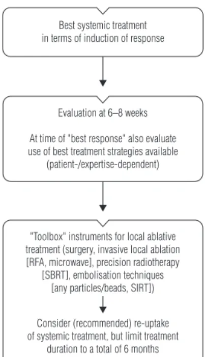
For patients with more extensive OMD involving more sites or lesions, e.g. primary, liver, lung, peritoneum, nodes, bones, brain, ovary and >4 organs, the value of a surgical approach is controversial. In these patients, surgery may contribute to long-term survival but is rarely curative [143]. For this group of patients, the consideration of localised interventions (LAT) becomes relevant, in combination with systemic therapy (as part of a multimodal therapy approach), following a careful MDT discussion and assessment. The goal for this group of patients is to achieve long-term disease control, potentially contributing to OS (and, although unlikely, potentially cure), with well-controlled sites of metastases, but without continued systemic therapy. Liver-directed therapy is probably the best established of the LAT interventions; however, the increasing use of the appropriate ablative treatment strategy from a ‘toolbox’ of options, including, for example, stereotactic ablative body radio-therapy (SBRT) and radiofrequency ablation (RFA) for visceral or nodal involvement, peritonectomy with or without hyperthermic intraperitoneal chemotherapy (HIPEC) for peritoneal disease, and nodal dissection, sees the management of this subgroup of patients becoming increasingly complex (Figure1). Furthermore, the potential still exists for isolated bone, pancreatic and brain metastases, but these are rare and likely to not have a defined treatment pathway.
Subcharacterisation of OMD according to site also impacts on the treatment options and the timing of treatment. Patients with liver and lung metastases have a much better prognosis than those with other metastatic disease locations. In fact, because lung
involvement is associated with better outcomes, it may be appro-priate to‘watch and wait’or at least employ a sequential approach [144,145]. The data showing different outcomes depending on the site(s) of OMD are likely to reflect molecular differences. For example, patients whose mCRC is associated withRAS and BRAF mutations have worse clinical outcomes, with RAS mutations shown to be associated with an increased incidence of lung, bone and brain metastases [146]. Moreover there are data to suggest that tumour TSexpression levels andRASmutation status are altered by site of metastasis compared with the primary [23–36,147].
recommendation 10: OMD.
• For patients with OMD, systemic therapy is the standard of
care and should be considered as the initial part of every treat-ment strategy (exception: patients with single/few liver or lung lesions, see below).
• The best local treatment should be selected from a‘toolbox’of procedures according to disease localisation, treatment goal (‘the more curative the more surgery’/higher importance of local/complete control), treatment-related morbidity and patient-related factors such as comorbidity/ies and age [IV, B].
liver metastases and surgical resection
For patients with colorectal liver metastases (CLM), the treat-ment strategy should be directed towards complete resection whenever possible, with both ‘oncological’ ( prognostic) and ‘technical’(surgical) criteria being considered when evaluating
patients for surgery [148]. However, prospective evaluations do not exist either for ‘oncological’or for‘technical’ criteria, and for many of these, there is no (international) consensus.
The ‘technical’ definitions of resectable CLM have evolved over time, with the current consensus proposing that disease
Toolbox of ablative treatments
Local treatments
Thermal devices
Radiofrequency ablation or cryoablation
Microwave ablation Radiotherapy withExternal Body high-precision RT
Chemoembolisation TACE/Beads Brachytherapy
electroporation
Radioembolisation SIRT
Non-thermal devices Embolic devices Local
chemotherapy
Locoregional treatments
Figure 1. Toolbox of ablative treatments. SIRT, selective internal radiation therapy; RT, radiation therapy; TACE, transarterial chemoembolisation.
| Van Cutsem et al.
at KU Leuven University Library on July 5, 2016
http://annonc.oxfordjournals.org/
should be considered technically resectable as long as complete macroscopic resection is feasible, while maintaining at least a 30% future liver remnant (FLR) or a remnant liver to body weight ratio >0.5 (e.g. >350 g of liver per 70 kg patient) [149– 151]. However, the concern remains that not all patients with technically resectable liver-limited metastases benefit from surgery, with approximately half developing widespread sys-temic disease within 3 years of resection [152].
The‘oncological’criteria provide prognostic information that predict a longer disease-free survival (DFS) or a higher likeli-hood of cure. These include, as strong parameters, the number of lesions, the presence (or suspicion) of extrahepatic disease and the criteria used in numerous retrospective evaluations and in the FONG score [153]. Thus, for some patients, neoadjuvant chemotherapy may be a better option than upfront surgery.
In practice, patients can be categorised into groups based on technological and oncological criteria as outlined in Figure 2 and according to the new system for deciding whether or not a patient is eligible for resection proposed by Adam et al. [148], and described in Table2.
imaging in the identification of resectable/ unresectable disease
Computed tomography (CT) scans are routinely used for primary staging and disease surveillance in patients with CRC. Although practice varies between treatment centres, the evi-dence suggests that the best methods for detection of liver metastases from CRC are CT and magnetic resonance imaging (MRI) [154]. However, many teams alternate liver ultrasonogra-phy (US) and CT for detection of disease to decrease the expo-sure of patients to the radiation resulting from repeated CT scans. For the characterisation of focal liver lesions, CT, con-trast-enhanced US (CEUS) and MRI can be used [155]. For lesions <10 mm in diameter, MRI is a more sensitive modality than CT [156] and specifically hepatobiliary MRI with specific
contrast enhancers (such as gadoxetate) which is associated with a higher accuracy of lesion detection [157].
For the detection of extrahepatic metastases and local recur-rence at the site of the initial colorectal surgery, CT and positron emission tomography (PET)/CT scans are used [158]. A pro-spective randomised trial evaluating high-quality CT and PET imaging involving 263 patients showed only a 7.6% change in management following PET [159], while a retrospective analysis reported a change in intended curative therapy to palliative therapy or vice versa in one-third of patients [160]. Also, a recently published meta-analysis of studies evaluating PET and PET/CT in patients with liver metastases reported PETfindings to result in changes in the management of a mean of 24% of patients, with a mean incidence of PET-based extrahepatic
Oncological criteria
(prognostic)
Bad
Good Perioperative FOLFOX
Preoperative FOLFOX
No preoperative therapy (adjuvant?)
Conversion with ‘best systemic therapy’
Excellent
Easy Difficult
Surgical criteria
(technical)
Figure 2. Categorisation of patients according to technical and oncological criteria. FOLFOX, infusional 5-fluorouracil, leucovorin, oxaliplatin. Table 2. Contraindications to hepatic resection in patients with CRC liver metastases (adapted from Adam et al. [148] with permission from AlphaMed Press)
Category Contraindication
Technical (A)
1. Absolute Impossibility of R0 resection with≥30% liver remnant Presence of unresectable extrahepatic disease
2. Relative R0 resection possible only with complex procedure (portal vein embolisation, two-stage hepatectomy, hepatectomy combined with ablationa)
R1 resection Oncological (B)
1. Concomitant extrahepatic disease (unresectable) 2. Number of lesions≥5
3. Tumour progression
Patients should be categorised as A1 or A2/B1, B2 or B3. a
All methods, including radiofrequency ablation.
doi:10.1093/annonc/mdw235 |
at KU Leuven University Library on July 5, 2016
http://annonc.oxfordjournals.org/


![Table 5. Historical ESMO groups for treatment stratification of fit patients with metastatic CRC [3]](https://thumb-eu.123doks.com/thumbv2/123dok_br/18804855.409415/17.918.92.839.103.413/table-historical-esmo-groups-treatment-stratification-patients-metastatic.webp)


