Introduction: Diferent low-level laser (LLL) irradiation protocols have been tested to accelerate orthodontic tooth movement (OTM). Nevertheless, divergent results have been obtained. It was suggested that the stimulatory action of low level laser irradia-tion occurs during the proliferairradia-tion and diferentiairradia-tion stages of bone cellular precursors, but not during later stages. Objective: The purpose of this study was to determine the efect of two protocols of LLL irradiation on experimental tooth movement: One with daily irradiations and another with irradiations during the early stages. Methods: Thirty-six rats were divided into control groups (CG1, CG2, CG3) and irradiated groups (IrG1, IrG2, IrG3) according to the presence of: experimental tooth movement, laser irradiation, type of laser irradiation protocol and date of euthanasia (3th or 8th day of experiment). At the end of experimental periods, a quantitative evaluation of the amount of OTM was made and the reactions of the periodontium were analyzed by de-scribing cellular and tissue reactions and by counting blood vessels. Results: The amount of OTM revealed no signiicant difer-ences between groups in the same experimental period (p < 0.05). Qualitative analysis revealed the strongest resorption activity in irradiated groups ater seven days, especially when using the daily irradiation protocol. There was a higher number of blood vessels in irradiated animals than in animals without orthodontic devices and without laser irradiation (p < 0.05). Conclusion: Moreover, angiogenesis was veriied in some of the irradiated groups. The irradiation protocols tested were not able to accelerate OTM and root resorption was observed while they were applied.
Keywords: Lasers. Low-level laser therapy. Tooth movement. Angiogenesis inducing agents.
Evaluation of two protocols for low-level laser application
in patients submitted to orthodontic treatment
Mariana Marquezan1, Ana Maria Bolognese2, Mônica Tirre de Souza Araújo3
How to cite this article: Marquezan M, Bolognese AM, Araújo MTS. Evaluation of two protocols for low-level laser application in patients submitted to orthodontic treatment. Dental Press J Orthod. 2013 Jan-Feb;18(1):33.e1-9.
Contact address: Mônica Tirre de Souza Araújo Av. Prof. Rodolpho Paulo Rocco 325 - Ilha do Fundão – Brazil CEP: 21.941-617 - Rio de Janeiro / RJ
E-mail: [email protected]
1 PhD student, Department of Orthodontics, UFRJ.
2 Full Professor, Department of Pediatric Dentistry and Orthodontics, UFRJ. 3 Associate Professor, Department of Pediatric Dentistry and Orthodontics, UFRJ.
Submitted: July 13, 2009 - Revised and accepted: April 27, 2010
» The author reports no commercial, proprietary or inancial interest in the prod-ucts or companies described in this article.
INTRODUCTION
In orthodontics, low-level laser (LLL) has been used
to relieve pain associated with tooth movement,23,26
ac-celerate bone regeneration during rapid maxillary
ex-pansion,21 as well as faster orthodontic tooth movement
(OTM).2,4,9 The latter is the main focus of studies on
laser therapy in orthodontics.
Methodological variations in studies evaluating the relationship between irradiation by LLL and the rate of OTM have generated conlicting results. Positive results
can be observed, with increase in movement rate,2,4,11 no
efect, when experimental and control groups did not
difer,13 and inhibitory efect in the irradiated groups.22
Although most of the protocols used for applying LLL during OTM are punctual, there are variations in the wavelengths, doses and the number of irradiation sessions. Some authors have used daily irradiations of
LLL,4,11,15 while others have set diferent intervals of laser
application.2,13,22 Considering that the efect of laser is
dose-dependent,6,10 greater attention should be given to
the irradiation protocols.
It has been suggested that the biostimulatory efect of LLL occurs during cellular proliferation and
diferentia-tion, and does not act in late stages.18 Furthermore,
stud-ies that observed an increase in OTM with daily laser ir-radiation have demonstrated that the rate of OTM rises between the second and third days, keeping a constant
diference between groups ater this period.4,11 Thus, it
is necessary to test protocols for LLL application which include irradiation only in the initial stage of OTM.
The aim of this study was to investigate the effect of two LLL irradiation protocols on OTM, one with a daily irradiation, and another in which the irradia-tion was given only in the earlier period. A macro-scopic quantitative analysis of OTM was performed, followed by a microscopic qualitative analysis of
cel-lular and tissue reactions of the periodontium and quantification of the number of blood vessels in the periodontal ligament (PDL).
MATERIAL AND METHODS
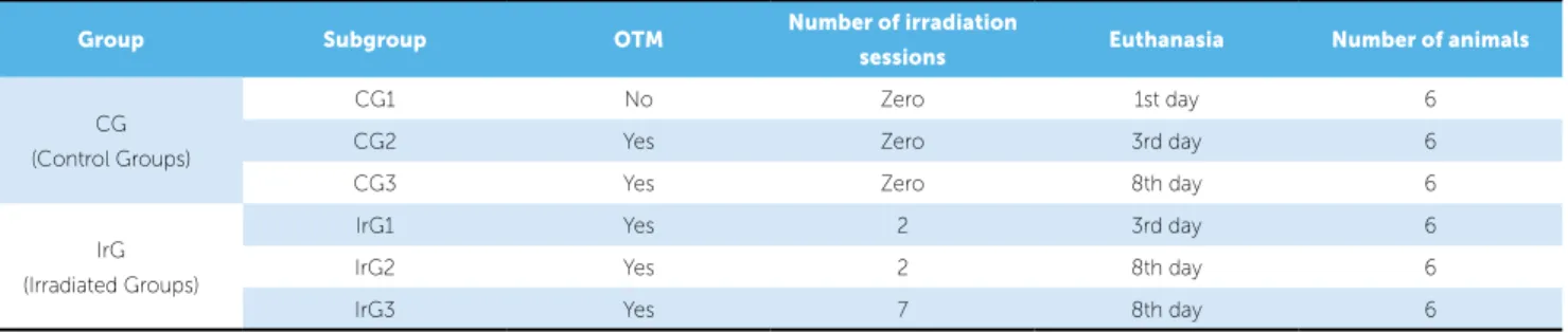
For the present in vivo experimental study, 36 adult
male Wistar rats, 12 weeks old, with a mean weight of 250 g were used. The rats were divided into 2 groups, Ir-radiated Group (IrG) and Control Group (CG), and then subdivided into 6 groups according the presence of ex-perimental tooth movement, number of laser irradiation sessions and euthanasia day (Table 1). The sample size calculation was made at a level of signiicance of 5% and power test of 80%. The rats were housed in cages inside a room with a 12 hours light/dark cycle and provided with powdered food to avoid damage to the orthodontic
ap-pliance and iltered water ad libitum. All procedures were
carried out under general anesthesia using intraperitoneal injection of a mixed solution of ketamine hydrochloride and xylazine hydrochloride. This protocol was reviewed and approved by the Ethics Commission on Animal Use in Scientiic Research at the Heath Center of the “Uni-versidade Federal do Rio de Janeiro”.
The orthodontic device was composed of a 7 mm nickel titanium closed coil spring (Dental Morelli Ltda, Sorocaba/SP) linked to the maxillary let irst molar by a stainless steel ligature wire (Dental Morelli Ltda, So-rocaba/SP), stretched to achieve 40 cN and then tied to the maxillary incisors with a second ligature wire. The orthodontic appliance was set with the rats sedated, ad-ministering intraperitoneal injection of the combination of ketamine (1.33 ml / kg) and xylazine (0.67 ml / kg). Composite resin (Transbond XT, 3M Unitek, Monro-via, California, USA) was used to cover the incisors to ensure maximum retention of the ligature and to im-prove the macroscopic analysis of OTM through the
Group Subgroup OTM Number of irradiation
sessions Euthanasia Number of animals
CG (Control Groups)
CG1 No Zero 1st day 6
CG2 Yes Zero 3rd day 6
CG3 Yes Zero 8th day 6
IrG (Irradiated Groups)
IrG1 Yes 2 3rd day 6
IrG2 Yes 2 8th day 6
IrG3 Yes 7 8th day 6
landmark created (Fig 1). Finally, the mandibular inci-sors were cut to avoid damage to the orthodontic appli-ance by occlusal trauma.
A continuous wave gallium-aluminum-arsenide (Ga-Al-As) diode laser (Thera Lase, DMC Equipamentos, São Carlos/SP) with a wavelength of 830 nm, power output
of 100 mW and spot area of nearly 0.003 cm2 was used
for this research. A luency of 6000 J/cm2 was applied.4,11
Irradiation was performed for 3 minutes at each of the three elected points (mesial, buccal and palatal) around the moved tooth, corresponding to a total energy of 54 J. The number of irradiation sessions was performed according to the group division (Table 1).
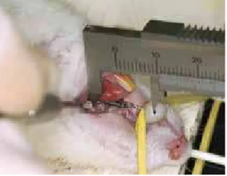
The distance between the mesial surface of the first molar and the landmark created in composite resin was measured by a single blind and calibrated examiner (ICC = 0.996), using an orthodontic caliper (Odin, Orthopli, Philadelphia, Pennsylvania, USA) immediately after the coil spring was placed, and before euthanasia (Fig 2). The subtraction was con-sidered the amount of OTM. The negative control group, CG1, was not included in this analysis because no OTM was performed.
The animals were euthanized by decapitation and maxillary bones were dissected. The left maxillary molars and their associated periodontium and sup-porting bone of each rat were fixed in 10% neutral buffered formalin, decalcified in Morse Solution and placed in paraffin blocks. Cross sections 5 μm thick were obtained from the cervical third of the root, and were hematoxylin and eosin stained (HE).
Qualitative analysis of cellular and tissue reactions of the periodontium of maxillary left first molar was performed on Nikon Eclipse E600 microscope at 40X, 100X and 400X magnifications. Quantitative analysis of the number of blood vessels in the PDL was also performed to verify the presence or absence of angiogenesis during OTM and the LLL irradia-tion. Photomicrographs at 400X magnification were obtained in four areas of the periodontal ligament of the mesial root (mesial, buccal, distal and palatal) and blood vessel counts were performed visually in the software Image Pro Plus 4.5 (Media Cybernetics, Sil-ver Spring, Maryland, USA). The ICC for counting was 0.841 showing good reliability.
The amount of OTM (recorded in millimeters) and the absolute number of vessels in each specimen were statistically analyzed using the Statistical Package for the Social Sciences (version 16, SPSS Inc., Chi-cago, Illinois, USA). The verification of normality and homogeneity was performed using the Shapiro-Wilk and Levene tests, respectively, at a significance level of 0.05. Having checked the normal distribution and homogeneity of the variables, analysis of variance (ANOVA) and Tukey’s multiple comparisons were applied to detect differences between groups.
RESULTS
Quantiication of OTM
Macroscopic analysis revealed no signiicant diference between irradiated and control groups in the evaluated peri-ods (3rd and 8th days) (p < 0.05) (Table 2).
Figure 1 - Schematic illustration of the LLL irradia-tion (red circles) on the ist upper left molar.
Figure 2 - Measurement of tooth movement:
Measure of the distance between the mesial sur-face of the irst upper left molar and the refer-ence point on the resin covering the upper inci-sors, with orthodontic caliper.
Group Mean (S.D.)
CG2 0.39 ± 0.04a
CG3 1.28 ± 0.10b
IrG1 0.40 ± 0.05a
IrG2 1.04 ± 0.06b
IrG3 1.25 ± 0.11b
Table 2 - Mean and standard deviation (S.D.) for
OTM (mm).
Diferent letters indicate statistical diference at
inserted perpendicularly in the cementum and alveolar bone. On the mesial surface, this parallelism was not so clear. Blood vessels of diferent sizes were observed throughout the extension of the PDL. In the mesial portion of alveolar bone, a line of bone apposition was observed, indicating recent osteogenesis. The bone ap-position was probably due to the normal eruptive pro-cess, which occurs in a distal direction. The marrow spaces were small in extent (Fig 3) and illed with he-matopoietic tissue and blood vessels.
CG2 with orthodontic appliance,
without LLL irradiation and euthanasia at 3rd day OTM changed the organization of cementoblasts, especially in the pressure zone. In these areas, a reduc-tion in the PDL space was observed, and iber bundles Qualitative analysis of cellular and
tissue reactions of the periodontium
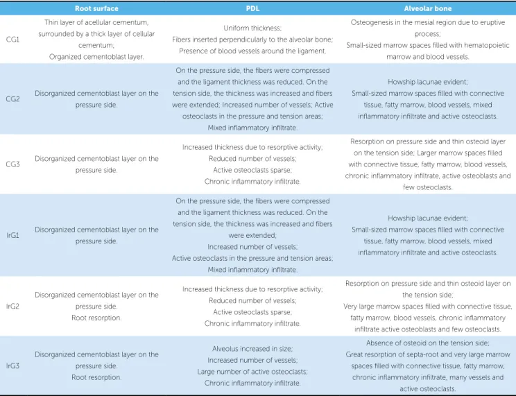
Analysis of cellular and tissue reactions are described below and summarized in Table 3.
CG1 without laser applications and without orthodon-tic appliance
These physiologically normal animals were consid-ered standard for comparison. In the mesial root, the presence of a thin layer of acellular cementum was ob-served, surrounded by a thin layer of cellular cementum in the buccal and mesial regions. There was a cemento-blast layer covering it and some cementoid areas. The PDL presented a relatively uniform thickness and its iber bundles were organized and surrounded by ibro-blasts. The distal segment showed parallel iber beams
Root surface PDL Alveolar bone
CG1
Thin layer of acellular cementum, surrounded by a thick layer of cellular
cementum; Organized cementoblast layer.
Uniform thickness;
Fibers inserted perpendicularly to the alveolar bone; Presence of blood vessels around the ligament.
Osteogenesis in the mesial region due to eruptive process;
Small-sized marrow spaces illed with hematopoietic marrow and blood vessels.
CG2 Disorganized cementoblast layer on the pressure side.
On the pressure side, the ibers were compressed and the ligament thickness was reduced. On the tension side, the thickness was increased and ibers were extended; Increased number of vessels; Active
osteoclasts in the pressure and tension areas; Mixed inlammatory iniltrate.
Howship lacunae evident;
Small-sized marrow spaces illed with connective tissue, fatty marrow, blood vessels, mixed inlammatory iniltrate and active osteoclasts.
CG3 Disorganized cementoblast layer on the pressure side.
Increased thickness due to resorptive activity; Reduced number of vessels;
Active osteoclasts sparse; Chronic inlammatory iniltrate.
Resorption on pressure side and thin osteoid layer on the tension side; Larger marrow spaces illed with connective tissue, fatty marrow, blood vessels, chronic inlammatory iniltrate, active osteoblasts and
few osteoclasts.
IrG1 Disorganized cementoblast layer on the pressure side.
On the pressure side, the ibers were compressed and the ligament thickness was reduced. On the tension side, the thickness was increased and ibers
were extended; Increased number of vessels;
Active osteoclasts in the pressure and tension areas; Mixed inlammatory iniltrate.
Howship lacunae evident;
Small-sized marrow spaces illed with connective tissue, fatty marrow, blood vessels, mixed inlammatory iniltrate and active osteoclasts.
IrG2
Disorganized cementoblast layer on the pressure side.
Root resorption.
Increased thickness due to resorptive activity; Reduced number of vessels;
Active osteoclasts sparse; Chronic inlammatory iniltrate.
Resorption on pressure side and thin osteoid layer on the tension side;
Very large marrow spaces illed with connective tissue, fatty marrow, blood vessels, chronic inlammatory
iniltrate active osteoblasts and few osteoclasts.
IrG3
Disorganized cementoblast layer on the pressure side.
Root resorption.
Alveolus increased in size; Increased number of vessels; Large number of active osteoclasts;
Chronic inlammatory iniltrate.
Absence of osteoid on the tension side; Great resorption of septa-root and very large marrow
spaces illed with connective tissue, fatty marrow, chronic inlammatory iniltrate, many vessels and
active osteoclasts.
areas of bone formation. The marrow spaces were still normal in size and were illed with connective tissue, fatty marrow, blood vessels, mixed inlammatory inil-trate and active osteoclasts (Fig 3).
CG3 with orthodontic appliance,
without LLL irradiation and euthanasia at 8th day The characteristics of cement were the same as in CG2. The ligament thickness was increased due to resorption. Active osteoclasts were scarce in pressure and tension sides, and in the marrow spaces.
were compressed and difuse. In tension areas the i-bers were stretched. There were blood vessels along the entire length of the ligament. These vessels were more numerous when compared to the group without orthodontic appliance (CG1). Active osteoclasts were observed in both pressure and tension sides, indicating the initial enlargement of the alveoli. Mixed inlamma-tory iniltrate was also observed, with mononuclear and polymorphonuclear inlammatory cells. In bone tissue, an irregular contour was observed, due to the presence of Howship lacunae. In the tension area, there were no
DL DL
ML ML
TB
TB
DV
DV INT
INT
M
M
Figure 3 - Photomicrograph of supporting tissues and irst upper molars undergone orthodontic movement in the diferent groups. M = mesial root; ML =
me-siolingual root; DL = distolingual root; INT = intermediate root; DV = distovestibular root; TB = trabecular bone (HE, 40X). DL
DL
ML
ML
TB
TB
DV
DV
INT
INT
M M
ML
TB
DV
INT
M
DL ML
TB
DV
INT
M DL
CG1 IrG1
CG2
CG3 IrG3
IrG2
1000µm
1000µm
1000µm
1000µm
1000µm
PDL vascularization remained well distributed, with little reduction in the number of blood vessels. Lymphoplasmacytic inflammatory infiltrate (chronic) was observed. In the alveolar bone, the compression side presented irregularities due to the resorptive pro-cess, while on the tension side narrow osteoid areas were observed. Marrow spaces were greater in extent than in CG2, filled with connective tissue, fatty mar-row, blood vessels, chronic inflammatory infiltrate, active osteoblasts and few osteoclasts (Fig 3).
IrG1 with orthodontic device,
two LLL applications and euthanasia at 3rd day The characteristics were similar to the CG2 (Fig 3).
IrG2 with orthodontic device, two LLL applications and euthanasia at 8th day
Bigger diferences were observed when teeth were moved in a period of seven days. Diferences in the char-acteristics of cement and marrow spaces were evident when compared with group CG3. The cement showed areas of resorption (Fig 4), and the marrow spaces were larger, with great reduction of the septa-root (Fig 3).
IrG3 with orthodontic device,
seven LLL applications and euthanasia at 8th day Root resorptions were also observed in this group (Fig 4) and further enlargement of the periodontal space was observed when compared with group IrG2 (Fig 3). The number of blood vessels was higher than in CG3.
Mononuclear inlammatory cells were abundant (chron-ic inlammatory iniltrate). The septa-root showed sig-niicant reduction, even disappearing in some animals. In the space between the roots, connective tissue, fat marrow, a high number of blood vessels, chronic in-lammatory iniltrate and active osteoclasts in the re-maining bone were observed.
In all of the groups that received an orthodontic ap-pliance on the irst molar, there were similar reactions in the periodontium of the second molar, but in lower ex-tension. It was possible to see areas of pressure and ten-sion in the PDL of the second molar as well as changes in the cells and size of marrow spaces.
Blood vessels counting
Means and standard deviations for each group are shown in Table 4. Statistically signiicant diference was found (p < 0.05) only among CG1 and groups CG2, IrG1 and IrG3, in which the number of vessels was higher (Fig 5).
DISCUSSION
Two diferent LLL irradiation protocols were tested during OTM in this study: One with daily irradiations and another in which the irradiation occurred only on the irst two days of OTM. The macroscopic evalua-tion of OTM showed no signiicant diference be-tween groups on the evaluated days (3rd and 8th). This means both tested protocols were unable to increase the amount of OTM (p < 0.05).
Figure 4 - Photomicrograph of mesial root resorption areas of irst molars of mice belonging to IrG2 (A) and IrG3 (B). D = dentin; C = cement; PL = periodontal ligament; black arrows = osteoclasts; (HE, 400X).
D
C
C PL
PL
A B
Figure 5 - Photomicrograph of periodontal ligament of rats’ mesial root belonging to CG1 (A) and IrG1 (B). Black arrows indicate blood vessels; D = dentin; C = cement; PL = periodontal ligament; AB = alveolar bone; (HE, 400X).
C C
D D
PL PL
AB AB
Group Mean ± S.D.
CG1 22.00 ± 3.16a
CG2 37.40 ± 9.91b
CG3 30.40 ± 5.41ab
IrG1 37.00 ± 6.89b
IrG2 27.66 ± 3.78ab
IrG3 38.00 ± 5.19b
Table 4 - Mean of induced tooth movement (mm) and standard deviation
(S.D.).
Diferent letters indicate statistical diference at α = 0,05% (ANOVA/Tukey).
This result is in agreement with the findings of
Limpanichkul et al,13 in which the irradiated group
and control group did not differ in the speed of OTM. Another study demonstrated the inhibitory effect of LLL, in other words, the amount of OTM
decreased.22 On the other hand, others have
demon-strated the effectiveness of LLL.2,4,11 It is believed that
LLL is dose-dependent and it can speed up or slow down biological processes depending on the fluency
applied and the irradiation protocol.1,6,9,10 In addition,
the mentioned studies used different subjects: Some of them used animals (rats or rabbits), and others used humans. Considering the body height and functional differences of these organisms, the interpretation of fluency as high or low may vary.
Two of the above-mentioned researches used the same animal model and the same luency as used in this study and obtained a positive result: The movement
rate increased when LLL was applied,4,11 disagreeing
with the results of the present study. This may be ex-plained by the diferent measurement method. They measured the distance between the occlusal surface of irst and second molars in digital dental casts under magniication. In the present study, an orthodontic caliper was used directly in the animal’s oral cavity, taking the measurement from the mesial surface of irst molar up to the incisors, as described by Drevenšek
et al.3 Although Kawasaki, Shimizu11 and Fujita et al4
used an indirect method for measurement associated
with magniication, which may have made it more sensitive, the choice of the second molar as a reference point does not seem adequate since this tooth position is inluenced by the irst molar movement through the
action of transeptal ibers.8,28
Another factor that difered between the studies of
Kawasaki, Shimizu11 and Fujita et al4 and this research
was the force applied. Both used 10 cN while in this
study 40 cN was used.7,12,14,15,24,25 This factor, however,
does not seem to explain the diference in the results
obtained, because Gonzales et al5 tested the strengths
of 10, 25, 50 and 100 cN for moving rat molars and observed that over a period of 14 days, the forces of 10, 25 and 50 cN did not difer in the amount of OTM.
B A
Moreover, the force used in this study was able to gener-ate bone remodeling without evidence of hyalinization, suggesting that it was appropriate.
Qualitative analysis of cellular and tissue reactions have been little explored in scientiic articles. The im-portance of quantifying data and submitting them to statistical analysis is known, however, the description of phenomena makes it easier for the reader to understand, and may arouse her/his curiosity to conduct further analysis and to quantify other data. In this research, in-teresting phenomena were observed. Root resorptions were evident during the eighth day of OTM in the irradiated groups (IrG2 and IrG3), while in the other groups it was not observed. Another interesting aspect was the efect on the alveolar bone tissue, speciically as regards the marrow spaces. Groups submitted to seven days of OTM and irradiated by laser, in particular those in which the irradiations were daily (IrG3), presented an increased resorptive activity, showing extremely large marrow spaces, including in the second molar region, as
a consequence of the action of transseptal ibers.28
Root resorptions were observed ater seven days in the mesial roots of irst molars undergoing OTM and irradiated by LLL. This inding disagrees with the
study conducted by Mendes,15 in which root
resorp-tion was considered similar between irradiated and unirradiated groups. The literature provides no infor-mation on the relationship between LLL and changes in the marrow spaces.
In this research, the aim was to quantify the blood vessels of the PDL, since and angiogenic efect of LLL
on wound repair has previously been reported.20,29
An-giogenesis is also beneicial to OTM by allowing greater movement of oxygen, nutrients and chemical mediators of inlammation, and by facilitating the iniltration of
repair cells.30 It also promotes the arrival of osteoclasts,
cells that arise from monocytes in the bone marrow and
are transported through the bloodstream.16
Groups CG2 and IrG1, submitted to two days of OTM, difered from CG1, conirming that there is an increase in the vascularity during the early periods
of OTM.17,20,28 These two groups, however, presented
similar number of vessels, showing that LLL was un-able to produce further increase in angiogenesis in the earlier period of OTM.
On the eighth day of OTM, vascularization tend-ed to return to normal, with no statistically signiicant diference between CG3, IrG2 and the other groups. Exception was found in IrG3, showing that daily ap-plications of laser were able to continue increasing the number of vessels. This result is in agreement with
Mendes,15 who observed an increase in the
vasculariza-tion of the periodontium during the 8th day of OTM when rats were irradiated daily.
Among the irradiated groups, only group IrG2 did not difer from CG1. This inding conirms the
conclusion of Saito and Shimizu.21 These authors
sug-gested that while applications of LLL during the initial stages of bone regeneration trigger stimulatory efects, applications in the late periods seem to have the func-tion of maintaining such an efect. The discontinua-tion of applicadiscontinua-tions appears to reduce the stimulus, as observed in this study.
CONCLUSION
Quantitative analysis demonstrated that OTM did not difer between control and irradiated groups in the equivalent experimental period, demonstrating that both LLL irradiation protocols tested were unable to in-crease the rate of OTM.
Qualitative analysis showed greater periodontal ab-sorptive activity, in both root and bone tissues, in irradi-ated groups on the eighth day of OTM (IrG2 and IrG3), especially when irradiations were carried out daily (IrG3).
The number of blood vessels in the PDL was in-creased by OTM and daily LLL irradiation. IrG1 and IrG3 presented a higher number of blood vessels in comparison with CG1 (device-free animals and not ir-radiated), showing that irradiations in later periods are necessary to maintain the stimulatory efect of LLL.
1. Coombe AR, Ho C-TG, Darendeliler MA, Hunter N, Philips JR, Chapple CC, et al. The efects of low laser irradiation on osteoblastic cells. Clin Orthod Res. 2001;4(1):3-14.
2. Cruz DR, Kohara EK, Ribeiro MS, Wetter NU. Efects of low-intensity laser therapy on the orthodontic movement velocity of human teeth: a preliminary study. Lasers Surg Med. 2004;35(2):117-20.
3. Drevensek M, Sprogar S, Boras I, Drevensek G. Efects of endothelin antagonist tezosentan on orthodontic tooth movement in rats. Am J Orthod Dentofacial Orthop. 2006;129(4):555-8.
4. Fujita S, Yamaguchi M, Utsunomiya T, Yamamoto H, Kasai K. Low-energy laser stimulates tooth movement velocity via expression of RANK and RANKL. Orthod Craniofac Res. 2008;11(3):143-55.
5. Gonzales C, Hotokezaka H, Yoshimatsu M, Yozgatian Jh, Darendeliler Ma, Yoshida N. Force magnitude and duration efects on amount of tooth movement and root resorption in the rat molar. Angle Orthod. 2008;78(3):502-9.
6. Hawkins DH, Abrahamse H. The role of laser luence in cell viability, proliferation, and membrane integrity of wounded human skin ibroblasts following Helium-Neon laser irradiation. Lasers Surg Med. 2006;38(1):74-83. 7. Hayashi H, Konoo T, Yamaguchi K. Intermittent 8-hour activation in
orthodontic molar movement. Am J Orthod Dentofacial Orthop. 2004;125(3):302-9.
8. Heller IJ, Nanda R. Afect of metabolic alteration of periodontal ibers on orthodontic tooth movement. An experimental study. Am J Orthod. 1979;75(3):239-58.
9. Houreld NN, Abrahamse H. Laser light inluences cellular viability and proliferation in diabetic-wounded ibroblast cells in a dose- and wavelength-dependent manner. Lasers Med Sci. 2008;23:11-8.
10. Karu T, Pyatibrat LV, Ryabykh TP. Nonmonotonic behavior of the dose dependence of the radiation efect on cells in vitro exposed to pulsed laser radiation at λ = 820 nm. Lasers Surg Med. 1997;21(5):485-92.
11. Kawasaki K, Shimizu N. Efects of low-energy laser irradiation on bone remodeling during experimental tooth movement in rats. Lasers Surg Med. 2000;26(3):282-91.
12. King GJ, Archer L, Zhou D. Later orthodontic appliance reactivation stimulates immediate appearance of osteoclasts and linear tooth movement. Am J Orthod Dentofacial Orthop. 1998;114(6):692-7.
13. Limpanichkul W, Godfrey K, Srisuk N, Rattanayatikul C. Efects of low-level laser therapy on the rate of orthodontic tooth movement. Orthod Craniofac Res. 2006;9(1):38-43.
14. Madan MS, Liu ZJ, Gu GM, King GJ. Efects of human relaxin on orthodontic tooth movement and periodontal ligaments in rats. Am J Orthod Dentofacial Orthop. 2007;131(1):8.e1-10.
15. Mendes OF. Inluência do laser de baixa potência na movimentação ortodôntica: estudo experimental em ratos [dissertação]. Rio de Janeiro (RJ): Universidade Federal do Rio de Janeiro; 2005.
REFERENCES
16. Mundy GR. Bone remodeling and its disorders. 2nd ed. London: Ed. Martin
Dunitz; 1999.
17. Murrel EF, Yen EHK, Johnson RB. Vascular changes in the periodontal ligament after removal of orthodontic forces. Am J Orthod Dentofacial Orthop. 1996;110(3):280-6.
18. Ozawa Y, Shimizu N, Kariya G, Abiko, Y. Low-energy laser irradiation stimulates bone module formation at early stages of cell culture in rat calvarial cells. Bone. 1998;22(4):347-54.
19. Rygh P, Bowling K, Hovlandsdal L, Williams S. Activation of the vascular system: a main mediator of periodontal iber remodeling in orthodontic tooth movement. Am J Orthod. 1986;89(6):453-68.
20. Rocha JCT. Terapia laser, cicatrização tecidual e angiogênese. Rev Bras Promoção Saúde. 2004;17(1):44-8.
21. Saito S, Shimizu N. Stimulatory efects of low-power laser irradiation on bone regeneration in midpalatal suture during expansion in the rat. Am J Orthod Dentofacial Orthop. 1997;111(5):525-32.
22. Seii M, Shafeei HA, Daneshdoost S, Mir M. Efects of two types of low-level laser wave lengths (850 and 630 nm) on the orthodontic tooth movements in rabbits. Lasers Med Sci. 2007;22(4):261-4.
23. Shimizu N, Yamaguchi M, Goseki T, Shibata Y, Takiguchi H, Iwasawa T, et al. Inhibition of prostaglandin E2 and interleukin 1-β production by low-power laser irradiation in stretched human periodontal ligament cells. J Dent Res. 1995;74(7):1382-8.
24. Stuani AS. Aspectos histológicos do periodonto no lado de pressão de dentes, quando submetidos à força ortodôntica [dissertação]. Rio de Janeiro (RJ): Universidade Federal do Rio de Janeiro; 2003.
25. Taweechaisupapong S, Srisuk N, Nimitpornsuko C, Vattraphoudes T, Rattanayatikul C, Godfrey K. Evening primrose oil efects on osteoclasts during tooth movement. Angle Orthod. 2005;75(3):356-61.
26. Turhani D, Scheriau M, Benesch T, Jonke E, Bantleon HP. Pain relief by single low-level laser irradiation in orthodontic patients undergoing ixed appliance therapy. Am J Orthod. 2006;130(3):371-6.
27. Vandevska-Radunovic V, Kristiansen Ab, Heyeraas Kj, Kvinnsland S. Changes in blood circulation in teeth and supporting tissues incident to experimental tooth movement. Eur J Orthod. 1994;16(5):361-9.
28. Verna C, Zafe D, Siciliani G. Histomorphometric study of bone reactions during orthodontic tooth movement in rats. Bone. 1999;24(4):371-9. 29. Walsh LJ. The current status of low level laser therapy in dentistry. Part 1. Soft
tissue applications. Aust Dent J. 1997;42(4):247-54.