DOI: 10.1590/0004-282X20150112 ARTICLE
Surgical treatment of non-functioning
pituitary macroadenomas by the endoscopic
endonasal approach in the elderly
Tratamento cirúrgico dos macroadenomas não secretores de hipófise por via endoscópica
endonasal em idosos
Horacio Armando Marenco1,3, Samuel Tau Zymberg1,2, Rodrigo de Paula Santos1,4, Cláuder Oliveira Ramalho1
Life expectancy has continued to increase in most devel-oped countries over the past two centuries. It is estimated that three-quarters of all infants born since 2000 will live to an age of 75 years, whereas more than half of these infants will reach 100 years1. he life expectancy in Brazil has increased
over the last century at a rate similar to that in most devel-oped countries2. Aging populations lead to more elderly
pa-tients presenting neurosurgical problems; pituitary
adeno-mas are a common inding in this age group, as has already
been observed in a series of autopsies of elderly individuals3.
Over the past three decades, several authors have pub-lished surgical series of elderly patients treated for pituitary adenomas, which account for 1.2% to 9% of all patients in the reported series4,5,6,7,8,9. his proportion increases to 14.6%
if only non-functioning pituitary adenomas are considered10.
Non-functioning pituitary adenomas are usually diagnosed as macroadenomas in elderly patients, at which point the adenomas become clinically relevant due to the local mass
efect9,10,11. In these cases, because surgery provides
decom-pression of the optic chiasm and may permit some degree of
1Universidade Federal de São Paulo, Programa de Pós-Graduação em Otorrinolaringologia e Cirurgia da Cabeça e Pescoço, Departamento de Otorrinolaringologia, Sao Paulo SP, Brazil;
2Universidade Federal de São Paulo, Departamento de Neurocirurgia, Sao Paulo SP, Brazil;
3Fundação Leonor de Barros Camargo, Hospital Augusto de Oliveira Camargo, Serviço de Neurocirurgia, Indaiatuba SP, Brazil;
4Universidade Federal de São Paulo, Departamento de Otorrinolaringologia, Setor de Rinologia, Sao Paulo SP, Brazil.
Correspondence: Horacio Armando Marenco; Av. Francisco de Paula Leite, 399; 13344-700 Indaiatuba SP, Brasil; E-mail: [email protected] Conflict of interest: There is no conlict of interest to declare
Received 19 March 2015; Received in inal form 17 April 2015; Accepted 07 May 2015. ABSTRACT
Over the past three decades, surgical series of elderly patients treated for pituitary adenomas have been published, all of which used the microscopic transsphenoidal or transcranial approach. The objective of this study was to retrospectively analyze the surgical results of our irst 25 elderly patients with non-functioning pituitary macroadenoma (NFPM) operated by the endoscopic endonasal approach (EEA). Preoperative visual loss was found in 92.8% of the cases, and 70.8% experienced visual improvement following surgery. Preoperative pituitary dysfunction was found in 69.2% of the cases and postoperative pituitary recovery occurred in 22.2% of them. Mean hospital stay was 6.7 days. The results of this study suggest that surgery remains the irst line of treatment for NFPM in the elderly. Because age alone is not a barrier for surgery, patients should be selected for surgical treatment based on their symptoms and clinical condition, as deined by comorbidities.
Keywords: aged, pituitary neoplasms, pituitary diseases, endoscopic surgical procedures.
RESUMO
Nas últimas três décadas, foram publicadas casuísticas de pacientes idosos operados por adenomas de hipóise, nas quais foram utilizadas as vias transcraniana ou transesfenoidal microcirúrgicas. O objetivo deste estudo foi analisar retrospectivamente os resultados dos nossos primeiros 25 pacientes idosos com macroadenomas não secretores de hipóise, operados pela via endoscópica endonasal. O déicit visual pré-operatório foi encontrado em 92,8% dos casos e 70,8% apresentaram melhora da visão depois da cirurgia. O hipopituitarismo pré-operatório foi encontrado em 69,2% dos casos e a sua recuperação ocorreu em 22,2% destes casos. A estadia hospitalar média foi de 6,7 dias. Este estudo sugere que a cirurgia permanece como o tratamento de primeira escolha para pacientes idosos com macroadenomas não secretores de hipóise e a idade por si só não é contraindicação para cirurgia.
endocrinological recovery, the irst line of treatment is sur -gery rather than radiotherapy or medical treatment6,10. Some
authors suggest that due to the elevated risks of surgery in the elderly, the sole indication for surgical treatment of pitu-itary adenomas in this population should be macroadenoma
with mass efect and visual impairment11.
In our series, radiotherapy was reserved for the treat-ment of the postoperative regrowth of tumor remnants.
After reviewing the current literature, we have identiied
three series dedicated exclusively to the surgical treatment of non-functioning pituitary macroadenomas (NFPM) in the el-derly10,12,13, all of which used the microscopic transsphenoidal
or transcranial approach. In this article, we review our
expe-rience with the irst 25 patients older than 65 years at time of
surgery who were operated upon for NFPM by the endoscop-ic endonasal approach (EEA).
METHOD
Between March 2001 and July 2013, 369 patients under -went surgery for sellar lesions in our Neurosurgery
depart-ment. Of these patients, 44 were identiied as being older than
65 years at the time of surgery (11.9%). For this study, we
select-ed all 25 patients who presentselect-ed with histologically conirmselect-ed
NFPM and who were treated with EEA. One patient with un-available medical records and three who were treated by the microscopic transsphenoidal approach were excluded.
he present study was approved by the Committee on
Ethics in Research of the Federal University of São Paulo
un-der the record 733.100 (July 30, 2014). All of the data from pa
-tients were carefully anonymized despite the informed con -sent form provided to all patients who remained in follow-up.
here were 14 (56%) female patients and 11 (44%) male patients; the mean age was 72.4 years. he physiological sta -tus of the patients was graded according to the American Society of Anesthesiologists (ASA) scale14.
All of the patients underwent detailed radiological studies
with preoperative sellar MRI and were classiied according
to the system proposed by Ludecke15. he irst postoperative
MRI, one to three months after surgery, determined the de-gree of resection. Data about tumor resection dede-gree were not available in two cases (Table).
he endocrinological evaluation of the patients included pre- and postoperative serum levels of GH, IGF-1, ACTH, cor
-tisol, PRL, TSH, FT4, FSH, LH and, for males, testosterone. he
specimens that were resected during surgery underwent mi-croscopic routine histological analysis, and most underwent immunohistochemical analysis using the HRP-StreptAvidin
Biotin method.
he patients had their visual deicits evaluated in our
Ophthalmology department both pre- and postoperatively; in some cases, campimetry was performed elsewhere. Two patients were operated upon twice due to tumor remnant
regrowth and a new visual deicit. A third patient was oper -ated upon twice due to tumor remnant regrowth on control
MRI without visual deicit. Subsequently, the sample in
-creased to 28 surgical procedures across 25 patients. he
follow-up time of our patients ranged from 11 to 132 months, with a mean duration of 47.2 months.
All of the data were analyzed using the software R Core Team (2014) (R Foundation for Statistical Computing,
Vienna, Austria). Normal distribution of obtained data was evaluated using Shapiro-Wilk test. Fisher’s exact test was used for analysis of the correlation between the categorical variables. Student t-test and Mann-Whitney test were used for the analysis of correlations between categorical and
nu-merical variables when appropriate. P-values < 0.05 were considered to be statistically signiicant.
RESULTS
Clinical presentation
Visual loss was the most common symptom, afecting 24 out of 25 patients prior to the irst surgical procedure and
two out of three patients who underwent a second surgery
(92.8%). he second most common symptom was headache,
which was present in 12 of the 28 cases (42.8%).
Less common symptoms included dizziness, weight loss, seizure and libido reduction. hree patients were
diagnosed with pituitary macroadenomas during investi-gation for dementia with magnetic resonance image (MRI)
Table.Patient details.
Characteristic No.
Total no. of patients 25
Total no. of surgical procedures 28
Male : Female 11 : 14
Age (years)
Mean ± SD 72.4 ± 4.7
Range 65 – 79
ASA score
1 1
2 19
3 8
4 / 5 0
Tumor size*
T1 0
T2 1
T3 9
T4 13
Range (mm) 17 – 48
Mean ± SD (mm) 34 ± 7.3
Invasion of adjacent structures 20 / 27 (74.1%)
Visual deicit 26 / 28 (92.8%)
Preoperative hypopituitarism 18 / 26 (69.2%)
or computerized tomography. One patient was admitted to
emergency care due to pituitary apoplexy with acute visual
loss and unilateral palpebral ptosis. he duration of the symp -toms varied from six months to eight years until diagnosis.
Chronic comorbid diseases were listed for each case
before surgery, and the ASA score was retrieved from the an-esthesiologist’s record. We found that most patients present-ed at least one chronic disease. Arterial hypertension was the most prevalent comorbidity, present in 92.9% of the patients. Diabetes was present in 35.7% of the patients, myocardial disease in 25%, dementia in 14.2%, chronic pulmonary dis-ease in 7.1%, and cancer in 7.1%.
According to the ASA scale, 3.6% of the patients were
clas-siied as grade 1, 67.8% as grade 2 and 28.6% as grade 3. here were no grade 4 or 5 classiied patients within this series.
Surgical outcome
All of the patients were operated upon by EEA, as described in detail previously16. Neuronavigation was not available for any case. he mean operative time was 146 ± 43 minutes, as
determined by the anesthesiologist’s records. Our multidis-ciplinary team performed all of the procedures. In addition, training neurosurgery and otorhinolaryngology residents and an observing endocrinology resident also participated; these residents would later assist the patient in the ward.
he results regarding visual deicits due to tumor com -pression of the optic pathways were available in 26 cases. Twenty-four cases presented visual loss preoperatively, and 17 (70.8%) of these patients experienced visual improvement following surgery; the other patients showed no change. Statistical analysis showed no relation between total tumor resection and postoperative visual improvement (p = 0.151).
here were no new cranial nerve dysfunctions or worsening visual deicits related to the surgical procedures. One patient
with preoperative unilateral VI nerve dysfunction and one with unilateral III nerve dysfunction improved after surgery.
Cerebrospinal luid (CSF) istulae occurred in only one
patient, who also presented with bacterial meningitis and
seizures and required antibiotic treatment. A second en -doscopic endonasal procedure was necessary to close the
istulae. his patient had a postoperative stay of 46 days, the longest hospitalization in our series, after which he was dis
-charged to his home. he mean hospitalization time was 6.7
days (range: 1 – 46 days).
One major bleeding event requiring transfusion, in the
same patient who presented CSF istulae, was the only intra -operative complication among our cases. Mild epistaxis
oc-curred in four cases during the irst three days after surgery,
although no special care was required. One patient present-ed moderate epistaxis ten days following surgery relatpresent-ed to
Ginkgo biloba use and required nasal endoscopic cauteriza -tion. Systemic postoperative complications included three cases of electrolyte imbalance, three cases of delirium and one case of symptomatic hypotension, all of them transient.
here was one death in our series. A 73-year-old woman
with a 45-mm pituitary adenoma was operated upon unevent-fully. However, this patient died due to the decompensation of
an existing cardiac insuiciency four days following surgery,
resulting in a 3.6% mortality rate in this series.
Total tumor resection was achieved in 30.8% of the cas-es, subtotal resection in 34.6% and partial resection in 34.6% (Figure 1). Total tumor resection was not related to tumor
size (p = 0.328) or invasion of the cavernous sinus (p = 0.145)
in this series.
he application of postoperative fractionated stereotactic
radiotherapy was reserved for patients who presented with recurrence of the NFPM and were not good candidates for
an additional surgical procedure. In our series, ive patients
with partial tumor resection and one with subtotal tumor resection presented regrowth of tumor remnants on MRI,
which we considered as recurrence. hree patients were op -erated upon again; no signs of recurrence were observed in
two of these patients after ive years of follow-up. One patient
who presented recurrence of the tumor seven months after the second surgery received radiotherapy. Another two pa-tients received radiotherapy to treat recurrences of a stable tumor remnant after three years of follow-up. One female
pa-tient who showed signs of recurrence on MRI ive years after
surgery without visual loss and at the age of 84 years refused further treatment.
Endocrinological outcome
Pituitary dysfunction was investigated prior to each
sur-gery. We found a deiciency of at least one pituitary axis
in 69.2% of the cases, and 11.5% of cases presented mild hyperprolactinemia. Details regarding preoperative anterior
pituitary dysfunction are speciied in Figure 2.
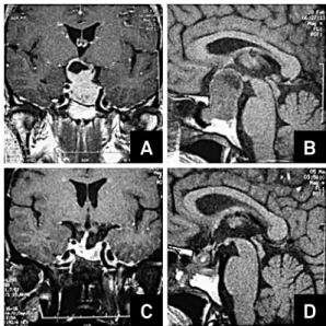
A
B
C
D
One to three months after each surgery, new serum hor-mone levels were obtained, and anterior pituitary function
was re-assessed. here was a new postoperative deiciency of IGF-1 and LH/FSH serum levels in one patient, TSH deicien
-cy in one patient and deicien-cy of LH/FSH in another pa -tient. All three patients with hyperprolactinemia had normal prolactin levels after the surgery, even those with only partial resection of their tumors.
Recovery from preoperative anterior pituitary insui -ciency occurred in four of the 18 cases, which accounted for 22.2% of the cases (Figure 3).
Postoperative diabetes insipidus occurred in 11.5% of the
cases, with none of these events lasting more than ive days.
Histological analysis
Microscopic histological analysis conirmed typical pitu -itary adenomas in all cases, with one being acidophilic and the rest being chromophobic. Immunohistochemical
analy-sis by the HRP-StreptAvidin Biotin method was available in
20 of the 25 patients. Ki-67 antigen labeling index positivity of 1 - 3% was found in most cases, and another two cases pre-sented up to 5% positivity.
According to the WHO classiication of pituitary tumors17,
the results of our histological and immunohistochemical analysis revealed 11 gonadotropin-producing adenomas, 8 null cell adenomas and 1 silent corticotroph adenoma.
DISCUSSION
Our data analysis demonstrates that the majority of the pa-tients were diagnosed with NFPM in a late stage of the disease. More than 56% of the tumors were diagnosed as T4 (31 mm or more), whereas 30% were diagnosed as giant adenomas (40 mm or more) at presentation. Preoperative MRI showed
74.1% of the patients with cavernous sinus invasion. he mean tumor size in our series was 34 mm. his result indicates that
we operated upon large invasive tumors in most cases; these
indings are similar to those reported by others10,13.
Preoperative evaluations demonstrated a 69.2%
preva-lence of hypopituitarism. Most of the afected pituitary axes
were gonadotrophic, with low LH/FSH levels in 61.5% of the cases, as well as somatotrophic, with low IGF-1 levels in 50%
of the cases. hese indings are similar to the other reports of
NFPM in elderly patients10,13. he adrenocorticotrophic and thyrotrophic axes were deicient in 26.9% and 23% of the cas
-es, respectively. Because pituitary dysfunction is related to the
compression of normal pituitary tissue by the tumor mass18, a
high incidence of hypopituitarism can be expected in a series of patients harboring large adenomas, such as is reported here.
he clinical manifestation of hypopituitarism is fre
-quently unrecognized in elderly patients because it may be
confused with the ageing process or masked by chronic con-ditions common to this age group5,9,13. his fact is of great
concern given that untreated hypothyroidism and hypocorti-solism are themselves life-threatening conditions, aside from
all of the symptoms that the patients sufer. Medical treat -ment with oral hormone replace-ment is mandatory in these cases because the symptoms and overall clinical condition can be improved despite the NFPM13. When appropriate,
pa-tients in this series received prednisone, levothyroxine and
testosterone injections to treat their hormone deicits. Because cataracts, retinal degeneration and amblyopia
are common visual disturbances in the elderly19, visual dei
-cits related to compression of optic pathways by the NFPM
can also go unrecognized in the elderly because they may be
attributed to other visual conditions9,13.
Considering that NFPMs in the elderly are primar -ily slow-growing lesions with a low proliferative index as
Number of patients
Normal Low High 25
20
15
10
5
0
GH IGF-1 TSH LH FSH ACTH PRL
Figure 2. Preoperative serum hormone levels.
Number of patients
Recovery
New deficit Unchanged 25
30
20
15
10
5
0
GH IGF-1 TSH LH FSH ACTH PRL
evaluated by the Ki-67 labeling index10,13, the long delay in
the diagnosis of NFPMs in the elderly may explain the high rate of large and invasive NFPMs found in this population. Some of our patients had symptomatic NFPMs that were di-agnosed almost a decade after the initial complaints.
In our patients, we found that 70.8% of those with visu-al loss improved after surgicvisu-al decompression of the chiasm,
irrespective of the duration of visual loss. Considering that
we did not pursue a radical tumor resection in each case, we achieved a high rate of visual recovery, similar to other au-thors who performed radical tumor resection7,9,10 and who
used neuronavigation20.
We found that our patients presented less postoperative
diabetes insipidus than other series with radical tumor re-section7,9,20 and those that were characterized by the same
surgical strategy6,13. New anterior pituitary deicits were low
-er in our patients than have been previously reported for se-ries consisting exclusively of NFPMs in elderly patients10,13. Neither cranial nerve lesion, even transitory, nor visual dei -cit worsening occurred in this series.
Comparing the postoperative hospital stay with a previ -ous series consisting exclusively of NFPM in the elderly10, our patients remained hospitalized less than half the time (6.7
days x 16.3 days). We believe that the EEA may be one de-termining factor for a shorter postoperative stay when com-pared with transsphenoidal microsurgery.
CSF istula is one of the main postoperative issues in
pituitary surgery. Despite an incidence as high as 15.9% of
postoperative CSF istulae in a large series of EEA21, we only
observed one incidence of this postoperative complication,
representing 3.6%. his was achieved by taking advantage of
others’ experience with closure techniques22, strictly
follow-ing the surgical technique as developed by our group16 and
not pursuing radical tumor excision.
One patient died in the early postoperative period from the
decompensation of a previous cardiac insuiciency. Although this patient was classiied as ASA grade 3 and was theoretically it for surgical treatment, this case highlights the impact of pa -tient comorbidities on surgical outcome and mortality, as was observed in the largest published analysis on pituitary surgery in the elderly, which found a 3.8% mortality rate23.
Recurrences were observed in only 21.4% of the patients despite a 69.2% rate of subtotal and partial tumor resection.
his result is in accordance with the low proliferative index of
the NFPMs found in our samples and with the observations of other authors, who have also reported low rates of tumor remnant regrowth in elderly patients7,9,10,13.
Due to the slow-growing nature of the NFPM in the el-derly, postoperative radiotherapy was not routinely indicated because it is associated with complications such as hypopi-tuitarism and optic neuropathy, even with modern applica-tion protocols24. his modality of treatment was reserved for documented recurrences in patients who were not it for an -other surgery.
he low rate of recurrence in our series suggests that not
pursuing radical tumor resection at all costs is a good strat-egy given that subtotal tumor resection can be “curative” in the case of an elderly patient with an NFPM while avoiding surgical morbidity.
In conclusion, this study suggests that surgery should
re-main the irst line of treatment for NFPMs in the elderly. Because age alone is not a barrier for surgery, patients should
be selected for surgical treatment based on their symptoms
and clinical condition, as deined by comorbidities. Local mass efect of the NFPM with visual loss is the best indication for surgical treatment. Pituitary insuiciency alone is not an indi -cation for surgical treatment because the postoperative recov-ery is poor and medical therapy can easily resolve this issue. Radical tumor resection should not be the goal in the case of
an elderly patient with an NFPM given that visual deicit im -provement is not related to total tumor resection and recur-rences are low even with incomplete resections. EEA provided
similar visual deicit outcomes as transsphenoidal microsur
-gery but with shorter hospital stays and better results for CSF istulae, diabetes insipidus and postoperative pituitary deicits.
ACKNOWLEDGEMENTS
We thank Mr. Matthew John Robbins for his helpful revi -sion of the manuscript, Prof. Gianni Mara Silva dos Santos from the Department of Statistics of the Federal University of São Paulo for her assistance with the statistical analysis
and Prof. Dr. Julio Zaki Abucham Filho and the Department
of Endocrinology of the Federal University of São Paulo for the caring of the patients included in this study.
References
1. Christensen K, Doblhammer G, Rau R, Vaupel JW. Ageing populations: the challenges ahead. Lancet. 2009;374(9696):1196-208.
doi:10.1016/S0140-6736(09)61460-4
2. Ramos LR, Veras RP, Kalache A. Envelhecimento populacional: uma realidade brasileira. Rev Saude Publica. 1987;21(3):211-24. doi:10.1590/S0034-89101987000300006
3. Kovacs K, Ryan N, Horvath E, Singer W, Ezrin C. Pituitary adenomas in old age. J Gerontol. 1980;35(1):16-22. doi:10.1093/geronj/35.1.16
4. Cohen DL, Bevan JS, Adams CB. The presentation and management of pituitary tumours in the elderly. Age Ageing. 1989;18(4):247-52. doi:10.1093/ageing/18.4.247
5. Turner HE, Adams CB, Wass JA. Pituitary tumours in the elderly: a 20 year experience. Eur J Endocrinol. 1999;140(5):383-9. doi:10.1530/eje.0.1400383
7. Ferrante L, Trillò G, Ramundo E, Celli P, Jaffrain-Rea ML, Salvati M et al. Surgical treatment of pituitary tumors in the elderly: clinical outcome and long-term follow-up. J Neurooncol. 2002;60 (2):185-91. doi:10.1023/A:1020652604014
8. Sheehan JM, Douds GL, Hill K, Farace E. Transsphenoidal surgery for pituitary adenoma in elderly patients. Acta Neurochir (Wien). 2008;150(6):571-74. doi:10.1007/s00701-008-1581-2
9. Hong J, Ding X, Lu Y. Clinical analysis of 103 elderly patients with pituitary adenomas: transsphenoidal surgery and follow-up. J Clin Neurosci. 2008;15(10):1091-5. doi:10.1016/j.jocn.2007.11.003
10. Kurosaki M, Lüdecke DK, Flitsch J, Saeger W. Surgical treatment of clinically nonsecreting pituitary adenomas in elderly patients. Neurosurgery. 2000;47(4):843-8. doi:10.1097/00006123-200010000-00009
11. Freda PU, Bruce JN. Surgery: risks of pituitary surgery in the elderly. Nat Rev Endocrinol. 2010;6(11):606-8. doi:10.1038/nrendo.2010.170
12. Pospiech J, Stolke D, Pospiech FR. Surgical treatment of pituitary adenomas in elderly patients. Acta Neurochir Suppl. 1996;65:35-6.
13. Del Monte P, Foppiani L, Ruelle A, Andrioli G, Bandelloni R, Quilici P et al. Clinically non-functioning pituitary macroadenomas in the elderly. Aging Clin Exp Res. 2007;19(1):34-40. doi:10.1007/BF03325208
14. Cullen SC. Cybernesthesia. Anesthesiology. 1963;24(1):110-111.
15. Lüdecke DK. Value of transcavernous surgery in the treatment of pituitary adenomas. Eur J Endocrinol. 1995;133(2):147-8. doi:10.1530/eje.0.1330147
16. Santos RP, Zymberg ST, Abucham Filho JZ, Gregório LC, Weckx LL. Endoscopic transnasal approach to sellar tumors. Braz J Otorhinolaryngol. 2007;73(4):463-75.
17. Asthagiri A, Lopes MBS. Neuropathological considerations of pituitary adenomas. In: Laws ER Jr, Sheehan JP, editors. Pituitary
surgery: a modern approach. frontiers of hormone research. Basel: Karger; 2006. (Frontiers of hormone research, Vol. 34). p. 206-35.
18. Arafah BM. Reversible hypopituitarism in patients with large nonfunctioning pituitary adenomas. J Clin Endocrinol Metab. 1986;62(6):1173-9. doi:10.1210/jcem-62-6-1173
19. Tielsch JM, Sommer A, Witt K, Katz J, Royall RM. Blindness and visual impairment in an American urban population: the Baltimore eye survey. Arch Ophthalmol. 1990;108(2):286-90. doi:10.1001/archopht.1990.01070040138048
20. Locatelli M, Bertani G, Carrabba G, Rampini P, Zavanone M, Caroli M et al. The trans-sphenoidal resection of pituitary adenomas in elderly patients and surgical risk. Pituitary. 2013;16(2):146-51. doi:10.1007/s11102-012-0390-z
21. Kassam AB, Prevedello DM, Carrau RL, Snyderman CH, Thomas A, Gardner P et al. Endoscopic endonasal skull base surgery: analysis of complications in the authors’ initial 800 patients. J Neurosurg. 2011;114(6):1544-68. doi:10.3171/2010.10.JNS09406
22. Hadad G, Bassagasteguy L, Carrau RL, Mataza JC, Kassam A, Snyderman CH et al. A novel reconstructive technique after endoscopic expanded endonasal approaches: vascular pedicle nasoseptal lap. Laryngoscope. 2006;116(10):1882-6. doi:10.1097/01.mlg.0000234933.37779.e4
23. Grossman R, Mukherjee D, Chaichana KL, Salvatori R, Wand G, Brem H et al. Complications and death among elderly patients undergoing pituitary tumour surgery. Clin Endocrinol (Oxf). 2010;73(3):361-8. doi:10.1111/j.1365-2265.2010.03813.x