121
ABSTRACT
BACKGROUND AND OBJECTIVES:Platelet-rich ibrin is a new and promising technique to accelerate repair, with possible analgesic efects; however, there is still a gap with regard to pe-ripheral nerve injury and the association with physical exercises. So, this study aimed at evaluating the efects of platelet-rich i-brin associated to physical exercises on nociception and edema in experimental median nerve compression model.
METHODS:hirty-six rats, all submitted to median nerve com-pression, were divided in six groups: G1: without additional ma-nipulation; G2: compression and treated with platelet-rich ibrin; G3: compression and treated with free swimming; G4: compres-sion and walking on a treadmill; G5: free swimming + platelet-rich ibrin; G6: walking on a treadmill + platelet-rich ibrin. Injury was induced by tying the median nerve with chrome plated catgut 4.0. Platelet-rich ibrin was obtained by centrifuging 1.5 mL of blood and positioning the ibrin clot directly on the compression region. Exercises were carried out during two weeks, between the 3rd and
14th postoperative days. Nociception and edema were evaluated,
respectively, by linch threshold and plethysmometer, in moments before injury and in the 3rd, 7th and 15th postoperative days.
RESULTS: here have been no diferences among groups, only among evaluations, showing increased nociception and edema, which has lasted or improved, respectively, over time.
CONCLUSION: Platelet-rich ibrin alone or associated to phys-ical exercises has not changed nociception and edema.
Keywords: Edema, Exercise therapy, Inlammation, Pain mea-surement.
Nociceptive evaluation of the association between physical exercises and
platelet-rich fibrin in Wistar rats submitted to median nerve compression
Avaliação nociceptiva da associação entre exercício físico e fibrina rica em plaquetas em ratos
Wistar submetidos ao modelo de compressão de nervo mediano
Jhenifer Karvat1, Camila Mayumi Martin Kakihata1, Ana Luiza Peretti1, Giovanni Ribeiro Bernardino1, José Luis da Conceição
Silva2, Gladson Ricardo Flor Bertolini3
1. Universidade Estadual do Oeste do Paraná, Departamento de Fisioterapia, Cascavel, PR, Brasil. 2. Universidade Estadual do Oeste do Paraná, Programa de Pós-Graduação em Ciências Farmacêuticas, Cascavel, PR, Brasil.
3. Universidade Estadual do Oeste do Paraná, Programa de Pós-Graduação de Biociências e Saúde, Cascavel, PR, Brasil.
Submitted in December 17, 2015. Accepted for publication in March 30, 2016.
Conlict of interests: none – Sponsoring sources: Conselho Nacional de Desenvolvimento Cientíico e Tecnológico (CNPq).
Correspondence to: Gladson Ricardo Flor Bertolini
Rua Universitária, 2069 – Jardim Universitário 85819-110 Cascavel, PR, Brasil.
E-mail: [email protected]
© Sociedade Brasileira para o Estudo da Dor
RESUMO
JUSTIFICATIVA E OBJETIVOS: Fibrina rica em plaquetas é uma técnica nova e promissora na aceleração do reparo, com possíveis efeitos analgésicos, contudo, ainda há uma lacuna com relação à lesão nervosa periférica, bem como com a associação com exercícios físicos. Assim, o objetivo deste estudo foi avaliar os efeitos da ibrina rica em plaquetas associada a exercício físico sobre a nocicepção e o edema, em modelo experimental de com-pressão do nervo mediano.
MÉTODOS: Foram utilizados 36 ratos, todos submetidos a compressão do nervo mediano e divididos em seis grupos: G1: sem manipulações adicionais; G2: compressão e tratado com i-brina rica em plaquetas; G3: compressão e tratado com natação livre; G4: compressão e exercício de caminhada em esteira; G5: natação livre + ibrina rica em plaquetas; G6: caminhada em es-teira + ibrina rica em plaquetas. O modelo de lesão foi realizado com amarria do nervo mediano, com io catgut 4.0 cromado. Para obtenção da ibrina rica em plaquetas, 1,5mL de sangue foi centrifugado e o coágulo de ibrina foi posicionado diretamente sobre a região da compressão. Os protocolos de exercício foram realizados durante 2 semanas, entre o 3º e 14º dias de pós-oper-atório. As avaliações nociceptivas e de edema ocorreram, respec-tivamente, pelo limiar de retirada de pata e pletismometria, nos momentos prévios à lesão, no 3º, 7º e 15º dias de pós-operatório. RESULTADOS: Não houve diferenças entre os grupos, apenas entre as avaliações, denotando que houve aumento da nocicep-ção e do edema, o qual perdurou ou foi decaindo, respectiva-mente, com o passar do tempo.
CONCLUSÃO: O uso isolado ou associado da ibrina rica em plaquetas com exercícios físicos não produziu alterações na no-cicepção e edema.
Descritores: Edema, Inlamação, Mensuração da dor, Terapia por exercício.
INTRODUCTION
Platelets play a critical role in angiogenesis regulation because they are responsible for activation and release of cytokines and growth factors which induce cell proliferation and activation1.
In addition, platelet concentrates have been used to speed tis-sue repair due to the high content of platelet-derived growth factor (PDGF), of transforming growth factor beta (TGF-β), of insulin growth factor (IGF) and of vascular endothelium
Rev Dor. São Paulo, 2016 apr-jun;17(2):121-4 ORIGINAL ARTICLE
122
Karvat J, Kakihata CM, Peretti AL, Bernardino GR, Silva JL and Bertolini GR
Rev Dor. São Paulo, 2016 apr-jun;17(2):121-4
growth factor (VEGF)2.
Platelet-rich fibrin (PRF) is a new autogenous biomaterial which may induce angiogenesis, immune control and in-creased circulating mesenchymal cells3. It has also great
po-tential for routine use to control postoperative (PO) pain and discomfort, being used both to repair bone tissue and soft tissue4. In addition, PRF is easy to use and has low cost.
There are however controversies about its results5,6, because
there are few evidences of its benefits4, such as, for example,
in peripheral neuropathies.
Most common upper extremity neuropathy is carpal tunnel syndrome (CTS). Such condition is responsible for substan-tial costs for society in terms of loss of productive capacity and treatment costs7. CTS is a condition affecting millions
of individuals, causing chronic pain, altered sensitivity and thenar atrophy8.
First treatment option for CTS is conservative, with the use of braces, local and oral steroids, in addition to physiothera-peutic resources such as physical exercises7. There is evidence
that exercise-induced analgesia is due to both increased pain threshold and increased blood endogenous opioid levels9.
Since new therapies are needed to treat peripheral neuropa-thies, this study aimed at evaluating the effect on nocicep-tion and edema of PRF associated to physical exercise in an experimental model of median nerve compression.
METHODS
Experiment was made up of 36 male Wistar rats, mean weight 363.4±59,6g and aged 12±2 weeks, kept in photo-period of 12h, 24±1 ºC, with free water and food. Animals were randomly divided in six groups, according to treat-ment:
G1 (n=8) – submitted to nerve compression; G2 (n=8) – nerve compression + PRF;
G3 (n=8) – nerve compression and free swimming;
G4 (n=8) – nerve compression and treadmill walking exer-cise;
G5 (n=8) – nerve compression and free swimming + PRF; G6 (n=8) – nerve compression and treadmill walking exer-cise + PRF.
Median nerve compression protocol
A tying model with chrome plated Catgut thread 4.0, in four points, with approximate distance of 1 mm in the me-dian nerve, in the proximal region of the right elbow was used to compress the median nerve10. For surgical median
nerve compression procedure animals were anesthetized with ketamine hydrochloride (85mg/kg) and xylazine hy-drochloride (10mg/kg).
The model induces painful symptoms and decreased motor function, which start around the third day and increase no-ciception around the 7th day.
Protocol to obtain platelet-rich fibrin
To prepare PRF, 1.5 mL of blood was removed from each
animal via cardiac puncture11, which is a safe volume
ac-cording to Ehrenfest et al.12. Immediately after blood
re-moval, sample was placed in sterile eppendorf-type tubes (without anticoagulant) for centrifugation in 3000rpm, with power of approximately 400G during 10 minutes. PRF was removed from the middle layer of the centrifuged sam-ple, between the red part (below) and plasma (above). Fibrin clot was then positioned directly on the compression region of the median nerve for G2, G5 and G6 animals.
Swimming protocol
One treatment modality for animals was low intensity swim-ming, without overload. For such, animals were placed in a water tank with capacity for 200L, depth of 60cm and water temperature between 30 and 32o C. G3 and G5 animals
re-ceived swimming stress five days before surgery (to get used to it), interrupting soon after surgery, and restarting in the 3rd until the 14th PO day, in a total of 12 days of therapy.
Both during training and during the swimming period, time was 10 minutes/day.
Treadmill walking protocol
Another therapy modality was walking on electric treadmill adapted to rats, with speed of 10m/min, for 10 minutes/day for G4 and G6. Similarly to swimming, there has also been a five-day training period before nerve compression procedure and then again from the 3rd to the 14th PO day.
Evaluation of nociception by flinching threshold
Nociception was evaluated by limb flinching threshold at mechanical stimulation. A Von Frey-type, Insight® brand
digital analgesimeter was used for painful sensitivity test. The equipment consists of a transducer arm, with a dispos-able polypropylene pointer with capacity of 0.1 to 1000g, connected to an amplifier, which measures pressure applied to animal’s surface.
Animals were manually contained and filament was applied in two regions: nerve compression and palmar regions. Poly-propylene pointer was applied perpendicularly to the area, with gradual pressure increase, and as soon as the animal removed the right foreleg, test was interrupted to record flinching threshold. There has been a three-day period for animals’ adaptation and training. Evaluations were per-formed before injury (AV1) and at beginning of treatment (3rd PO – AV2), 7th (AV3) e 15th PO day (one day after
therapy completion – AV4).
Evaluation of edema
Edema formation was evaluated in the distal compression region (distal to wrist) because it may influence clinical ani-mal evolution. For such, a plethysmometer (Insight ®) was used always after flinching threshold evaluation in the same days. At 15th PO day, at the end of evaluations, animals were
123
Nociceptive evaluation of the association between physical exercises and platelet-rich ibrin in Wistar rats submitted to median nerve compression
Rev Dor. São Paulo, 2016 apr-jun;17(2):121-4
Statistical analysis
Results were expressed and analyzed by means of descriptive and inferential statistics. Data normality was analyzed with Kolmogorov-Smirnov test and then with mixed Analysis of Variance to decrease the possibility of type II errors by ex-cess of tests; in all cases significance level was 5%.
Project was carried out according to international standards for animal experiment ethics and was approved by the Ani-mal Experiment and Practical Lessons Ethics Committee of the institution under protocol 05612 of 2012.
RESULTS
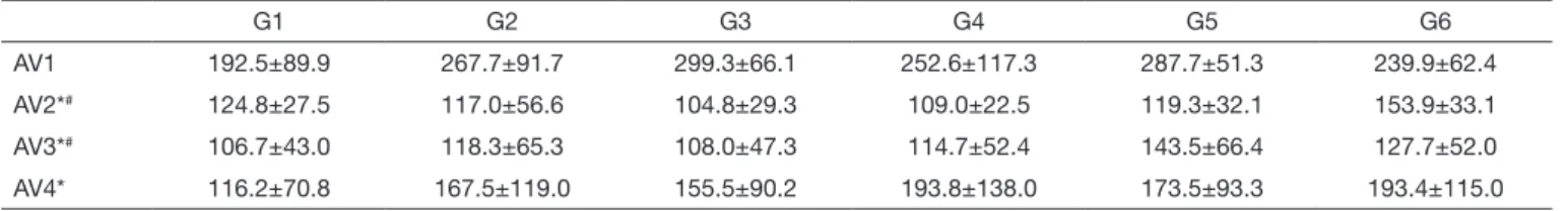
During nociception evaluation there have been signiicant diferences at nerve compression site (F(2.3; 69.8)=49.8, p<0.001). Diferences were not among groups (p>0.05) but rather among evaluations being AV1 higher than remaining evaluations (p<0.001). AV4 was signiicantly higher than AV2 (p=0.038) and AV3 (p=0.002) (Table 1).
here have also been signiicant diferences in nociceptive evalu-ation of the palmar region (F(1.9; 55.9)=14.1, p<0.001). Similar to what has been observed at compression site, diferences were not among groups (p>0.05) but rather among evaluations, being AV1 higher than remaining evaluations (p<0.001) (Table 2). here have been signiicant diferences in edema evalua-tion (F(3; 90)=11.3, p<0.001). Again, diferences were not among groups (p>0.05) but rather among evaluations, being AV1 lower than AV2 (p=0.005) and AV2 higher than AV3 (p=0.003) and AV4 (p>0.001) (Table 3).
DISCUSSION
PRF has been especially studied and used in dentistry, due to its efects on speeding repair by the release of growth factors13,
and also for being easy to use and prepare14. Our study aimed
at looking for other ways to use it, such as peripheral nerve injuries; however, regardless of the group, PRF alone or as-sociated to physical exercise has not changed nociception and edema. Lack of positive results was also observed in a study evaluating PRF to cover gingival recesses in the central region of jaws of 10 volunteers15. his was also observed with regard
to inefective bone regeneration in chronic periodontitis pa-tients16. On the other hand, other authors have reported PRF
eicacy on bone regeneration of patients with intraoral bone defects after cystic enucleation17.
In a study with PRF associated or not to ceramics for cal-varium bone repair of rabbits, authors have reported that PRF had positive efect on bone formation when used alone or in combination2. Other authors, however, when evaluating
the efects of PRF on rotator cuf insertion regeneration of rats submitted to bilateral tenotomy and supraspinous repair, have observed more tensile strength on animals submitted to PRF. Repair characteristic, however, was compatible with i-brovascular tissue, suggesting a possible inhibitory efect of rotator cuf healing18. A study evaluating PRF in cavities due
to third molar extraction has shown no diferences on bone tissue repair, however pain reduction may suggest soft tissue repair4. Another study, also with third molar extractions, has
shown improved edema without however decreased pain
in-Table 1. Mean and standard deviation of linching when pressure (gf) was applied to nerve compression site
G1 G2 G3 G4 G5 G6
AV1 192.5±89.9 267.7±91.7 299.3±66.1 252.6±117.3 287.7±51.3 239.9±62.4
AV2*# 124.8±27.5 117.0±56.6 104.8±29.3 109.0±22.5 119.3±32.1 153.9±33.1
AV3*# 106.7±43.0 118.3±65.3 108.0±47.3 114.7±52.4 143.5±66.4 127.7±52.0
AV4* 116.2±70.8 167.5±119.0 155.5±90.2 193.8±138.0 173.5±93.3 193.4±115.0
*Signiicant difference as compared to AV1. # Signiicant difference as compared to AV4.
Table 2. Mean and standard deviation of linching when pressure (gf) was applied to palmar region
G1 G2 G3 G4 G5 G6
AV1 188.6±65.8 243.2±55.2 255.6±44.8 278.5±43.1 308.4±43.5 252.0±44.2
AV2* 235.5±70.9 175.4±44.7 203.9±59.9 205.8±30.2 154.8±30.5 202.7±52.1
AV3* 181.4±29.8 175.0±47.5 175.7±18.9 178.7±28.4 164.4±27.1 164.4±40.5
AV4* 176.2±51.0 189.9±106.5 184.9±92.9 162.7±80.8 161.3±47.7 208.0±88.1
*Signiicant difference as compared to AV1.
Table 3. Mean and standard deviation of limb edema, by plethysmometry (mL)
G1 G2 G3 G4 G5 G6
AV1 1.56±0.30 1.66±0.26 1.47±0.24 1.34±0.24 1.39±0.46 1.29±0.21
AV2* 1.77±0.23 1.93±0.33 1.81±0.34 1.93±0.61 1.50±0.41 1.73±0.25
AV3# 1.60±0.57 1.39±0.43 1.56±0.13 1.33±0.25 1.32±0.54 1.39±0.35
AV4# 1.36±0.16 1.43±0.36 1.47±0.09 1.39±0.08 1.08±0.48 1.22±0.20
124
Karvat J, Kakihata CM, Peretti AL, Bernardino GR, Silva JL and Bertolini GR
Rev Dor. São Paulo, 2016 apr-jun;17(2):121-4
tensity19. Although mean linching in G1 being lower than
those for other groups, statistical analysis has shown no difer-ences among groups, but only among evaluations.
Our study had several limitations, such as the use of interme-diate PRF fraction, which may not have the highest amount of available growth factors20; centrifugation time might have
been short to be able to potentiate growth factors release, as shown by a recent study which has pointed to advantages of centrifuging for 12 minutes as compared to 10 minutes21, in
addition to the evaluation of just two signs of the inlamma-tion/repair process. It has also to be stressed that PRF associ-ated or not to other modalities is still a new technique with in vitro22 and in vivo15-17 studies showing contradictory results,
thus requiring further studies with regard to its efects.
CONCLUSION
O uso isolado ou associado da FRP com exercícios físicos não produziu alterações na nocicepção e no edema, no modelo de compressão de nervo mediano.
PRF alone or associated to physical exercises has not changed nociception and edema in a median nerve compression model.
REFERENCES
1. Martínez CE, Smith PC, Palma Alvarado VA. he inluence of platelet-derived pro-ducts on angiogenesis and tissue repair: a concise update. Front Physiol. 2015;6:290. 2. Acar AH, Youlcu Ü, Gül M, Keles A, Erdem NF, Altundag Kahraman A. Micro-com-puted tomography and histomorphometric analysis of the efects of platelet-rich ibrin on bone regeneration in the rabbit calvarium. Arch Oral Biol. 2015;60(4):606-14. 3. Choukroun J, Diss A, Simonpieri A, Girard MO, Schoeler C, Dohan SL, et al.
Platelet-rich ibrin (PRF): a second-generation platelet concentrate. Part IV: Clini-cal efects on tissue healing. Oral Surg Oral Med Oral Pathol Oral Radiol Endod. 2006;101(3):e56-60.
4. Kumar YR, Mohanty S, Verma M, Kaur RR, Bhatia P, Kumar VR, et al. Platelet-rich ibrin: the beneits. Br J Oral Maxillofac Surg. 2015;54(1):57-61.
5. Moraschini V, Barboza ED. Use of platelet-rich ibrin membrane in the
treat-ment of gingival recession: a systematic review and meta-analysis. J Periodontol. 2016;87(3)281-90.
6. di Lauro AE, Abbate D, Dell’Angelo B, Iannaccone GA, Scotto F, Sammartino G. Soft tissue regeneration using leukocyte-platelet rich ibrin after exeresis of hyperplastic gingival lesions: two case reports. J Med Case Rep. 2015;9:252.
7. Bickel KD. Carpal tunnel syndrome. J Hand Surg. 2010;35(1):147–52.
8. Gupta R, Rowshan K, Chao T, Mozafar T, Steward O. Chronic nerve compression induces local demyelination and remyelination in a rat model of carpal tunnel syndro-me. Exp Neurol. 2004;187(2):500-8.
9. Koltyn KF. Analgesia following exercise. A review. Sport Med. 2000;29(2):85-98. 10. Chen JJ, Lue JH, Lin LH, Huang CT, Chiang RP, Chen CL, et al. Efects of
pre--emptive drug treatment on astrocyte activation in the cuneate nucleus following rat median nerve injury. Pain. 2010;148(1):158-66.
11. Ding XG, Li SW, Zheng XM, Hu LQ, Hu WL, Luo Y. he efect of platelet-rich plasma on cavernous nerve regeneration in a rat model. Asian J Androl. 2009;11(2):215-21. 12. Ehrenfest DM, Lemo N, Jimbo R, Sammartino G. Selecting a relevant animal model
for testing the in vivo efects of Choukroun’s platelet-rich ibrin (PRF): rabbit tricks and traps. Oral Surg Oral Med Oral Pathol Oral Radiol Endod. 2010;110(4):413-6. 13. Kobayashi E, Flückiger L, Fujioka-Kobayashi M, Sawada K, Sculean A, Schaller B,
et al. Comparative release of growth factors from PRP, PRF, and advanced-PRF. Clin Oral Investig. 2016. Epub ahead of print.
14. Khorshidi H, Raooi S, Bagheri R, Banihashemi H. Comparison of the mechanical properties or early leuckocyte- and Platelet-Rich Fibrin versus PRGF/Endoret mem-branes. Int J Dent. 2016;1849207.
15. Rajaram V, hyegarajan R, Balachandran A, Aari G, Kanakamedala A. Platelet Rich Fibrin in double lateral sliding bridge lap procedure for gingival recession coverage: an original study. J Indian Soc Periodontol. 2015;19(6):665-70.
16. Gamal AY, Ghafar KAA, Alghezwy OA.Crevicular luid growth factors release proile following the use of Platelets Rich Fibrin (PRF) and Plasma Rich Growth Factors (PRGF) in treating periodontal intrabony defects (randomized clinical trial). J Perio-dontol. 2016. [Epub ahead of print].
17. Meshram V, Lambade P, Meshram P, Kadu A, Tiwari M. he autologous platelet rich ibrin: A novel approach in osseous regeneration after cystic enucleation: A pilot study. Indian J Dent Res. 2015;26(6):560-4.
18. Hasan S, Weinberg M, Khatib O, Jazrawi L, Strauss EJ. he efect of platelet-rich i-brin matrix on rotator cuf healing in a rat model. Int J Sport Med. 2016;37(1):36-42. 19. Ozgul O, Senses F, Er N, Tekin U, Tuz HH, Alkan A, et al. Eicacy of platelet rich ibrin in the reduction of the pain and swelling after impacted third molar surgery : randomized multicenter split-mouth clinical trial. Head Face Med. 2015;11(1):37. 20. Nishimoto S, Fujita K, Sotsuka Y, Kinoshita M, Fujiwara T, Kawai K, et al. Growth
factor measurement and histological analysis in platelet rich ibrin: a pilot study. J Maxillofac Oral Surg. 2015;14(4):907-13.
21. Eren G, Gürkan A, Atmaca H, Dönmez A, Atilla G. Efect of centrifugation time on growth factor and MMP release of an experimental platelet-rich ibrintype product. Platelets. 2016. [Epub ahead of print].