ISSN/$–see front matter © 2013 Sociedade Brasileira de Ortopedia e Traumatologia. Published by Elsevier Editora Ltda. All rights reserved. Rev Bras Ortop. 2013;48(1):104-107
www.rbo.org.br/
*Corresponding author at: Hospital Geral de Fortaleza, Serviço de Radiologia e Diagnóstico por Imagem, Rua Ávila Goulart, 900, Papicu, Fortaleza, CE, Brazil. CEP: 60115-290.
E-mail: pacheco.licia@gmail.com A RT I C L E I N F O
Article history:
Received September 19 2009 Approved October 5 2012
Keywords: Syndrome Hand Injuries Ultrasonography
Magnetic Resonance Imaging
Case Report
Hypothenar hammer syndrome: case report and literature
review
Márcia Maria Muniz de Queiroz,
1Lícia Pachêco Pereira,
2*Clarissa Gondim Picanço,
3Rodrigo de Castro Luna,
2Fabrício da Silva Costa,
4Cláudio Régis Sampaio Silveira
21Resident Physician (R2) in Nephrology, University Hospital of Brasília, Brasília, DF, Brazil.
2Radiologist in the Radiology Service, General Hospital of Fortaleza, and Titular Member of the Brazilian College of Radiology,
Fortaleza,CE, Brazil.
3Resident Physician (R1) in Medical Genetics, University of São Paulo, Ribeirão Preto, SP, Brazil.
4PhD in Gynecology and Obstetrics from the University of São Paulo (USP); Postdoctoral Attending Physician in the Royal Women’s
Hospital, Melbourne, Australia.
Work performed in the Imaging Diagnostics Service, General Hospital of Fortaleza, Fortaleza, CE, Brazil.
a b s t r a c t
Case report of a 69 year-old patient, with history of repetitive trauma events in the wrist, clinically simulating tenosynovitis, being held with Doppler Ultrasound and Magnetic Nuclear Resonance, which showed ulnar artery thrombosis. The accurate diagnosis of the hammer hypothenar disease through those tests enable an early intervention, improving the prognosis of patients affected by this rare disease.
© 2013 Sociedade Brasileira de Ortopedia e Traumatologia. Published by Elsevier Editora Ltda. All rights reserved.
Rev Bras Ortop. 2013;48(1):104-107
105
Introduction
Thrombosis of the ulnar artery, a rare condition that is often secondary to trauma at the hypothenar eminence, may be attributable to a single traumatic event or to repeated trauma in this region. In the latter case, it is named hypothenar hammer syndrome,1,2 and it results in degeneration and thrombosis of the ulnar artery at the level of the hamate.3
It was first described by Von Rosen in 1934, and has been reported in cyclists, tennis players, golf players and practitioners of a variety of other sports.2 It may also be associated with fractures of the hamate, arteritis, use of walking sticks and anomalous muscles.4,5
Because of the trauma mechanism, the cases reported in the literature have predominantly occurred in males4 and in smokers.6
The signs and symptoms are very variable. There may be intolerance of cold, cyanosis, hyperalgesia, Raynaud’s phenomenon, ulnar neuropathy, a mass in the hypothenar region or even necrosis of the fingers.4 Demonstration of a positive Allen test for occlusion of the ulnar artery is highly suggestive of this condition.4
It is fundamentally important to make a differential diagnosis in relation to Raynaud’s disease, thromboangiitis obliterans, scleroderma, giant cell arteritis, thoracic outlet syndrome, ulnar neuropathy and thromboembolic phenomena.4
The preferred examination for confirming a clinical suspicion is arteriography, although Doppler ultrasonography (US) and nuclear magnetic resonance (NMR) may be useful in making the diagnosis.
To avoid traumatic etiological factors, anticoagulants, vasodilators and thrombolytic agents have been used in therapy for this condition. Blockade of the sympathetic nerves, sympathectomy and, in acute and severe cases, embolectomy with a catheter have been reported. Surgical treatment can be performed, consisting of resection and vein grafting using a microsurgical technique.
Case report
The patient was a 66-year-old man who was a smoker and hypertensive, with a history of regularly suffering small traumatic events. In particular, 15 days before the current symptoms appeared, he had suffered bruising trauma on the distal third of the right forearm, and started to present increased volume and pain in this region. His condition was clinically diagnosed as post-traumatic tenosynovitis, but even after clinical treatment, physiotherapy and anti-inflammatory drug administration, he did not present any improvement. He was then referred to our service for NMR and Doppler US to be performed on his wrist.
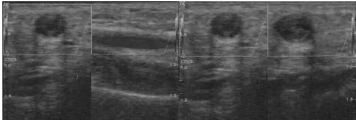
Doppler US showed that the ulnar artery presented significant parietal thickening, starting in the middle third of the forearm. In the distal third, echogenic content was observed inside the forearm, and no flow was seen in this
region. Distally to the thrombus in the palmar region, flow in the caudal-cranial direction was noted, probably relating to collateral circulation (Fig. 1).
NMR showed that the ulnar artery presented increased caliber and thickened walls. There was a heterogenous signal with predominance of hypersignal in T1, hypersignal in T2 and parietal highlighting after injection of paramagnetic contrast medium. The ulnar artery was around 5 cm long, with a diameter of 1 cm (Fig. 2).
The patient underwent conservative treatment, with recommendations to stop smoking, avoid further local trauma, control the hypertension and use anticoagulants. At a control echographic examination performed around three months after the diagnosis, on an occasion when the patient was asymptomatic, chronic thrombosis was also seen, and the vascularization of the palmar arch remained collateral. The patient refused to undergo any further control imaging examinations, but in a telephone contact 12 months later, he said that the symptoms had resolved spontaneously, without recurrences during this period.
Fig. 1 - Doppler ultrasound on the ulnar artery. It was found to be dilated, with thickened walls, although with flow present, in the middle third of the forearm (A), while in the distal third (B) there was echogenic material inside the vessel (thrombus) and absence of flow.
106
Rev Bras Ortop. 2013;48(1):104-107professional sports players can also return to their activities sooner through surgical treatment.12 Previous studies have also described nonsurgical approaches for patients with ischemia of the fingers, consisting of combined conservative treatments with intra-arterial infusion of vasodilators,13 distal microcatheterization and embolization.14,15 Surgical treatments include excision of the segment of the ulnar artery with venous grafting,16 resection of the aneurysm with end-to-end anastomosis, thoracic sympathectomy and, if this is the case, amputation of fingers presenting necrotic ulcers.11 Temming et al.17 recently described autologous use of the descending branch of the lateral femoral circumflex artery as an option for venous grafting in reconstruction of the ulnar artery, with positive results. Marie et al.18 reported that the rate of symptom recurrence within 12 months (mean of 11 months) was 27.7% in a retrospective study on 47 patients, among whom the majority underwent conservative treatment, thus emphasizing the importance of adequate medium and long-term control among these patients.
Conclusion
Thrombosis of the ulnar artery, which is a condition that is difficult to clinically diagnose in isolation because of its nonspecific clinical characteristics, is associated with intense morbidity in the patients affected, when not adequately diagnosed and not treated early on.
Demonstration of thrombosis in the distal ulnar artery by means of ultrasonography or NMR enables precise diagnosis of hypothenar hammer disease, when considered in combination with the frequent clinical manifestations of this affection, thereby enabling early intervention and improving the prognosis for patients affected by this rare condition.
Conflicts of interest
The authors declare that there was no conflict of interests in conducting this study.
R E F E R E N C E S
1. Troum SJ, Floyd WE 3rd, Saap J. Ulnar artery thrombosis: a 6-year experience. J South Orthop Assoc. 2001;10(3):147-54. 2. Cooke RA. Hypothenar hammer syndrome: a discrete
syndrome to be distinguished from hand-arm vibration syndrome. Occup Med (Lond). 2003;53(5):320-4.
3. Okereke CD, Knight S, McGowan A, Coral A. Hypothenar hammer syndrome diagnosed by ultrasound – case report. Injury. 1999;30(6):448-9.
4. Fernandes CH, Tinós MS, Meirelles LM. Trombose da artéria ulnar por digitação: relato de caso. Rev Bras Ortop. 1998;33(11):911-3.
5. Mueller LP, Mueller LA, Degreif J, Rommens PM. Hypothenar hammer syndrome in a golf player – a case report. Am J Sport Med. 2000;28(5):741-5.
Discussion
Approximately 50% of the cases of ulnar thrombosis are not diagnosed initially, as was also the case with our patient. This restricts the therapeutic arsenal and worsens the prognosis for these patients.7 The clinical condition varies greatly in intensity and form,4 and Raynaud’s phenomenon may be presented, in which pallor and cyanosis may appear, while hyperemia has not yet been described.8 There may be intolerance of cold conditions, cyanosis, pain, ischemia of the fingers, symptoms of ulnar neuropathy4 and a mass or heat in the hypothenar region,4 and the condition my reach irreversible gangrene in the fingers.9
The Allen test should be used for clinically confirming the diagnosis,4 which may be negative in 17% of the cases.2 This consists of simultaneously occluding the radial and ulnar arteries and subsequently releasing the pressure on either of the two arteries, which should cause immediate filling of this artery and return of coloration to the hand. The test is said to be positive when the color does not return to the whitened hand.
The imaging diagnosis is generally done by means of arteriography, Doppler US or NMR. The preferred examination for confirming the diagnosis is arteriography, which shows occlusion or aneurysm of the distal ulnar artery.2,4 Doppler US has been shown to provide excellent diagnostic sensitivity, and to enable perfect viewing of the radial and ulnar arteries and the palmar arch, thus making it possible to identify aneurysms,10 Doppler US is capable of distinguishing hypothenar hammer syndrome from other causes of ischemia of the fingers.2 This examination is useful for evaluating reperfusion after surgical reconstitution6 and for following up these patients. Furthermore, it has many advantages due to its easy accessibility, relatively low cost, safety and noninvasiveness.3 NMR is also a noninvasive examination, which uses relatively safe paramagnetic contrast without irradiation, in a multiplanar manner with high resolution for soft tissues. This enables detailed viewing of the vascular structures and their alterations, such as intraluminal thrombi, and it is also very useful for detecting anatomical variations in the small palmar muscles. However, because of its high cost, it is not used routinely.
Histopathological examination generally hyperplasia of the tunica intima, with fragmentation along the internal elastic lamina and luminal occlusion with organized thrombi, with or without formation of aneurysms.10
Rev Bras Ortop. 2013;48(1):104-107
107
6. De Monaco D. de, Fritsche E, Rigoni G, Schlunke S, von Wartburg U. Hypothenar hammer syndrome – retrospective study os nine cases. J Hand Surg Br. 1999;24(6):731-4. 7. Mehlhoff TL, Wood MB. Ulnar artery thrombosis and the role
of interposition vein grafting: patency with microsurgical technique. J Hand Surg Am. 1991;16(2):274-8.
8. Pineda CJ, Weisman MH, Bookstein JJ, Saltzstein SL.
Hypothenar hammer syndrome: from of reversible Raynaud’s phenomenon. Am F Med. 1985;79(5):561-70.
9. Conn J Jr, Bergan J, Bell J. Hypothenar hammer syndrome: posttraumatic digital ischemia. Surgery. 1970;68:1122-8. 10. Dethmers RS, Houpt P. Surgical management of hypothenar
and thenar hammer syndromes: a retrospective study of 31 instances in 28 patients. J Hand Surg Br. 2005;30(4):419-23. 11. Yuen JC, Wright E, Johnson LA, Culp WC. Hypothenar hammer
syndrome: an update with algorithms for diagnosis and treatment. Ann Plast Surg. 2011;67(4):429-38.
12. Swanson KE, Bartholomew JR, Paulson R. Hypothenar hammer syndrome: a case and brief review. Vasc Med. 2012;17(2):108-15.
13. Sharma R, Ladd W, Chaisson G, et al. Images in cardiovascular medicine: hypothenar hammer syndrome. Circulation. 2002;105(13):1615-6.
14. Bakhach J, Chahide N, Conde A. Hypothenar hammer syndrome: management of distal embolization by intraarterial fibrinolytics. Chir Main. 1998;17(3):215-20. 15. Abdel-Gawad EA, Bonatti H, Housseini AM, Maged IM,
Morgan RF, Hagspiel KD. Hypothenar hammer syndrome in a computer programmer: CTA diagnosis and surgical and endovascular treatment. Vasc Endovascular Surg. 2009;43(5):509-12.
16. Lifchez SD, Higgins JP. Long-term results of surgical treatment for hypothenar hammer syndrome. Plast Reconstr Surg. 2009;124(1):210-6.
17. Temming JF, van Uchelen JH, Tellier MA. Hypothenar hammer syndrome: distal ulnar artery reconstruction with autologous descending branch of the lateral circumflex femoral artery. Tech Hand Up Extrem Surg. 2011;15(1):24-7.