Is quantitative diffusion-weighted MRI a valuable technique
for the detection of changes in kidneys after extracorporeal
shock wave lithotripsy?
_______________________________________________
Elif Hocaoglu1, Ercan Inci1, Sibel Aydin3, Dilek Hacer Cesme1 , Nadir Kalfazade2
1Department of Radiology, Bakirkoy Dr. Sadi Konuk Research and Training Hospital, Istanbul, Turkey;
2Department of Urology, Bakirkoy Dr. Sadi Konuk Research and Training Hospital, Istanbul, Turkey;
3Department of Radiology, Haydarpasa Numune Research and Training Hospital, Istanbul, Turkey;
ABSTRACT ARTICLE INFO
______________________________________________________________ ______________________
Objective: The aim of this study was to evaluate the capability and the reliability of diffusion-weighted imaging (DWI) in the changes of kidneys occurring after extracor-poreal shock wave lithotripsy (ESWL) treatment for renal stones.
Materials and Methods: A total of 32 patients who underwent ESWL treatment for renal stone disease between June and December 2011 were enrolled in this prospective study. Color Doppler ultrasonography (CDUS) and DWI were performed before and within 24 hours after ESWL. DWI was obtained with b factors of 0, 500 and 1000 s/ mm² at 1.5 T MRI. Each of Resistive index (RI) and ADC values were calculated from the three regions of renal upper, middle and lower zones for both of the affected and contralateral kidneys. Paired sample t test was used for statistical analyses.
Results: After ESWL, the treated kidneys had statistically significant lower ADC values in all different regions compared with previous renal images. The best discriminative parameter was signal intensity with a b value of 1000 s/mm2. The changes of DWI after ESWL were noteworthy in the middle of the treated kidney (p<0.01). There were no significant difference between RI values in all regions of treated and contralateral kidneys before and after treatment with ESWL (p>0.05).
Conclusion: DWI is a valuable technique enables the detection of changes in DWI after ESWL treatment that may provide useful information in prediction of renal damage by shock waves, even CDUS is normal.
Key words:
Lithotripsy; Kidney; Calculi; Urinary Tract
Int Braz J Urol. 2015; 41: 139-46
_____________________
Submitted for publication: March 10, 2013
_____________________
Accepted after revision: June 30, 2014
INTRODUCTION
The prevalence of stone disease shows an increase in the developing world. The invention and development of extracorporeal shock wave li-tothripsy (ESWL) in the last 25 years has brought an effective perspective to the treatment of urinary stone diseases as a non-invasive method. It is an optimal technique that may save time and resour-ces and decrease the suffering of patients
kid-ney. The side effects of shock waves are intra renal hematomas, subcapsular hematomas, subcapsular fluid collections, perinephric soft tissue stranding, cardiac arrhythmias, fascial thickening and loss of corticomedullary junction. Histopathologic chan-ges are observed in the form of diffuse interstitial fibrosis, focal calcification, glomerular hyaniliza-tion and sclerosis, damaged capillary vessels and acellular scars ranged from cortex to medulla. The-se changes results in 0.01-20% loss of renal func-tion (1-4). Color Doppler ultrasonography (CDUS) has been used to detect the effects of ESWL on the kidneys as a noninvasive technique (5). Also, the kidney is a suitable organ for diffusion studies be-cause of its high blood flow and its fluid trans-port function. Besides, it’s a noninvasive technique with no need of contrast agents. Recent ultrafast sequences made diffusion studies more applicable with shorter examination times and fewer motion artifacts (6, 7). The purpose of our study was to de-tect the changes of kidneys after ESWL treatment by diffusion weighted imaging (DWI) and CDUS and to evaluate whether DWI has advantages in prediction of renal damage due to shock waves.
MATERIALS AND METHODS
The study protocol was approved by the local Ethics Committee. Written informed consents were obtained from all participants. During the pe-riod of June to December 2011, a total of 32 conse-cutive patients 23-68 years old (mean age, 41±0.6 years old), diagnosed with nephrolithiasis by abdo-minal X-ray and US, underwent ESWL and were enrolled in the study group. Exclusion criteria in-cluded urinary system infection, marked collecting system dilatation, renal parenchymal disease such as diabetes mellitus and hypertension.
ESWL Protocol
ESWL was performed by piezoelectric li-thotriptor, Wolf Piezolith 3000 with F3 triple fo-cus (2.5 x 16 mm / 4.0 x 25 mm / 6.0 x 30 mm). Outline X-ray and inline ultrasound was used for stone location. The stones ranged in size from 9 x 6 mm to 18 x 14 mm. Three sessions of ESWL (with interval of 3 days) were applied to all pa-tients and the number of pulses ranged between
1600 and 2000 (mean 1880). Main supply of li-thotriptor was 220-240 V/ 50-60 Hz with power consumption 1000 VA. Energy density and peak pressure applied to all patients ranged from 0.84 to 1.14 mJ / mm² (mean 0.98 mJ / mm²) and 36 to 112 MPa (mean 88 MPa), respectively. DWI and CDUS were performed before and within 24 hours after 3 sessions of ESWL in all patients.
Color Doppler Ultrasound (CDUS)
All the Doppler sonographic measure-ments were performed by the same operator using a Toshiba Aplio SSA-770A/80 ( Toshiba Medi-cal Systems Corporation, Tokyo, Japan ) scanner using with Convex array probe PVT-375AX (1.9-6 mhz) before and after ESWL in the first day. The resistive index ( RI ) measurements were taken at an interlobar or arcuate artery from the three re-gions of kidney (upper, middle and lower zones) for both the affected and contralateral kidneys.
Diffusion Weighted Imaging (DWI)
DW images were obtained with a 1.5 T whole body system (Avanto; Siemens, Erlangen, Germany) with a 33 mT/m maximum gradient ca-pability using an eighteen channel phased-array body coil. No specific preparatory measures were required such as fasting or drinking prior to the examination. Also, no oral or intravenous contrast agent were administered. Axial diffusion weighted single-shot spin-echo echo-planar sequence with chemical shift selective fat-suppression technique ( TR/TE 4738/80; matrix 192 x 192; slice numbers 36; slice thickness 5 mm; interslice gap 30%; FOV 40 cm; averages 5; acquisition time approximately 4 minutes; PAT factor 2; PAT mode generalized au-tocalibrating partially parallel acquisition –GRA-PPA-) was performed. DW images were obtained with b-factors of 0, 500 and 1000 sec/mm². The phase encode direction was set antero-posteriorly in both sequences. All slices were acquired from the superior pole to the bottom of the kidneys. All images were obtained without restriction of fluid intake and without breath-holding.
Imaging Interpretation
24 hours after ESWL. Measurements were taken three times for each region repeatedly in the three different regions of affected and contralateral kid-neys ( middle zone, upper, and lower poles). Ave-raged values were recorded for each region as the RI value.
DW images were interpreted by two readers at random order in consensus. All were of diag-nostic quality with no exclusion. The DWI datasets were transferred to an independent workstation (Leonardo console, software version 2.0; Siemens AG Medical Solutions, Forchheim, Germany) for postprocessing, and the ADC maps were recons-tructed. The kidneys were prospectively evaluated quantitatively with the DW sequences before and after ESWL. We measured DWI signal intensity to quantify visual conspicuity and the level of de-tectability. A large circular region of interest (ROI) was placed at the corticomedullary junction for the measurement of ADC values. ADC values were obtained on the different sites of kidneys. For each ADC calculation, 3 ROI measurements were taken and the average value was accepted. All measure-ments were repeated at different b values (0, 500, 1000 s/mm²). Each patient’s ADC values were re-corded in square milimeters per second quantitati-vely (Figures 1a, 1b, 2a, 2b, 2c and 2d).
Statistical analysis
Statistical analysis was performed with the NCSS (Number Cruncher Statistical System) 2007&PASS 2008 Statistical Software (Utah, USA) packages program. The paired sample t-test was used to compare RI and ADC values before and after ESWL. A p value of less than 0.05 was con-sidered statistically significant.
RESULTS
Color Doppler Ultrasound
Comparison of the mean RI values before and after ESWL showed no statistically significan-ce for both ipsilateral and contralateral kidneys (p>0.05). The mean RI values pre and post ESWL were 0.58±0.05 and 0.59±0.04 in the ipsilateral kidney, 0.58±0.05 and 0.59±0.05 in the contra-lateral kidney, respectively. The changes in the RI
Figure 1 - 56 years old male patient. Apparent diffusion coefficient (ADC) maps of treated and contralateral kidneys before (Figure-1a) and after (Figure-1b) treatment with
ESWL. The ADC value is decreased from 2.29x10⁻³ mm2/sn
to 1.92x10⁻³ mm2/sn in treated kidney. The ADC values of
the contralateral kidney are 2.30x10⁻³ mm2/sn and 2.23x10⁻³
mm2/sn before and after ESWL treatment, respectively.
values before and after ESWL in certain regions are shown in Table-1.
Diffusion weighted imaging
The mean ADC values of ipsilateral kid-neys with b=0, b=500 and b=1000 values before and after ESWL treatment are shown in Table-2.
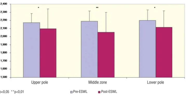
Ipsilateral kidneys had statistically signi-ficant lower ADC values in all different regions compared to contralateral kidneys after ESWL (Figure-3). The changes after ESWL were
conspi-A
cuous in the middle zone (p<0.01). The best discri-minative parameter was signal intensity with a b value of 1000 (Figure-4).
In contralateral kidneys, minimum and maximum ADC values of renal parenchyma ran-ged from 1.98 to 2.47x10⁻³ mm2/sn.
DISCUSSION
ESWL has dominated the treatment of re-nal stone disease since its introduction in 1980. It
has been a major advance in Urology which is the fragmentation of stone by means of acoustic sho-ck waves created by an extracorporeal source. It is non-invasive, effective and very well tolerated. The shock waves generated by ESWL cause frag-mentation of renal calculi by exerting on the brit-tle calculi mechanical stresses sufficient to exceed the tensile strength of the stone (1-4). Although the focal point of the shock wave is centered on the renal stone, the waves must pass through the soft tissues of the back and the renal
parenchy-Figure 2 - 44 years old male patient. Apparent diffusion coefficient (ADC) maps and signal intensities (with b values of 1000 s/mm²) of treated (left) and contralateral kidneys before (Figures 2a and 2b) and after (Figures 2c and 2d) treatment with
ESWL. The ADC value decreased from 2.17x10⁻³ mm2/sn (Figure-2a) to 1.64x10⁻³ mm2/sn (Figure-2c) in the treated kidney.
Signal intensity of the treated kidney with b values of 1000 s/mm² increased from 27.0 s/mm2 (Figure-2b) to 46.6 s/mm2
(Figure-2d). The ADC values of the contralateral kidney are 2.16x10⁻³ mm2/sn (Figure-2a) and 2.21x10⁻³ mm2/sn (Figure-2c)
before and after ESWL treatment, respectively. Signal intensities of the contralateral kidney with b values of 1000 s/mm² are 27.7 s/mm2 (Figure-2b) and 30.7 s/mm2 (Figure-2d) before and after ESWL treatment, respectively.
A
C
B
Table 1 - The RI values of treated kidneys before and after ESWL in certain regions.
RI p
Pre-ESWL Post-ESWL
Mean±SD Mean±SD
Upper Pole 0.58±0.05 0.59±0.04 0.375
Middle Zone 0.58±0.05 0.59±0.04 0.204
Lower Pole 0.58±0.04 0.59±0.04 0.307
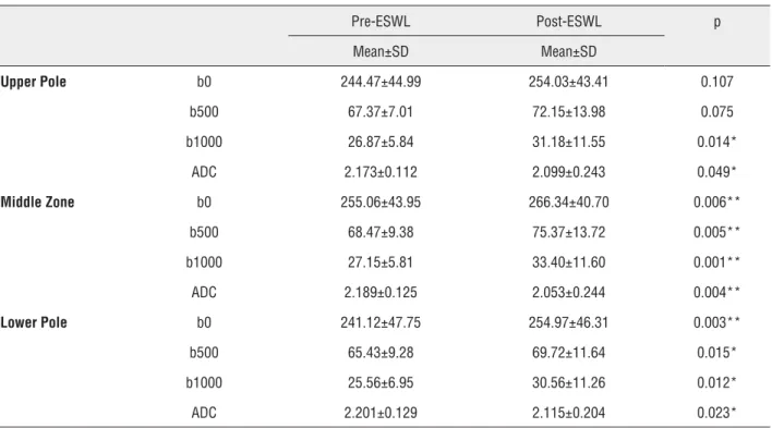
Table 2 - The mean ADC values of ipsilateral kidneys with b-0, b-500 and b-1000 values before and after ESWL treatment.
Pre-ESWL Post-ESWL p
Mean±SD Mean±SD
Upper Pole b0 244.47±44.99 254.03±43.41 0.107
b500 67.37±7.01 72.15±13.98 0.075
b1000 26.87±5.84 31.18±11.55 0.014*
ADC 2.173±0.112 2.099±0.243 0.049*
Middle Zone b0 255.06±43.95 266.34±40.70 0.006**
b500 68.47±9.38 75.37±13.72 0.005**
b1000 27.15±5.81 33.40±11.60 0.001**
ADC 2.189±0.125 2.053±0.244 0.004**
Lower Pole b0 241.12±47.75 254.97±46.31 0.003**
b500 65.43±9.28 69.72±11.64 0.015*
b1000 25.56±6.95 30.56±11.26 0.012*
ADC 2.201±0.129 2.115±0.204 0.023*
ADC (x10-3 mm2/sn)
*p<0.05; **p<0.01
ma before reaching the calculus. Therein lies the potential for damage to these tissues. Kaude and co-workers (8) hypothesized that the spherical sho-ck waves passing through the kidney during ESWL produce renal contusions. Lang et al. (9) have defi-ned renal contusion as interstitial extravasation of small amounts of urine and blood and interstitial edema. However, the treatment with shock waves carries the risk of acute injury with the potential for long-term adverse effects (8-12). Many techniques have been used to show the effects of ESWL on the kidneys, such as US, CT, MRI, laboratory findings.
Figure 3 - The mean ADC values before and after ESWL treatment in upper pole, middle zone and lower pole of treated kidneys.
Figure 4 - Signal intensities with b=1000 s/mm2 before and after ESWL treatment in upper pole, middle zone and lower pole
of treated kidneys.
mean+SD
mean+SD
p<0,05 **p<0,01
p<0,05 **p<0,01
Pre-ESWL
Pre-ESWL
Post-ESWL
Post-ESWL
The values of ADC
The values of DW-MRI with b1000 Middle zone
Middle zone
Lower pole
Lower pole Upper pole
RI values in the treated region within 3 hours after ESWL were significantly higher in the group of patients aged >60 years.
Baumgartner et al. (10) showed the presence of abnormality in 74% of patients studied after ESWL by MR imaging. According to their results, MR ima-ging may be a very sensitive method to image these pathologic alterations occurring in the kidney.
The results of diffusion-weighted MRI in the kidney are still preliminary, and more resear-ch should be done (16). DWI provides information on perfusion and diffusion simultaneously in any organ; it can be used to differentiate normal and abnormal structures of tissues and it might help in the characterization of various abnormalities. Thus, calculating the ADC of low and high b va-lues separately provides more specific information on kidney function (17). Thoeny et al. (18) showed the decrease in the ADC values of kidneys in pa-tients with chronic renal failure and pyelonephri-tis. Chan et al. studied DWI to differentiate betwe-en hydronephrosis and pyonephrosis (7). Powers et al. (19) used a spin-echo diffusion-weighted se-quence with respiratory triggering in dog kidneys, and found a drop in ADC in the unilateral renal ar-tery stenosis that correlated with renal blood flow. Müller et al. (20) demonstrated that acute ureteral obstruction shows a quick decrease in ADC.
Researches have been processing about the long term side affects after ESWL. These possible adverse effects include a decrease in renal func-tion, an increased rate of new stone formation and an increase in systemic blood pressure. Yokoya-ma and friends (21) have witnessed 1.5% of new initiative hypertension on normotensive patients after one year of ESWL. It is realized that retros-pectively more than 8% of patients have suffered hypertension within the following two years (11). Uozumi et al. (22) determined a decrease of renal blood flow and delay of radionuclide urinary cle-arance in the affected kidney from ESWL.
In our study, renal changes after ESWL treatment were demonstrated with DWI in spite of a normal CDUS. After ESWL session, ipsilate-ral kidneys had statistically significant lower ADC values in all certain regions compared to contra-lateral kidneys. This condition can be explained by the decrease of renal blood flow or renal
mi-crocontusion arising from shock waves. ESWL centers routinely apply shock wave dosages in the range of 1500 to 2500 per treatment session. Since the possibility of an adverse effect of ESWL on the blood pressure and the lack of evidence that effi-cacy is enhanced by the utilization of such high doses of shock wave energy, such practices should be discouraged.
Our data suggest that DWI may be a very useful method for imaging pathologic altera-tions occurring in the kidney after ESWL therapy. Additionally, to our knowledge there has been no previous report relating the DWI findings in the kidneys treated by ESWL. One of the limitations of our study is that all images were obtained wi-thout any specific preparation of the patients such as fasting or drinking. However it’s known that hydration has some effects on the ADC levels even though minimal.
CONCLUSIONS
DWI is a valuable technique for detection of post-ESWL changes in the kidneys even while the Doppler US is normal. Further studies specifically investigating long-term effects are warranted.
CONFLICT OF INTEREST
None declared.
REFERENCES
1. Chaussy C, Schmiedt E, Jocham D, Brendel W, Forssmann B, Walther V. First clinical experience with extracorporeally induced destruction of kidney stones by shock waves. J Urol. 1982;127:417-20.
2. Tomomasa H, Kaneko S, Ogawa K, Satoh S, Muramatsu H, Satoh M, et al. Results of extracorporeal shock wave lithotripsy for the treatment of upper urinary tract stones. Hinyokika Kiyo. 2007;53:771-6.
3. Knoll T, Michel MS, Köhrmann KU, Alken P. Urologic interventional therapy of kidney calculi (I)--extracorporeal shockwave lithotripsy. Ther Umsch. 2003;60:98-102. 4. D’Addessi A, Vittori M, Racioppi M, Pinto F, Sacco E, Bassi
5. Derchi LE, Martinoli C, Pretolesi E, Mancini G, Bottino P, Germinale F, et al. Renal changes from extracorporeal shock-wave lithotripsy: evaluation using Doppler sonography. Eur Radiol 1994;4:41-4.
6. Zhang H, Prince MR. Renal MR angiography. Magn Reson Imaging Clin N Am. 2004;12:487-503.
7. Chan JH, Tsui EY, Luk SH, Fung SL, Cheung YK, Chan MS, et al. MR diffusion-weighted imaging of kidney: differentiation between hydronephrosis and pyonephrosis. Clin Imaging. 2001;25:110-3.
8. Kaude JV, Williams CM, Millner MR, Scott KN, Finlayson B. Renal morphology and function immediately after extracorporeal shock-wave lithotripsy. AJR Am J Roentgenol. 1985;145:305-13.
9. Lang EK, Sullivan J, Frentz G. Renal trauma: radiological studies. Comparison of urography, computed tomography, angiography, and radionuclide studies. Radiology. 1985;154:1-6.
10. Baumgartner BR, Dickey KW, Ambrose SS, Walton KN, Nelson RC, Bernardino ME. Kidney changes after extracorporeal shock wave lithotripsy: appearance on MR imaging. Radiology. 1987;163:531-4.
11. Knapp PM, Kulb TB, Lingeman JE, Newman DM, Mertz JH, Mosbaugh PG, et al. Extracorporeal shock wave lithotripsy-induced perirenal hematomas. J Urol. 1988;139:700-3. 12. Labanaris AP, Kühn R, Schott GE, Zugor V. Perirenal
hematomas induced by extracorporeal shock wave lithotripsy (ESWL). Therapeutic management. ScientificWorldJournal. 2007;7:1563-6.
13. Bedük Y, Erden I, Gögüs O, Sarica K, Aytac S, Karalezli G. Evaluation of renal morphology and vascular function by color flow Doppler sonography immediately after extracorporeal shock wave lithotripsy. J Endourol. 1993;7:457-60.
14. Aoki Y, Ishitoya S, Okubo K, Okada T, Maekawa S, Maeda H, et al. Changes in resistive index following extracorporeal shock wave lithotripsy. Int J Urol. 1999;6:483-92.
15. Janetschek G, Frauscher F, Knapp R, Höfle G, Peschel R, Bartsch G. New onset hypertension after extracorporeal shock wave lithotripsy: age related incidence and prediction by intrarenal resistive index. J Urol. 1997;158:346-51.
16. Yamashita Y, Tang Y, Takahashi M. Ultrafast MR imaging of the abdomen: echo planar imaging and diffusion-weighted imaging. J Magn Reson Imaging. 1998;8:367-74.
17. Le Bihan D, Turner R, Douek P, Patronas N. Diffusion MR imaging: clinical applications. AJR Am J Roentgenol. 1992;159:591-9.
18. Thoeny HC, De Keyzer F, Oyen RH, Peeters RR. Diffusion-weighted MR imaging of kidneys in healthy volunteers and patients with parenchymal diseases: initial experience. Radiology. 2005;235:911-7.
19. Powers TA, Lorenz CH, Holburn GE, Price RR. Renal artery stenosis: in vivo perfusion MR imaging. Radiology. 1991;178:543-8.
20. Müller MF, Prasad PV, Bimmler D, Kaiser A, Edelman RR. Functional imaging of the kidney by means of measurement of the apparent diffusion coefficient. Radiology. 1994;193:711-5. 21. Yokoyama M, Shoji F, Yanagizawa R, Kanemura M, Kitahara
K, Takahasi S, et al. Blood pressure changes following extracorporeal shock wave lithotripsy for urolithiasis. J Urol. 1992;147:553-7; discussion 557-8.
22. Uozumi J, Ueda T, Naito S, Ogata N, Yasumasu T, Koikawa Y, et al. Clinical significance of urinary enzymes and beta 2-microglobulin following ESWL. Int Urol Nephrol. 1994;26:605-9.
______________________ Correspondence address:
Elif Hocaoglu, MD Department of Radiology Bakirkoy Dr. Sadi Konuk Research and Training Hospital
Tevfik Sa lam Cad. No:11 Zuhuratbaba 34147 Bakirköy