Added value of diffusion-weighted MRI in detection of cervical cancer
recurrence: comparison with morphologic and dynamic
contrast-enhanced MRI sequences
Rita Lucas
João Lopes Dias
Teresa Margarida Cunha
C
ervical cancer is the fourth most frequent cancer in women worldwide (1). Early stage disease is treated with surgery or chemoradiotherapy and has a good prog-nosis. However, around 30% of all patients treated for cervical carcinoma develop progressive or recurrent tumors (2).Recurrent cervical cancer is deined as local tumor regrowth or the development of dis-tant organ/lymph node metastases at least six months after regression of the initial lesion. Approximately two-thirds of recurrences appear within the irst two years following initial treatment, with 90% recurring by ive years post-treatment (3). Risk factors for recurrence include histopathologic features, depth of tumor invasion, and nodal status (4).
Pelvic recurrence can be located centrally (cervix, uterus, vagina, parametria, ovaries, bladder, or rectum) or in the pelvic sidewalls. Extrapelvic recurrence most commonly in-volves the para-aortic lymph nodes, lungs, liver, or bone (4–6).
Treatment of recurrent cancer depends on the primary treatment approach, location, and extension. Patients with locally recurrent disease can be ofered salvage treatments with curative potential (chemoradiotherapy, if not given previously, or pelvic exenteration in pa-tients who already received chemoradiotherapy). Distant metastases, however, are nearly always incurable (3).
In patients who successfully completed primary treatment, surveillance has been advocat-ed to detect the residual or recurrent disease at curable stages (7). The use of imaging studies such as magnetic resonance imaging (MRI) is indicated on the basis of clinical suspicion (8).
T2-weighted (T2W) imaging is the reference sequence for cervical cancer staging (9). Re-current tumors are known to show high signal intensity on T2W MRI, contrasting with the low signal intensity of the cervical stroma. However, some benign conditions such as necrosis, From the Department of Radiology (R.L.
[email protected]), Hospital de Santo António dos Capuchos, CHLC, Lisboa, Portugal; the Department of Radiology (J.L.D.), Hospital de São José, CHLC, Lisboa, Portugal; the Department of Radiology (T.M.C.), Instituto Português de Oncologia de Lisboa Francisco Gentil, Lisbon, Portugal.
Received 11 October 2014; revision requested 27 November 2014; inal revision received 11 February 2015; accepted 10 March 2015.
Published online 21 July 2015 DOI 10.5152/dir.2015.14427
Diagn Interv Radiol 2015; 21: 368–375
© Turkish Society of Radiology 2015
A B D O M I N A L I MAG I N G
O R I G I N A L A R T I C L EPURPOSE
We aimed to evaluate the added value of difusion-weighted imaging (DWI) to standard magnetic resonance imaging (MRI) for detecting post-treatment cervical cancer recurrence. The detection accuracy of T2-weighted (T2W) images was compared with that of T2W MRI combined with either dynamic contrast-enhanced (DCE) MRI or DWI.
METHODS
Thirty-eight women with clinically suspected uterine cervical cancer recurrence more than six months after treatment completion were examined with 1.5 Tesla MRI including T2W, DCE, and DWI sequences. Disease was conirmed histologically and correlated with MRI indings. The diag-nostic performance of T2W imaging and its combination with either DCE or DWI were analyzed. Sensitivity, positive predictive value, and accuracy were calculated.
RESULTS
Thirty-six women had histologically proven recurrence. The accuracy for recurrence detection was 80% with T2W/DCE MRI and 92.1% with T2W/DWI. The addition of DCE sequences did not signii-cantly improve the diagnostic ability of T2W imaging, and this sequence combination misclassiied two patients as falsely positive and seven as falsely negative. The T2W/DWI combination revealed a positive predictive value of 100% and only three false negatives.
CONCLUSION
inlammation, and edema may also increase signal intensity on T2W images, representing a potential challenge to the radiologist, par-ticularly after radiotherapy (10–13).
Moreover, post-treatment changes can result in areas of ibrosis that are also dif-icult to diferentiate from recurrence (14). MRI has proven to be superior to computed tomography (CT) in distinguishing ibrosis and scarring from active disease, but imag-ing indimag-ings are sometimes indeterminate, complicating the evaluation of recurrent disease (3).
In recent years, the functional MRI techniques such as dynamic multiphase contrast-enhanced (DCE) MRI and dif-fusion-weighted imaging (DWI) have emerged as fundamental tools in female pelvic imaging evaluation (15). Although DCE was shown to be more accurate than T2W alone for tumor recurrence identiica-tion, the use of both sequences is recom-mended (10).
Recently, DWI has been added to pelvic MRI protocols to increase diagnostic accu-racy in tumor staging. This technique is a functional tool that relies on tissue water dis-placement to create a contrasted image. For correct evaluation and avoidance of pitfalls, the generated images must be interpreted alongside anatomical sequences. The appar-ent difusion coeiciappar-ent (ADC) map is also needed to reduce image misinterpretation, for example due to the T2 shine-through efect (15). In highly cellular tissues, water movement is restricted and such lesions ap-pear bright at high b-values (1000 s/mm2)
and have low ADC value, appearing dark gray on ADC maps in contrast to areas of freely
moving water such as urine in the bladder (14). Some recent studies have suggested that DWI and ADC maps can be potentially useful in oncologic follow-up (14, 16).
The purpose of this study was to compare the accuracy of T2W/DWI with that of con-ventional anatomical sequences alone and T2W/DCE imaging sequences in the evalua-tion of recurrent disease in patients treated for uterine cervical carcinoma.
Methods
Patient selection
This is a retrospective, institutional re-view board-approved single-center study. From May 2013 until July 2014, a total of 38 female patients (median age, 58 years; range, 28–85 years) with clinically suspect-ed cervical cancer recurrence underwent standard pelvic examinations and pelvic MRI. All patients included in the study were women who had undergone treatment for cervical cancer, had completed their ther-apeutic regimens at least six months prior to study entry, and had clinical suspicion of tumoral recurrence.
As primary treatment, nine patients un-derwent hysterectomy, one received radio-therapy only, and the remaining 28 patients received combined radiochemotherapy.
Clinical suspicion of recurrence was as-sociated with particular symptoms such as pelvic pain (n=25), metrorrhagia (n=2), he-maturia (n=1), and fecaluria (n=1). The oth-er nine patients had macroscopic lesions detected during gynecologic follow-up ex-aminations.
Thirty-four women underwent biopsy of the suspicious lesion and four women un-derwent surgery for lesion excision.
MRI protocol
All MRI studies were performed on a 1.5 Tesla body scanner unit (Philips Intera Pul-sar) with a pelvic phased-array coil (Syner-gy). Peristalsis was suppressed with 40 mg of N-butylscopolamine bromide. Superior and anterior saturation bands were used.
The pelvis was examined using axial tur-bo spin-echo T1-weighted sequence (TR/ TE, 400 ms/10 ms; matrix size, 512×512; slice thickness, 4 mm; gap, 0.4 mm) and a set of spin-echo T2-weighted sequence (TR/ TE, 5000 ms/102 ms; matrix size, 512×512; number of excitations, 2; slice thickness, 4 mm; gap, 0.4 mm) in the sagittal and axial planes. In women who did not undergo surgery as primary treatment, an
addition-al T2W (slice thickness, 5 mm; gap, 0.5 mm) axial oblique (perpendicular to the cervical canal) sequence was performed. An axial spin-echo T2W sequence (slice thickness, 6 mm; gap, 1 mm) of the abdomen extending from the pelvic brim to the left renal vein was also performed.
Fat-suppressed T1 3D gradient echo ac-quisitions of the pelvis (frequency-selective suppression SPAIR—Spectral Attenuated Inversion Recovery) were obtained in the sagittal or axial plane (accordingly to the individual preferences of the performing radiologist) after a bolus injection of gad-oteric acid (Dotarem, Laboratoire Guerbet) at a dose of 0.1 mmol/kg of body weight followed by a rapid infusion of normal sa-line solution (10 mL). Injection started after the irst acquisition, and scanning was con-ducted at 30, 60, 90, 120, and 150 s after the injection. We also obtained a late (taken at 5 min) T1W spectral presaturation inversion recovery (SPIR) sequence in the axial plane.
In three patients, contrast-enhanced imaging was impossible due to impaired renal function (glomerular iltration rate <30 mL/min).
DWI was performed for both the abdo-men and pelvis using single-shot echo-pla-nar imaging and the array spatial sensitivity encoding technique (SENSE) (TR/TE, 3100 ms/53 ms; matrix size, 256×256; number of excitations, 3; slice thickness, 4 mm; gap, 1 mm; R factor, 2; lip angle, 90º) in the axial plane with a b-value of 0, 600, and 1000 s/ mm2. The image software automatically
generated ADC maps.
Image analysis
Two radiologists (with 4 and 19 years of experience in interpreting pelvic MRI) per-formed a consensus interpretation of all images while blinded to the histopathology reports to determine whether lesions were recognizable on T2W and DCE imaging, and also on DWI (with b=1000).
Four separate sets of images were ana-lyzed: anatomical images only (T2W), DCE sequences, T2W and DCE sequences (T2W/ DCE), and T2W and DWI sequences (T2W/ DWI). To minimize any recall bias, the ex-ams were sorted randomly and a two-week interval was present was present between interpretation sessions. T1-weighted imag-es were also analyzed in each set to exclude potential pitfalls such as hemorrhage.
DWI sequences were not evaluated alone because of the lack of anatomical referenc-es. In the combined sets of sequences, DCE Main points
•
Recurrent cervical cancer is deined as tumor re-growth or the development of metastases, at least six months after regression of the initial lesion.•
Recurrent tumors are known to show high signalintensity on T2W imaging, but also necrosis, inlammation and edema. Post-treatment changes can result in areas of ibrosis that are also diicult to diferentiate from recurrence.
•
The addition of DCE MRI improves T2W imaging sensitivity, however, sometimes it is not possible to administer contrast, e.g., due to kidney function impairment.•
The rates of tumoral detection were higher with the combination of T2W/DWI sequences.•
The initial MRI protocol for the detection of cervicaland DWI sequences were synchronized with T2W images for better lesion localiza-tion and the avoidance of pitfalls.
DWI (with b=1000) was analyzed qual-itatively according to the signal intensity of uterine cervical cancer, as determined by visually comparing the signal intensity with the myometrium signal. ADC maps were also analyzed qualitatively. T2 shine-through artifacts (high signal on DWI with normal or high-signal ADC) were not con-sidered as true restricted difusion.
For DCE MRI, all acquisitions were an-alyzed together, and the early enhance-ments (30–90 s) of any abnormal structure, as well as isolated enhancing areas, were registered. Each observer individually re-corded the presence and location of lesions in each set of images (T2W, DCE, and DWI), assigning each as: (a) “suspicious for recur-rent disease” (in patients exhibiting lesions with a high signal intensity on T2W imaging compared with muscle, early contrast up-take on DCE sequence, or bright and dark areas on DWI and ADC maps); (b) “unappar-ent disease” (when no lesion was id“unappar-entii- identii-able); or (c) “ill-deined/equivocal disease” (when there was only a slight signal eleva-tion in an ill-deined area on T2W image, delayed contrast uptake on DCE image, or only a discrete signal elevation on b=1000 or a discrete hyposignal on the ADC map).
In the combination sets, when at least one technique was suspicious for malig-nancy the global result was considered sus-picious for recurrent disease.
Histopathology examination
All biopsy results were reviewed by geni-tourinary pathologists blinded to the imag-ing indimag-ings.
Statistical analysis
MRI and histopathology results were cor-related for each patient. A general descrip-tive analysis was performed for each vari-able. Using the histology results as a gold standard, we calculated sensitivity, positive predictive value, and accuracy for T2W and DCE imaging alone and for sequences com-binations (T2W/DCE and T2W/DWI). The ac-curacy of the T2W sequence was compared with that of the DCE sequence and the se-quence combinations (T2W/DCE and T2W/ DWI) using a nonparametric test (McNemar two-tailed test). Then, a similar statistical comparison was made between the two se-quence combinations. A P ≤ 0.05 was con-sidered statistically signiicant. All statistical
analyses were performed using SPSS Statis-tics for Windows, Version 17.0 (SPSS Inc.).
Results
Of the 38 suspicious lesions on MRI, 36 were proven to be recurrent disease on his-topathologic examination and two showed no signs of neoplastic disease. The sites of recurrent disease were the cervix (n=13), vaginal vault (n=9), vaginal wall (n=4), uter-ine body (n=1), posterior bladder wall (n=1), and retroperitoneal lymph nodes (n=10).
Patient demographics, histology of the primary treated tumor, FIGO stage at di-agnosis, and histology and location of the suspicious lesion are summarized in Table 1.
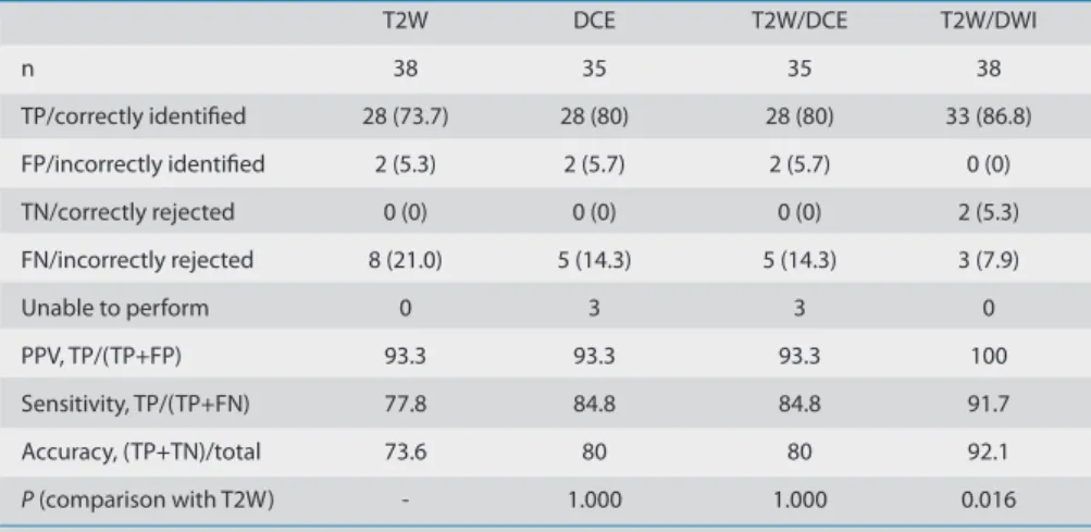
Table 2 depicts the concordance be-tween the MRI analyses and the histology results. For each image set, the sensitivity, positive predictive value, and accuracy for lesion detection are also presented.
T2W sequences correctly depicted 28 tu-moral recurrences via hyperintensity criteria. Four lesions were misclassiied as “unappar-ent disease” because they were isointense/
slightly hypointense and four were misclas-siied as “ill-deined/equivocal disease” be-cause there was only a slight signal elevation that was diicult to diferentiate from the normal myometrium (Fig. 1). Two patients were erroneously classiied as having “recur-rent disease” due to focal cervical (Fig. 2) and retroperitoneal lymph node hyperintensity.
Only 35 patients underwent DCE se-quence since a contrast-enhanced study was impossible in three patients due to kidney function impairment. Considering all DCE acquisitions for each patient, 28 malignant lesions were identiied. Of these, 27 lesions had also been depicted on T2W imaging. The last lesion was only deined after contrast ad-ministration. Four malignant lesions showed no signiicant early contrast uptake and were classiied as “unapparent disease,” and another with just discrete contrast uptake was considered equivocal. Two lesions with early contrast enhancement were benign on histopathologic work-up (false positive ind-ings). There was no statistically signiicant
Table 1. Patient characteristics
Number of patients 38
Histology of primary tumor, n (%)
Squamous cell carcinoma 34 (89.5)
Adenocarcinoma 4 (10.5)
FIGO stage at diagnosis, n ( %)
IA 1 (2.6)
IB 2 (5.2)
IIA 7 (18.4)
IIB 23 (60.5)
IIIA 1 (2.6)
IIIB 4 (10.5)
Histology of suspicious lesion, n (%)
Squamous cell carcinoma 34 (89.5)
Adenocarcinoma 2 (5.25)
Benign 2 (5.25)
Median age at recurrence diagnosis (years) 58
Location of the lesion suspicious of recurrence, n (%)
Cervix 13 (34.2)
Vaginal vault 9 (23.7)
Vaginal wall 4 (10.5)
Uterine body 1 (2.6)
Posterior bladder wall 1 (2.6)
Retroperitoneal lymph nodes 10 (26.3)
diference between the accuracy of T2W and DCE imaging (P > 0.05).
The T2W/DCE imaging combination cor-rectly classiied 28 lesions as malignant. The
addition of contrast allowed for the correct identiication of one additional lesion in a patient with an enhancing focal area not seen on the morphologic (T2W) sequence.
However, one patient with a suspicious le-sion on T2W imaging could not be evaluat-ed with DCE MRI (due to contraindications to contrast administration) and was there-fore not included in the statistical analysis of this combination. Combined T2W/DCE imaging was unable to exclude malignancy in two women with clinical suspicion of re-current disease (both complaining of pelvic pain) but only benign changes on patho-logic analysis. There was no statistically sig-niicant diference between the accuracies of T2W and T2W/DCE imaging (P > 0.05).
When evaluating the combined imaging indings of T2W/DWI, 33 of the recurrent tu-mors were correctly identiied (Fig. 3).
The two disease-free women who were misclassiied using the T2W/DCE sequences were correctly categorized as “unapparent disease” with this combination. Three le-sions were unapparent (Fig. 4). There was no statistically signiicant diference in ac-curacy between the T2W/DCE and T2W/ DWI combinations (P > 0.05). However, the diference in diagnostic accuracy between T2W imaging and T2W/DWI was statistically signiicant (P = 0.016).
These results support the assumption that the rates of tumor recurrence detec-tion are similar between a complete multi-parametric MRI study (combination of mor-phologic sequences with DCE and DWI) and a combination of T2W/DWI.
Discussion
In our study, the combination T2W/DWI had a positive predictive value of 100% and an accuracy of 92.1% for recurrent disease detection, while both T2W imaging alone and the combination T2W/DCE MRI regis-tered values of 93.3% and 80%, respective-ly. Also there were no false positive indings with T2W/DWI.
The identiication of a recurrent tu-mor has a major impact on the survival outcomes of patients treated for cervical cancer. Imaging is undertaken only in the presence of suspicious clinical symptoms. However, residual disease is diicult to eval-uate and the accuracy of MRI depends on the time elapsed since the end of therapy, as local inlammatory or ibrotic phenom-ena may hamper accurate diagnosis (7, 11, 14, 17, 28).
Conventional MRI has a high sensitivity but low speciicity for recurrent disease de-tection. Thus, the added value provided by functional MRI is becoming increasingly
im-Figure 1. a–d. MRI in the axial plane of the pelvis denoting vaginal recurrence of endometrioid adenocarcinoma of the cervix (initial stage IIB) treated with chemoradiotherapy. On T2-weighted (a) and dynamic contrast-enhanced (DCE) (b) MRI sequences, no obvious lesion was identiied. On difusion-weighted imaging (DWI) with b=1000 s/mm2(c), the tumor is clearly deined with a hypersignal and low signal on the apparent difusion coeicient (ADC) map (d).
c a
d b
Table 2. Performance of each MRI technique separately and in combination
T2W DCE T2W/DCE T2W/DWI
n 38 35 35 38
TP/correctly identiied 28 (73.7) 28 (80) 28 (80) 33 (86.8)
FP/incorrectly identiied 2 (5.3) 2 (5.7) 2 (5.7) 0 (0)
TN/correctly rejected 0 (0) 0 (0) 0 (0) 2 (5.3)
FN/incorrectly rejected 8 (21.0) 5 (14.3) 5 (14.3) 3 (7.9)
Unable to perform 0 3 3 0
PPV, TP/(TP+FP) 93.3 93.3 93.3 100
Sensitivity, TP/(TP+FN) 77.8 84.8 84.8 91.7
Accuracy, (TP+TN)/total 73.6 80 80 92.1
P (comparison with T2W) - 1.000 1.000 0.016
Data are presented as n (%).
portant in oncologic diagnosis, particularly in gynecologic cancers, where newer appli-cations have been gaining ground (18, 19).
In DCE MRI, a dynamic image acquisition is performed after the administration of an intravenous bolus of gadolinium-based con-trast agent. The paramagnetic concon-trast mol-ecules lead to changes in local ield strength and thus relect the tumor microvascular net-work (20). Typically, cervical tumors enhance avidly in the early dynamic phase compared with the slight enhancement of the cervical epithelium and stroma, which may allow for the distinction of recurrent tumors from radiation ibrosis (21). However, the use of gadolinium-based contrast media is limited in patients with kidney impairment (patients with glomerular iltration rates <30 mL/min, patients on dialysis, and patients with acute
kidney insuiciency), as well as in cases of al-lergy requiring medical treatment, pregnan-cy, or patient refusal (22).
DWI is a noninvasive technique based on molecular difusion that, combined with conventional T2W imaging, enables the as-sessment of morphologic and physiologic changes in a single examination. It also al-lows for a quantitative evaluation of ADC from images with diferent b-values (15, 23, 24). DWI can provide excellent tissue con-trast, making it an excellent choice in cases where contrast administration is not possi-ble. Furthermore, the additional scanning time is relatively short.
Numerous studies have described the use of DWI for the diagnosis and staging of cervical cancer (3, 9, 14–16, 23, 25, 26). However, only few studies have analyzed
the utility of DWI in assessing early tumor response to treatment (4, 17, 24, 27). To our knowledge, there are no published data on the use of DWI for cervical tumor recurrence detection and no studies have compared DCE MRI and DWI in this ield.
We recognize that pelvic DWI is an extremely motion-sensitive sequence plagued by artefacts related to arterial pul-sation, bowel peristalsis, and susceptibility efects. However, advances in hardware and sequences have contributed to the minimization of these efects, increasing the use of DWI in pelvic pathology (26). It is also well known that high b-value imag-es have low anatomical detail, but this can be overcome by fusing the images with anatomical sequences to optimize anatom-ical correlation. These fused images can be displayed in inverted grey-scale or arbitrary color-coded scales (15).
In our study, T2W imaging alone could identify recurrent tumors with a sensitivi-ty of 77.8%. Histopathology examinations of the false negative patients revealed squamous cell carcinoma cells involved by abundant ibrous tissue. This may justify the absence of a high signal on T2W imaging, especially considering that six of these pa-tients have had chemoradiotherapy, which is known to decrease tumor signal intensity on MRI (28, 29).
These results are concordant with the work of Hricak et al. (12), who found that tumors with intense desmoplastic reactions demonstrate a low signal on T2W imaging. In addition, Kinkel et al. (10) found that the presence of both ibrosis and tumor cells in the same lesion could justify the lower sig-nal intensity on T2W imaging, with a higher reported T2W imaging sensitivity (91.4%). However, this study included other gyne-cologic cancers besides cervical cancer, which can explain this diference. On the other hand, all the false negative lesions in their study were from patients treated for cervical cancer, relecting the diiculties in establishing this diagnosis.
In this study, in cases where the morpho-logic sequences failed to identify tumor re-currence, DCE MRI could correctly identify only one additional lesion. This was in a pa-tient treated with chemoradiotherapy who showed an early enhancing area in the vag-inal wall that was ill-deined and diicult to depict on T2W imaging, probably due to the presence of radiation-induced ibrosis precluding higher signal intensity on T2W imaging.
Figure 2. a–e. Pelvic MRI of a 42-year-old patient who underwent chemoradiotherapy for cervical cancer that ended one year ago (initial stage IIB). On sagittal (a) and axial (b) T2-weighted images an irregular cervical nodular lesion is identiied with necrotic center and ill-deined intermediate signal on the periphery. On sagittal DCE MRI (c) an early heterogeneous peripheral enhancement is seen. On axial DWI with b=1000 s/mm2(d) the lesion shows difuse hyperintense signal, mainly due to central necrosis and there is no peripheral low signal on the correspondent ADC map (e) denoting T2 shine-through efect, without evidence of restricted difusion. Histopathology examination revealed only inlammatory changes.
d a
e b
Three patients could not undergo a con-trast-enhanced study due to kidney impair-ment (in one of these patients a tumoral lesion was readily identiied on T2W imag-ing) and ive patients remained incorrectly diagnosed (false negative). This did not represent a signiicant diference in sensi-tivity between these two sequences. Sala et al. (15) also concluded that DCE MRI was not necessary during staging, as it could not improve accuracy compared with T2W imaging alone, which increased our coni-dence. In the Kinkel et al. study (10) based on subtraction DCE MRI, there was also no increment in sensitivity with contrast-en-hanced studies compared with anatomical sequences only. However, tumor identii-cation accuracy was superior with contrast, with maximum tumor enhancement oc-curring between 45 and 90 s after contrast administration (10). In our study, all patients with early enhancement (30–90 s) were his-tologically proven.
Among lesions that did not show early enhancement (false negative), two were recurrences in the vaginal vaults of hyster-ectomized patients who received neoadju-vant chemotherapy and three were cervical lesions in patients who completed chemo-radiotherapy at least two years ago. On pathology, all these lesions revealed abun-dant ibrous stroma with scant microscopic
tumor foci. The former may be related to post-surgical ibrosis, whereas the latter may be due to post-radiotherapy vasculop-athy, which afects the microvasculature of irradiated ields and thereby limits the early enhancing capacity of these lesions (12).
We found that the two false-positive indings on T2W imaging remained false positive after the addition of DCE MRI. Histo-pathologic examination of these patients re-vealed inlammatory changes, necrosis, and hyperemia in a cervical lesion of a patient who completed chemoradiotherapy one year ago and an enlarged retroperitoneal lymph node with inlammatory changes and increased vessel density in a patient who underwent hysterectomy 18 months prior. Engin (28) reviewed postirradiation changes in pelvic organs and found that sometimes the tumoral signal decrease typical of radia-tion therapy may be delayed, and a high-sig-nal-intensity stroma may persist longer (28). In the study by Hricak et al. (12), there was also nonspeciic enhancement of the cervix after radiation therapy related to benign changes such as postirradiation ibrosis, in-lammation, and necrosis. Furthermore, in another paper by Hawighorst et al. (13), the visual inspection of a lesion’s contrast en-hancement alone was considered unhelpful in establishing a malignancy diagnosis be-cause of nonspeciic tissue changes that can
show similar enhancement. In contrast with these works, Kinkel el al. (10) found that DCE MRI is helpful in improving the speciicity and accuracy of tumor recurrence detection. They also proved that in the irst ive months after radiation therapy, induced inlammato-ry changes are known to be responsible for early enhancement that mimics recurrence. We believe that this efect may be prolonged in some patients.
In our study, we observed that the combi-nation of T2W imaging and DWI could pre-dict tumor recurrence with a higher sensi-tivity than the combination of T2W and DCE MRI (91.7% vs. 84.8%). The addition of DWI to T2W imaging allowed ive patients to be reclassiied as having a recurrent tumor.
On the other hand, the two false-positive patients with bright lesions on T2W imag-ing and early enhancement after contrast administration did not show water restric-tion on DWI, denoting the absence of high cellularity of these benign changes (Fig. 3). These results are in concordance with those of Chen et al. (26), who found that patients without disease after treatment showed no residual hyperintense lesions in the cervical areas on DWI.
The three cases that remained falsely negative on all sequences represented bi-opsy-proven local recurrences of well-dif-ferentiated squamous carcinoma that did
Figure 3. a–f. Pelvic MRI of a 55-year-old patient who underwent chemoradiotherapy for cervical cancer (initial stage IIIB) with central recurrent disease after treatment. On axial T2-weighted image performed four years after treatment (a), no lesion was promptly identiiable, although DWI with b=1000 (b) showed a small focal area of hyperintensity on the right posterior bladder wall with a discrete hyposignal on the ADC map (c) that was not promptly considered relevant by the clinicians. After this examination, a targeted biopsy was performed conirming recurrence of cervical squamous cell carcinoma (this irst evaluation was the one included in our study). The patient refused treatment for personal reasons and one year later, a hyperintense heterogeneous cervical mass with right posterior bladder invasion is readily visible on T2-weighted image (d) along with a high signal on DWI with b=1000 s/mm2(e) and a low signal on the ADC map (f).
d a
e f
not show marked restricted difusion, prob-ably due to their low cellularity (30).
Globally, in this study, the accuracy was 73.6% for T2W imaging, 80% for DCE MRI, 80% for combined T2W/DCE MRI, and 92.1% for combined T2W/DWI.
Our study had several limitations. First, our population was small and had a high prevalence of recurrence, which could lead to the overestimation of sensitivity and render the estimation of speciicity im-possible. Second, DWI was assessed only qualitatively by inspecting signal intensity, as there is no established ADC value that is diagnostic for neoplastic tissue (because imaging parameters change from center to center, making standardization almost impossible). Third, contrast could not be administered in three patients, thus the
entire patient sample could not undergo evaluation with the T2W/DCE MRI combi-nation. However, this is also a limitation in everyday practice, which reinforces the role of DWI as an alternative to contrasted studies in tumoral recurrence follow-up. Fourth, the patient group is not homoge-neous with regard to therapeutic options that may have conditioned the appear-ance of pelvic structures on follow-up MRI. This point warrants further studies with larger samples. Finally, there is inhomo-geneity regarding recurrence site, but this actually represents the pattern of cervical cancer recurrence, which can have a local or regional distribution in the form of pel-vic or para-aortic adenopathy (28).
In conclusion, although we recognize that our data are presented on the basis of
preliminary observations that are sustained by histopathologic conirmation and a sig-niicant diference on statistical analysis, we hypothesize that in follow-up study of patients treated for cervical cancer, contrast administration can be omitted from the ini-tial protocol. We hereby support the notion that an initial follow-up protocol for treated cervical cancer can consist of a T2W/DWI combination, which can save both time and money. DCE MRI would only be performed if the radiologist remained uncertain re-garding the diagnosis.
Conlict of interest disclosure
The authors declared no conlicts of interest.
References
1. Ferlay J, Soerjomataram I, Ervik M, et al. GLOB-OCAN 2012 v 1.0, Cancer incidence and mortal-ity worldwide: IARC cancer base no. 11: Inter-national Agency for Research on Cancer. Lyon, France, 2013. Available at: http://globocan.iarc. fr. Accessed on June 8, 2014.
2. Long HJ. Management of metastatic cervical cancer: review of the literature. J Clin Oncol 2007; 10:2966–2974. [CrossRef]
3. Liyanage SH, Roberts CA, Rockall AG. MRI and PET scans for primary staging and detection of cervical cancer recurrence. Womens Health (Lond Engl) 2010; 6:251–267. [CrossRef] 4. Schieda N, Malone SC, Dandan OA,
Ram-chandani P, Siegelman ES. Multi-modality organ-based approach to expected imaging indings, complications and recurrent tumour in the genitourinary tract after radiotherapy. Insights Imaging 2014; 5:25–40. [CrossRef] 5. Gadducci A, Tana R, Cosio S, Cionini L.
Treat-ment options in recurrent cervical cancer (Re-view). Oncol Lett 2010; 1:3–11.
6. Fulcher AS, O’Sullivan SG, Segreti EM, Kavana-gh BD. Recurrent cervical carcinoma: typical and atypical manifestations. Radiographics 1999; 19:103–116. [CrossRef]
7. Salani R, Backes FJ, Fung MF, et al. Posttreatment surveillance and diagnosis of recurrence in wom-en with gynecologic malignancies: Society of Gy-necologic Oncologists recommendations. Am J Obstet Gynecol 2011; 204:466–478. [CrossRef] 8. Koh WJ, Greer BE, Abu-Rustum NR, et al.
Cer-vical cancer. J Natl Compr Canc Netw 2013; 11:320–343.
9. Togashi K, Nishimura K, Sagoh T, et al. Carci-noma of the cervix: staging with MR imaging. Radiology 1989; 171:245–251. [CrossRef] 10. Kinkel K, Ariche M, Tardivon AA, et al.
Diferen-tiation between recurrent tumor and benign conditions after treatment of gynecologic pel-vic carcinoma: value of dynamic contrast-en-hanced subtraction MR imaging. Radiology 1997; 204:55–63. [CrossRef]
11. Antunes D, Cunha TM. Recurrent cervical can-cer: how can radiology be helpful. OMICS J Ra-diology 2013; 2:138.
12. Hricak H, Swift PS, Campos Z, Quivey JM, Gilden-gorin V, Göranson H. Irradiation of the cervix uteri: value of unenhanced and contrast-enhanced MR imaging. Radiology 1993; 189:381–388. [CrossRef]
Figure 4. a–d. Pelvic MRI of a 59-year-old patient who underwent chemoradiotherapy for cervical cancer that ended two years ago (initial stage IIB). No lesion is depicted on either sagittal T2-weighted (a) or DCE (b) images. In addition, no hyperintense lesion is seen on axial DWI with b=1000 s/mm2(c) or on the corresponding ADC map (d). However, due to on-going pelvic pain, the patient underwent a cervical biopsy that was positive for recurrence of cervical squamous cell carcinoma.
c a
13. Hawighorst H, Knapstein PG, Schaefer U, et al. Pelvic lesions in patients with treated cervical carcinoma: eicacy of pharmacokinetic analysis of dynamic MR images in distinguishing recur-rent tumors from benign conditions. AJR Am J Roentgenol 1996; 166:401–408. [CrossRef] 14. Nougaret S, Tirumani SH, Addley H,
Pand-ey, H, Sala E, Reinhold C. Pearls and pitfalls in MRI of gynecologic malignancy with difu-sion-weighted technique. AJR Am J Roentge-nol 2013; 200:261–276. [CrossRef]
15. Sala E, Rockall A, Rangarajan D, Kubik-Huch RA. The role of dynamic contrast-enhanced and difusion weighted magnetic resonance imaging in the female pelvis. Eur J Radiol 2010; 76:367–385. [CrossRef]
16. Thoeny HC, De Keyzer F. Extracranial applica-tions of difusion-weighted magnetic reso-nance imaging. Eur Radiol 2007; 17:1385–1393. [CrossRef]
17. Vincens E, Balleyguier C, Rey A, et al. Accuracy of magnetic resonance imaging in predicting residual disease in patients treated for stage IB2/II cervical carcinoma with chemoradiation therapy: correlation of radiologic indings with surgicopathologic results. Cancer 2008; 113:2158–2165. [CrossRef]
18. Mahajan M, Kuber R, Chaudhari KR, Chaudhari P, Ghadage P, Naik R. MR imaging of carcinoma cervix. Indian J Radiol Imaging 2013; 23:247– 252. [CrossRef]
19. Kaur H, Silverman PM, Iyer RB, Verschraegen CF, Eifel PJ, Charnsangavej C. Diagnosis, staging and surveillance of cervical carcinoma. AJR Am J Roentgenol 2003; 180:1621–1632. [CrossRef] 20. O’Connor JPB, Tofts PS, Miles KA, Parkes LM,
Thompson G, Jackson A. Dynamic contrast-en-hanced imaging techniques: CT and MRI. Br J Radiol 2011; 84:S112–S120. [CrossRef] 21. Hricak H, Swift PS, Campos Z, Quivey JM,
Gild-engorin V, Goranson H. Irradiation of the cervix uteri: value of unenhanced and contrast-en-hanced MR imaging. Radiology 1993; 189:381– 388. [CrossRef]
22. European Society of Urogenital Radiology. ESUR guidelines on contrast media. version 8.1 2013. Available at: http://www.esur.org/es-ur-guidelines/. Accessed Jan 2014.
23. Namimoto T, Awai K, Nakaura T, Yanaga Y, Hirai T, Yamashita Y. Role of difusion-weighted im-aging in the diagnosis of gynecological diseas-es. Eur Radiol 2009; 19:745–760. [CrossRef]
24. Levy A, Caramella C, Chargari C, et al. Accuracy of difusion-weighted echo-planar MR imaging and ADC mapping in the evaluation of residual cervical carcinoma after radiation therapy. Gy-necol Oncol 2011; 123:110–115. [CrossRef] 25. McVeigh PZ, Syed AM, Milosevic M, Fyles A,
Haid-er MA. Difusion-weighted MRI in cHaid-ervical cancHaid-er. Eur Radiol 2008; 18:1058–1064. [CrossRef] 26. Chen J, Zhang Y, Liang B, Yang Z. The utility of
difusion-weighted MR imaging in cervical can-cer. Eur J Radiol 2010; 74:e101–106. [CrossRef] 27. Harry VN, Semple SI, Gilbert FJ, Parkin DE. Dif-fusion-weighted magnetic resonance imaging in the early detection of response to chemora-diation in cervical cancer. Gynecol Oncol 2008; 111:213–220. [CrossRef]
28. Engin G. Cervical cancer: MR imaging indings before, during, and after radiation therapy. Eur Radiol 2006; 16: 313–324. [CrossRef] 29. Addley HC, Vargas HA, Moyle PL, Crawford R, Sala
E. Pelvic imaging following chemotherapy and radiation therapy for gynecologic malignancies. Radiographics 2010; 30:1843–1856. [CrossRef] 30. Whittaker CS, Coady A, Culver L, Rustin G,