J Bras Pneumol. 2013;39(6):753-756
Sarcomatoid carcinoma of the lung with brain metastases
Carcinoma sarcomatoide de pulmão com metástases cerebrais
Matheus Fernandes de Oliveira, Sílvia Conde Watanabe, Mara Patrícia Guilhermina de Andrade, José Marcus Rotta,
Fernando Campos Gomes Pinto
To the Editor:
We report the case of a 61-year-old Brazilian female who presented with cough, hemoptysis, and fever for seven days prior to evaluation. Her previous medical history revealed that she was a heavy smoker (80 pack-years) and had systemic arterial hypertension. No occupational exposures were reported. Physical examination was unremarkable. Blood workup revealed leukocytosis (white blood cell count, 15,000). Chest X-ray was also unremarkable, and the initial management was antibiotic treatment. A CT of the chest was requested, which revealed a spiculated tumor mass of 5 cm in diameter in the right upper lobe, as well as mediastinal lymphadenopathy. The patient was then submitted to bronchoscopy, which was negative for neoplasms. The decision was to perform pulmonary lobectomy.
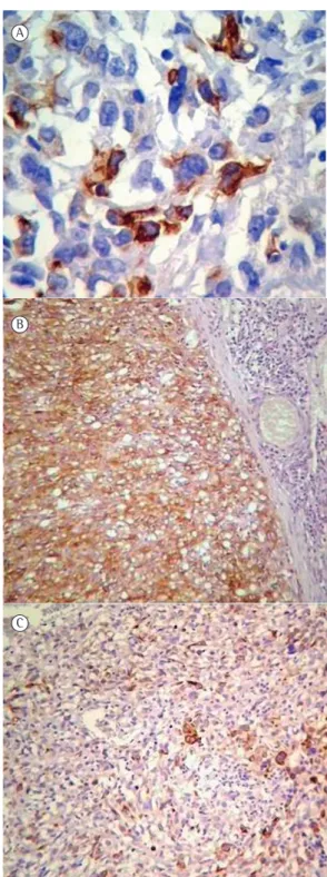
Gross pathology showed that the right upper lobe weighed 300 g and measured 18.5 × 12.0 × 3.5 cm. The cut surface revealed a poorly circumscribed, tan-gray, hemorrhagic lesion measuring 7.0 × 6.0 × 3.8 cm that infiltrated into the visceral pleura. Histologically, the tumor was composed of spindle-shaped cells with marked pleomorphism and abnormal mitosis. These tumor cells displayed a sarcoma-like growth pattern with scarce multinucleated giant cells. Multiple foci of necrosis were present. Squamous cell carcinoma or adenocarcinoma areas were not observed. Immunohistochemical studies were performed using the avidin-biotin-peroxidase complex method. The tumor cells were positive for pan-cytokeratin (AE1/AE3), CK7, and epithelial membrane antigen (Figure 1) but negative for other antibodies. The pathology report revealed sarcomatoid carcinoma of the lung, with spindle cell features. No lymph nodes were involved. The tumor-node-metastasis staging classification, based on the findings, was T2bN0Mx.
Immediately after the surgery, the oncological evaluation was completed. Abdominal CT and bone scintigraphy resulted normal. Chest CT scans
revealed pneumothorax in the right upper lobe in the site of the resected tumor. Radiotherapy and chemotherapy were proposed. However, the patient was lost to follow-up, and no adjuvant treatment was performed.
After four months without any adjuvant therapy, the patient returned to the hospital presenting with progressive, complete right hemiparesis and impaired verbal expression/aphasia for 10 days. At admission to the neurosurgery ward, the patient was alert and cooperative, with a Glasgow Coma Scale score of 15, the pupils were isochoric and reactive to light, and the patient presented with a grade-4 right hemiparesis in association with aphasia.
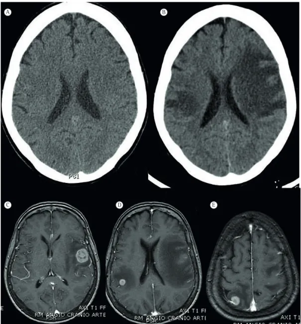
The patient was promptly submitted to a neuroimaging study, which revealed three images (one in the left inferior frontal gyrus and two in the right parietal cortex) of lesions with different dimensions in the brain parenchyma that resembled tumor invasion (Figure 2).
The neurosurgery staff decided to perform microsurgery for the resection of the larger lesion in the left frontal gyrus and adjuvant radiosurgery of the other lesions.
The resected specimen was sent to pathology. The microscopic findings were similar to the sarcomatoid carcinoma of the lung, characterized by the proliferation of pleomorphic spindle-shaped cells. An immunohistochemical study was performed and revealed focal positivity of the tumor cells for CK7 and diffuse positivity for epithelial membrane antigen. The microscopic and immunohistochemical findings were consistent with metastatic sarcomatoid carcinoma.
The patient was discharged without any postoperative complications; however, she was rehospitalized one week later with progressive worsening of her general health status and, two months after the surgery, she died, prior to performing any adjuvant therapy.
754 Oliveira MF, Watanabe SC, Andrade MPG, Rotta JM, Pinto FCG
J Bras Pneumol. 2013;39(6):753-756
Figure 1 - Photomicrographs of pathological and immunohistochemical studies. In A, pan-cytokeratin cytoplasmic positivity of the tumor cells (AE1/AE3 immunostaining; magnification, ×400). In B, tumor cells showing diffuse positivity for CK7 (CK7 immunostaining; magnification, ×100). In C, carcinoma cells showing strong positivity for epithelial membrane antigen in contrast with inflammatory adjacent non-tumoral tissue (immunostaining; magnification, ×100).
Sarcomatoid carcinoma of the lung is a rare tumor, showing a male-to-female ratio of 7.25:1.00. The mean and median age of the patients is 65 years, and the tumor accounts for 0.1-0.3% of all
lung cancers.(1-4) It arises from the central airway
in two-thirds of the patients and exhibits the
morphology of polypoid airway lesions.(3)
Sarcomatoid carcinoma most often presents as solitary masses in the upper lobes, with an
average size of 7 cm in diameter.(4,5) Parenchymal
masses appear as cavities with marked central necrosis and peripheral rim. The association with
smoking is strong.(1-4) The treatment is essentially
performed with surgical resection, chemotherapy and radiotherapy being used as adjuvant therapy or in cases of poor surgical conditions, since there
seems to be little benefit.(1-4,6,7) The five-year
survival for patients with sarcomatoid carcinoma is approximately 20% (compared with 50% for those with non-small cell lung cancer), and the
median duration of survival is three months.(1,2,5,7)
The probable pathogenesis of sarcomatoid carcinoma includes malignant transformation of hamartoma, simultaneous malignant transformation of epithelial elements and stroma, malignant transformation of cancer-derived stroma, sarcomatous change in carcinoma, and
carcinomatous change in sarcoma.(2,3)
Although there are reports of systemic metastases, such as in the skin, stomach, pancreas, esophagus, jejunum, rectum, kidneys, bones, and
adrenal glands,(4-10) to our knowledge, only one
report documented brain metastases,(1) especially
because of the aggressiveness and low survival time. Few reports have presented and discussed the presentation of metastatic sarcomatoid carcinoma of the lung in the brain. In our case, we illustrate the presentation of brain metastases in a patient who had been previously treated for sarcomatoid carcinoma of the lung and highlight the need to consider the differential diagnosis of pulmonary cancer and the aggressive features of sarcomatoid carcinoma. The treatment, in accordance with current therapies in metastatic brain disease, consisted of the surgical resection of the larger lesion and complementary radiosurgery in the surgical cavity, with the resection of the two smaller lesions.(1,2,10) However, due to the progressive
worsening of the general status of the patient, the radiosurgical treatment was not performed, and the patient died two months after being diagnosed with brain metastases.
A
B
Sarcomatoid carcinoma of the lung with brain metastases
J Bras Pneumol. 2013;39(6):753-756
755
A B
C D E
Prando Cau, Maria do Carmo Cruvinel, and Ester Nei Aparecida Martins Coletta for their invaluable help with the present manuscript.
Matheus Fernandes de Oliveira Resident in Neurosurgery, Neurosurgery
Residency Program, Department of Neurosurgery, Hospital do Servidor Público Estadual de São Paulo,
São Paulo, Brazil
Figure 2 - Neuroimaging scans. In A, a CT scan of the skull taken when the pulmonary mass was discovered four months prior to the appearance of neurological symptoms, which is apparently normal. In B, a CT scan of the skull taken four months after readmission, when the patient presented with neurological symptoms. In C-E, magnetic resonance imaging scans revealing details of the metastatic spread and intense vasogenic edema.
The early diagnosis and proper surgical and adjuvant therapy of the primary disease might lead to prolonged survival, and physicians should be aware of the appearance of metastatic symptoms of sarcomatoid carcinoma, such as neurological symptoms, which can be prominent and misleading to other differential diagnoses.
Acknowledgments
756 Oliveira MF, Watanabe SC, Andrade MPG, Rotta JM, Pinto FCG
J Bras Pneumol. 2013;39(6):753-756
3. Ito K, Oizumi S, Fukumoto S, Harada M, Ishida T, Fujita Y, et al. Clinical characteristics of pleomorphic carcinoma of the lung. Lung Cancer. 2010;68(2):204-10. http:// dx.doi.org/10.1016/j.lungcan.2009.06.002 PMid:19577320 4. Mochizuki T, Ishii G, Nagai K, Yoshida J, Nishimura M,
Mizuno T, et al. Pleomorphic carcinoma of the lung: clinicopathologic characteristics of 70 cases. Am J Surg Pathol. 2008;32(11):1727-35. http://dx.doi.org/10.1097/ PAS.0b013e3181804302 PMid:18769330
5. Rossi G, Cavazza A, Sturm N, Migaldi M, Facciolongo N, Longo L, et al. Pulmonary carcinomas with pleomorphic, sarcomatoid, or sarcomatous elements: a clinicopathologic and immunohistochemical study of 75 cases. Am J Surg Pathol. 2003;27(3):311-24. http://dx.doi.org/10.1097/00000478-200303000-00004 PMid:12604887
6. Park JY, Kim HS, Zo JI, Lee S, Choi SW. Initial presentation of lung sarcomatoid carcinoma as a metastatic lesion in the mandibular gingiva. J Periodontol. 2006;77(4):734-7. http://dx.doi.org/10.1902/jop.2006.050137 PMid:16584358 7. Robbins JR, Ryu S, Kalkanis S, Cogan C, Rock J, Movsas
B, et al. Radiosurgery to the surgical cavity as adjuvant therapy for resected brain metastasis. Neurosurgery. 2012;71(5):937-43. http://dx.doi.org/10.1227/ NEU.0b013e31826909f2 PMid:22806080
8. Terada T. Sarcomatoid carcinoma of the lung presenting as a cutaneous metastasis. J Cutan Pathol. 2010;37(4):482-5. http://dx.doi.org/10.1111/j.1600-0560.2009.01292.x PMid:19602061
9. Travis WD, Brambilla E, Noguchi M, Nicholson AG, Geisinger KR, Yatabe Y, et al. International association for the study of lung cancer/american thoracic society/ european respiratory society: international multidisciplinary classification of lung adenocarcinoma. J Thorac Oncol. 2011;6(2):244-85. http://dx.doi.org/10.1097/ JTO.0b013e318206a221 PMid:21252716
10. Yendamuri S, Caty L, Pine M, Adem S, Bogner P, Miller A, et al. Outcomes of sarcomatoid carcinoma of the lung: a Surveillance, Epidemiology, and End Results Database analysis. Surgery. 2012;152(3):397-402. http:// dx.doi.org/10.1016/j.surg.2012.05.007 PMid:22739072
Sílvia Conde Watanabe Resident in Pathology, Pathology Residency Program, Department of Pathology, Hospital do Servidor Público Estadual de São Paulo, São Paulo, Brazil
Mara Patrícia Guilhermina de Andrade Pathologist, Department of Pathology,
Hospital do Servidor Público Estadual de São Paulo, São Paulo, Brazil
José Marcus Rotta Neurosurgeon, Department of Neurosurgery, Hospital do Servidor
Público Estadual de São Paulo, São Paulo, Brazil
Fernando Campos Gomes Pinto Neurosurgeon, Department of Neurosurgery, Instituto de Assistência
Médica ao Servidor Público Estadual, São Paulo, Brazil
References
1. Franks TJ, Galvin JR. Sarcomatoid carcinoma of the lung: histologic criteria and common lesions in the differential diagnosis. Arch Pathol Lab Med. 2010;134(1):49-54. PMid:20073605
2. Goto T, Maeshima A, Tajima A, Kato R. A resected case of pulmonary carcinosarcoma. Ann Thorac Cardiovasc Surg. 2010;16(3):190-3. PMid:20930681