Anatomy of spinal blood supply
Anatomia da circulação medular
Alexandre Campos Moraes Amato1,2
*
, Noedir Antônio Groppo Stolf2
Abstract
he intricate three-dimensional vascular anatomy of the spinal cord is still not completely understood, and its terminology varies between studies. In view of its importance in spinal ischemia, an analysis is needed of the anatomic vocabulary used to describe the spinal cord blood supply to improve understanding of the subject. he main supply is the Adamkiewicz artery, also known as great anterior radicular artery. he literature was reviewed to equate the diferent nomenclatures employed and an accurate description of current knowledge on spinal cord vascularization was prepared.
Keywords: spinal cord; anatomy; spine; aorta.
Resumo
A intrincada anatomia tridimensional da irrigação medular é frequentemente explanada na literatura com diferentes nomenclaturas e devido a sua alta relevância no estudo da isquemia medular, o estudo da terminologia se faz necessário para melhor compreensão do tema. A artéria de Adamkiewicz, também chamada de artéria radicular magna, é a via principal. Foi realizada a revisão da literatura com equiparação das nomenclaturas utilizadas e elaboração de descrição acurada e sumarizada do conhecimento atual sobre a vascularização medular.
Palavras-chave: medula espinhal; anatomia; coluna vertebral; aorta.
1Universidade de Santo Amaro – Unisa, São Paulo, SP, Brazil. 2Universidade de São Paulo – USP, São Paulo, SP, Brazil.
Financial support: None.
Conlicts of interest: No conlicts of interest declared concerning the publication of this article. Submitted: February 05, 2015. Accepted: June 30, 2015.
INTRODUCTION
Spinal blood supply was irst studied by Albert
Wojciech Adamkiewicz [AFI: ʔadamkiɛviʧ],
1,2a
Polish pathologist, in 1881.
3-6The great radicular
artery is also known eponymously as the Adamkiewicz
artery (AKA).
4Knowledge of the blood supply to the spinal cord is
important when planning treatment of diseases of the
aorta. However, the vasculature involved is complex
and dificult to study because of the small caliber of
arteries, which make up an intricate three-dimensional
network with a large degree of anatomic variation.
7The lack of a gold standard imaging exam also makes
it dificult to compare existing imaging methods.
8This study is intended to clarify the anatomic
presentation of the spinal vasculature and propose
a standardization of the terms for use in Portuguese.
REVIEW OF THE LITERATURE
The intricate three-dimensional anatomy of the
spinal blood supply is often explained in the literature
using different terminology
9and merits review in order
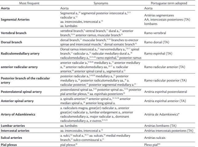
to clarify the standard that should be used (Table 1).
The intercostal and lumbar arteries that supply
the spinal marrow originate in the aorta, as do the
subclavian and hypogastric branches. The intercostal
and lumbar arteries divide three times before reaching
the spinal cord. The irst branch is the spinal branch,
which divides into the anterior and posterior radicular
arteries and, farther on, bifurcates into the dorsal and
vertebral branches. The last bifurcation of the spinal
branch is constant for anterior and posterior supply
of the vertebral canal, of the nerve roots and of the
dura mater, at some levels only, and the anterior and
posterior radicular arteries pass through the dura mater
and reach the marrow. Only some (2-14, a mean of 6)
of these segmental branches remain into adulthood.
The anterior spinal artery (ASA) is crucial to
vascularization of the marrow and anterior and lateral
funiculi and is basically an anastomotic channel
between the ascending and descending branches of
the adjacent anterior radicular arteries (Figure 1).
Generally, one of the anterior radicular arteries
is dominant in terms of caliber and is known as the
great anterior radicular artery or Adamkiewicz artery
(Figure 2). The posterior radicular artery follows a
Table 1. Terms found in the literature and Portuguese terms proposed.
Most frequent Synonyms Portuguese term adopted
Aorta Aorta Aorta
Segmental Arteries
Segmental a.,10 segmental posterior intercostal a.,9,11
radicular a.12
aa. intercostales, intercostal a.13
aa. lumbales
Artérias segmentares
AA. intercostais posteriores (TA) lombares
Vertebral branch vertebral branch,
9 ventral branch,11 dural a.,12 anterior
branch,13,14 anterior ramus, muscular branch15 Ramo vertebral
Dorsal branch dorsal branch,
11 muscular branch,14,16 branches to erector
spinae and intercostal muscle,17 dorsal somatic branch15 Ramo dorsal (TA)
Radiculomedullary artery
Dorsal ramus intercostal a.,12 nervomedullary a.,7,9,11 spinal
branch,11 radicular a.,12 radicular medullary dural a.,18
radiculomedullary a.,13,14,16 ramo espinhal,19 posterior ramus
Ramo espinhal (TA)
anterior radicular artery
anterior radicular a.,9,10,20 medullary a.,12 anterior medullary
a.,18 anterior radiculomedullary aa.,16,17 a. radicular
anterior,19 anterior spinal canal a., segmental a.21
Ramo radicular anterior (TA)
Posterior branch of the radicular artery
posterior radicular a.,9,10,20 medullary a.,12 posterior
medullary a.,18 posterior radiculomedullary aa.,16 a.
radicular posterior,19 posterior segmental medullary a.22
Ramo radicular posterior (TA)
Posterolateral spinal artery posterolateral spinal aa.,
9,18 posterior spinal aa.,12,16 posterior
pial arterilar plexus,10 aa. espinhais posteriores19 Artéria espinhal posterolateral
Anterior spinal artery a. spinalis anterior;
5,6 anterior spinal a.,12-14,18 anterior
median spinal a.,10 anterior long spinal a. Artéria espinhal anterior (TA)
Artery of Adamkiewicz
a. radicularis magna, great(er) radicular a., anterior great(er) radicular a., lumbar enlargement a., anterior radiculomedullary a., major radicular a., dominant radiculomedullary a., e outros.9,11,23
Artéria de Adamkiewicz*
Lumbar arteries aa. lumbales Artérias lombares (TA)
Intercostal arteries aa. intercostales, intercostal a.13 Artérias intercostais posteriores (TA)
Sulcal arteries a. sulci,
5,6 sulcal a.,10,11 aa. sulcais,19 medial medullary
branch,22 sulco-commissural a.21 Artérias sulcais
Pial plexus pial plexus18 Plexo pial**
TA: Term adapted from the Brazilian Anatomic Society’s reference work Anatomic Terminology.24 *Eponym adopted in view of frequent use in the literature.23 **Term
similar pattern, but gives rise to two longitudinal
anastomotic channels: the posterolateral spinal
arteries. Arteries that supply the spine are divided
between a central system, fed by the sulcal arteries,
and a peripheral system, the pial plexus, which gives
origin to perforant branches (Figure 2).
7,25-27Spinal drainage is no less controversial, and
its principal characteristics are the posterior great
radicular vein, in the shape of a “coat-hook”, the
posterior spinal vein and the anterior spinal vein.
15The anatomic importance of venous drainage with
relation to this article, dedicated to the spinal arteries,
lies in anatomic differentiation of the arterial system
(Figure 3) and the subject will not be dealt with in
depth. Posteriorly, there is just one posterior spinal
vein, rather than two smaller posterolateral veins, and
this is frequently of smaller caliber than the anterior
median vein.
28Although there is a single identifiable artery
supplying the spine at the thoracic height, this is not
the only source of medullary blood supply. Griepp et al.
recently reined the conceptualization of the collateral
circulation network for spinal blood supply,
29providing details of its vascular redundancy, but the
importance of the AKA has not yet been suficiently
elucidated. There is an axial network of small arteries
in the spinal canal, in paravertebral tissues and in
paraspinal muscles that anastomose with each other
and with the arteries supplying the spinal marrow;
the entry to this network includes segmental vessels
(intercostal and lumbar arteries), subclavian arteries,
hypogastric arteries and their branches (Figure 4).
30,31In addition to these multiple entry routes, there is also
an extensive network of epidural arterial and small
vessels that supply the paraspinal musculature. All of
these vessels are interconnected and anastomose with
the subclavian arteries cranially and the hypogastric
arteries caudally.
31Figure 1. Schematic drawing of the blood supply of the spinal
marrow.
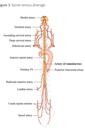
Figure 2. Anatomy of the spinal arterial supply, showing the
Adamkiewicz artery.
Figure 3. Spinal venous drainage.
Figure 4. Collateral network: subclavian, hypogastric, intercostal
This collateral network can provide compensatory
low to the spinal cord in the event of occlusion of the
larger caliber routes,
31and the low from one source
can increase when another is reduced; or vice versa:
low can reduce if a low resistance alternative route
is opened, i.e. in cases of arterial steal.
29According
to Adamkiewicz’s theory of partial low, the low in
the anterior spinal artery originates from the radicular
arteries, arriving at the spinal cord in two currents,
a cranial and a caudal, and so pressure changes, or
occlusion of a route in the collateral network, can
invert the low in the anterior spinal artery.
11INFLUENCE OF POSTOPERATIVE SPINAL
CORD ISCHEMIA
A recent retrospective study using a risk model to
analyze a database of results from 19 European centers
with 2,235 patients registered found that 38 (1.7%)
patients exhibited symptomatic spinal ischemia,
providing evidence that endovascular exclusion of
the intercostal arteries combined with interruption
of another collateral route of spinal blood supply is a
risk factor for this event. The mathematical algorithm
employed identiied intraoperative hypotension and
simultaneous exclusion of at least two spinal supply
territories as relevant to the genesis of ischemia, and it
was concluded that extensive exclusion of the intercostal
arteries alone was not associated with symptomatic spinal
ischemia.
30Notwithstanding, retrospective assessment
of 457 patients and their intrahospital complications
demonstrated that paraplegia and paraparesis had a
signiicant relationship with endovascular exclusion
of more than 20 cm of the aorta,
32which corroborates
the importance of the segmental arteries to spinal
blood supply. Yingbin et al.
33demonstrated the
importance of identifying the AKA to selection of
long endoprostheses for aortic dissection.
An article on interruption of the AKA during
spondylectomy
34suggests that the AKA is not the
only important route of spinal blood supply.
The mechanism of spinal ischemia after endovascular
repair of thoracic aorta aneurysms has not been
entirely elucidated and is apparently related to an
intricate mechanism of several different factors, and
not exclusively to permanent interruption of supply via
the segmental artery.
9,35The collateral network concept
described by Griepp et al. proposes the existence of
extensive redundant spinal blood supply. However, in
acute situations, such as surgical procedures, spinal
perfusion is dependent on the gradient of arterial blood
pressure and of cerebrospinal luid.
35Spinal cord
ischemia is therefore correlated with perioperative
episodes of hypotension and exclusion of the hypogastric
artery as part of the collateral network.
28CONCLUSIONS
Despite the great variation in terminology found in
the literature, studies are in agreement with relation
to the anatomy of the spinal circulation and the
existence of a large network of collateral circulation.
Standardization of the terminology is necessary and
the suggestions for use in Portuguese made in this
study are based on current anatomic terminology.
The clinical importance of anatomic knowledge of
this region lies in planning for endovascular surgery
procedures on the aorta, in order to minimize the risk
of ischemia, avoiding unnecessary occlusion of the
spinal blood supply.
REFERENCES
1. Forvo. Palavra: adamkiewicz. Pronúncia em polonês. [citado 2012 set 30]. http://pt.forvo.com/word/adamkiewicz/.
2. Zeldes A. Automatic phonetic transcription and syllable analysis. [citado 2012 out 10]. http://web.archive.org/web/20140122114850/ http://korpling.german.hu-berlin.de/~amir/phon.php. 3. Milen MT, Bloom DA, Culligan J, Murasko K. Albert Adamkiewicz
(1850-1921)--his artery and its significance for the retroperitoneal surgeon. World J Urol. 1999;17(3):168-70. http://dx.doi.org/10.1007/ s003450050126. PMid:10418091.
4. Skalski JH, Zembala M. Albert Wojciech Adamkiewicz: the discoverer of the variable vascularity of the spinal cord. Ann Thorac Surg. 2005;80(5):1971-5. http://dx.doi.org/10.1016/j. athoracsur.2005.06.022. PMid:16242505.
5. Adamkiewicz A. Die blutgefäße des menschlichen rückenmarkes. I theil. Sitzungsber. Kaiserl. Akad. Wiss., Wien, Math.-Naturwiss. Cl. 1881;84(3):469-502.
6. Adamkiewicz A. Die blutgefässe des menschlichen rückenmarkes. II theil. Sitzungsber. Kaiserl. Akad. Wiss., Wien, Math.-Naturwiss. Cl. 1882;85(2):101-35.
7. Melissano G, Civilini E, Bertoglio L, Calliari F, Campos Moraes Amato A, Chiesa R. Angio-CT imaging of the spinal cord vascularisation: a pictorial essay. Eur J Vasc Endovasc Surg. 2010;39(4):436-40. http://dx.doi.org/10.1016/j.ejvs.2009.11.026. PMid:20034815. 8. Valenstein PN. Evaluating diagnostic tests with imperfect standards.
Am J Clin Pathol. 1990;93(2):252-8. PMid:2405632.
9. Chiesa R, Melissano G, Bertoglio L, et al. The risk of spinal cord ischemia during thoracic aorta endografting. Acta Chir Belg. 2008;108(5):492-502. PMid:19051455.
10. Toole JF, Patel AN. Cerebrovascular disorders. United States of America: McGraw-Hill; 1967.
11. Thron AK, Rossberg C. Vascular anatomy of the spinal cord: neuroradiological investigations and clinical syndromes. Springer; 1988.
12. Dickman C, Fehlings M, Gokaslan Z. Spinal cord and spinal column tumors: principles and practice. New York: Thieme; 2006.
14. Yoshioka K, Niinuma H, Ehara S, Nakajima T, Nakamura M, Kawazoe K. MR angiography and CT angiography of the artery of Adamkiewicz: state of the art. Radiographics. 2006;26(Supl 1):S63-73. http://dx.doi.org/10.1148/rg.26si065506. PMid:17050520.
15. Takase K, Akasaka J, Sawamura Y, et al. Preoperative MDCT evaluation of the artery of Adamkiewicz and its origin. J Comput Assist Tomogr. 2006;30(5):716-22. http://dx.doi.org/10.1097/01. rct.0000228150.35410.45. PMid:16954917.
16. Yoshioka K, Niinuma H, Ohira A, et al. MR angiography and CT angiography of the artery of Adamkiewicz: noninvasive preoperative assessment of thoracoabdominal aortic aneurysm. Radiographics. 2003;23(5):1215-25. http://dx.doi.org/10.1148/rg.235025031. PMid:12975511.
17. Etz CD, Kari FA, Mueller CS, et al. The collateral network concept: a reassessment of the anatomy of spinal cord perfusion. J Thorac Cardiovasc Surg. 2011;141(4):1020-8. http://dx.doi.org/10.1016/j. jtcvs.2010.06.023. PMid:21419903.
18. Bowen BC, DePrima S, Pattany PM, Marcillo A, Madsen P, Quencer RM. MR angiography of normal intradural vessels of the thoracolumbar spine. AJNR Am J Neuroradiol. 1996;17(3):483-94. PMid:8881243.
19. Machado ABM. Neuroanatomia funcional. 2. ed. São Paulo: Atheneu; 2002.
20. Mauney MC, Blackbourne LH, Langenburg SE. Prevention of spinal cord injury after repair of the thoracic or thoracoabdominal aorta. Ann Thorac Surg. 1995;59:245-52.
21. Charles YP, Barbe B, Beaujeux R, Boujan F, Steib JP. Relevance of the anatomical location of the Adamkiewicz artery in spine surgery. Surg Radiol Anat. 2011;33(1):3-9. http://dx.doi.org/10.1007/ s00276-010-0654-0. PMid:20589376.
22. Manjila S, Haroon N, Parker B, Xavier AR, Guthikonda M, Rengachary SS. Albert Wojciech Adamkiewicz (1850-1921): unsung hero behind the eponymic artery. Neurosurg Focus. 2009;26(1):E2. http://dx.doi.org/10.3171/FOC.2009.26.1.E2. PMid:19119888.
23. Cech P, Kachlik D, Liskovec T, Musil V. Frekvence eponym s příjmením adamkiewicz a neeponymních alternativ pro pojmenování hlavní tepny hřbetní míchy v článcích vedených v databázi medline na počátku 21. Století. Plzen Lek Sb. 2009; (S82):149-55.
24. Sociedade Brasileira de Anatomia. Terminologia anatômica internacional. São Paulo: Manole; 2001.
25. Thron AK. Vascular anatomy of the spine. Oxford: Oxford University Press; 2002.
26. Thron AK, Rossberg C. Vascular anatomy of the spinal cord: neuroradiological investigations and clinical syndromes. New York: Springer; 1988.
27. Thron AK. Vascular anatomy of the spinal cord: neuroradiological investigations and clinical syndromes. New York: Springer-Verlag; 1989.
28. Melissano G, Chiesa R. Advances in imaging of the spinal cord vascular supply and its relationship with paraplegia after aortic
interventions. A review. Eur J Vasc Endovasc Surg. 2009;38(5):567-77. http://dx.doi.org/10.1016/j.ejvs.2009.07.011. PMid:19713133.
29. Griepp RB, Griepp EB. Spinal cord perfusion and protection during descending thoracic and thoracoabdominal aortic surgery: the collateral network concept. Ann Thorac Surg. 2007, Feb;83(2):S865-9. 30. Czerny M, Eggebrecht H, Sodeck G, et al. Mechanisms of symptomatic spinal cord ischemia after TEVAR: insights from the European Registry of Endovascular Aortic Repair Complications (EuREC). J Endovasc Ther. 2012;19(1):37-43. http://dx.doi.org/10.1583/11-3578.1. PMid:22313200.
31. Griepp EB, Di Luozzo G, Schray D, Stefanovic A, Geisbüsch S, Griepp RB. The anatomy of the spinal cord collateral circulation. Ann Cardiothorac Surg. 2012;1(3):350-7. PMid:23977520. 32. Fattori R, Nienaber CA, Rousseau H, et al. Results of endovascular
repair of the thoracic aorta with the talent thoracic stent graft: The talent thoracic retrospective registry. J Thorac Cardiovasc Surg. 2006;132(2):332-9.
33. Yingbin J, Jiefei M, Jian L, et al. Evaluation of the thoracic aortic dissection treated by endografts covering a longer distance of aorta according to the location of the Adamkiewicz artery. Thorac Cardiovasc Surg. 2013;61(7):569-74. PMid:22956338.
34. Murakami H, Kawahara N, Tomita K, Demura S, Kato S, Yoshioka K. Does interruption of the artery of Adamkiewicz during total
en bloc spondylectomy affect neurologic function? Spine.
2010;35(22):E1187-92.
35. Chiesa R, Melissano G, Marrocco-Trischitta MM, Civilini E, Setacci F. Spinal cord ischemia after elective stent-graft repair of the thoracic aorta. J Vasc Surg. 2005;42(1):11-7. http://dx.doi.org/10.1016/j. jvs.2005.04.016. PMid:16012446.
*
Correspondence
Alexandre Campos Moraes Amato Av. Brasil, 2283 - Jardim América CEP 01431-001 - São Paulo (SP), Brazil E-mail: [email protected]
Author information
ACMA - Professor of Vascular Surgery, Universidade de Santo Amaro (Unisa); School of Medicine, Universidade de São Paulo (USP). NAGS - Emeritus Professor of Cardiovascular Surgery, School of Medicine, Universidade de São Paulo (USP).
Author contributions
Conception and design: ACMA, NAGS Analysis and interpretation: ACMA, NAGS Data collection: ACMA, NAGS Writing the article: ACMA, NAGS Critical revision of the article: ACMA, NAGS Final approval of the article*: ACMA, NAGS Statistical analysis: N/A. Overall responsibility: ACMA, NAGS