Mast Cells Modulate Acute Toxoplasmosis in Murine
Models
Bo Huang
1,2☯, Shiguang Huang
3☯, Ying Chen
1,2, Huanqin Zheng
1,2, Jilong Shen
4, Zhao-Rong Lun
2,5, Yong
Wang
6, Lloyd H. Kasper
7, Fangli Lu
1,2*1 Department of Parasitology, Zhongshan School of Medicine, Sun Yat-sen University, Guangzhou, Guangdong, China, 2 Key Laboratory of Tropical Disease Control (Sun Yat-sen University), Ministry of Education, Guangzhou, Guangdong, China, 3 Department of Periodontology, School of Medicine, Jinan University, Guangzhou, Guangdong, China, 4 The Anhui Provincial Laboratory of Pathogen Biology, Anhui Medical University, Hefei, Anhui, China, 5 Center for Parasitic Organisms, State Key Laboratory of Biocontrol, School of Life Sciences, Sun Yat-sen University, Guangzhou, Guangdong, China, 6 Department of Pathogen Biology, Nanjing Medical University, Nanjing, Jiangsu, China, 7 Department of Microbiology, Immunology, Dartmouth Medical School, Lebanon, New Hampshire, United States of America
Abstract
The role of mast cells (MCs) in Toxoplasma gondii infection is poorly known. Kunming outbred mice were infected intraperitoneally with RH strain T. gondii, either treated with compound 48/80 (C48/80, MC activator) or disodium cromoglycate (DSCG, MC inhibitor). Compared with infected controls, infected mice treated with C48/80 exhibited significantly increased inflammation in the liver (P < 0.01), spleen (P < 0.05), and mesentery (P < 0.05) tissues, higher parasite burden in the peritoneal lavage fluids (P < 0.01), and increased levels of mRNA transcripts of T. gondii tachyzoite surface antigen 1 (SAG1) gene in the spleen and liver tissues (P < 0.01), accompanied with significantly increased Th1 cytokine (IFN- , IL-1βp40, and TNF-α) (P < 0.01) and decreased IL-10 (P < 0.01) mRNA expressions in the liver, and increased IFN- (P < 0.01) and IL-1βp40 (P < 0.01) but decreased TNF-α (P < 0.01) and IL-4 (P < 0.01) in the spleens of infected mice treated with C48/80 at day 9-10 p.i. Whereas mice treated with DSCG had significantly decreased tissue lesions (P < 0.01), lower parasite burden in the peritoneal lavage fluids (P < 0.01) and decreased SAG1 expressions in the spleen and liver tissues (P < 0.01), accompanied with significantly increased IFN- (P < 0.01) and IL-1βp40 (P < 0.05) in the liver, and decreased IFN- (P < 0.05) and TNF-α (P < 0.01) in the spleens; IL-4 and IL-10 expressions in both the spleen and liver were significantly increased (P < 0.01) in the infected mice treated with DSCG. These findings suggest that mediators associated with the MC activation may play an important role in modulating acute inflammatory pathogenesis and parasite clearance during T. gondii infection in this strain of mice. Thus, MC activation/inhibition mechanisms are potential novel targets for the prevention and control of T. gondii infection.
Citation: Huang B, Huang S, Chen Y, Zheng H, Shen J, et al. (β01γ) Mast Cells Modulate Acute Toxoplasmosis in Murine Models. PLoS ONE 8(10): e77γβ7. doi:10.1γ71/journal.pone.0077γβ7
Editor: Markus M. Heimesaat, Charité, Campus Benjamin Franklin, Germany
Received May 18, β01γ; Accepted August γ0, β01γ; Published October 16, β01γ
Copyright: © β01γ Huang et al. This is an open-access article distributed under the terms of the Creative Commons Attribution License, which permits unrestricted use, distribution, and reproduction in any medium, provided the original author and source are credited.
Funding: This work was supported in part by grants from the National Basic Research Program of China (97γ Program) [grant number β010CB5γ0001] and the Natural Science Foundation of China to F.L. [grant numbers 81071γ87 and 81β71854]. The funders had no role in study design, data collection and analysis, decision to publish, or preparation of the manuscript.
Competing interests: The authors have declared that no competing interests exist. * E-mail: fanglilu@yahoo.com
☯ These authors contributed equally to this work.
Introduction
Toxoplasma gondii is a common and significant obligate intracellular pathogen of humans and animals, which infects nearly one third of the human population and is found in an extraordinary range of vertebrate hosts [1]. The immune/ inflammatory response to T. gondii infection is essential to control parasite replication and tissue spread but also can cause tissue damage, being decisive to pathogenesis [β]. Mast cells (MCs) are abundant in tissues exposed to the external environment including the skin, intestinal tract, and trachea,
innate immune responses, wound healing, and inflammatory disease [7]. Recent studies indicate that MCs are also involved in immune regulation [8,9].
MCs are recognized as effector cells involved in clearance of diverse parasites, including Trichinella spiralis and
Trichomonas vaginalis [10,11]. MC deficient mice infected with
Leishmania major develop larger lesion load with increased parasitemia [1β]. Although MCs are involved during the acute phase of the inflammatory response to T. gondii infection in animal model [1γ], the mechanism by which MC alter the immune response during T. gondii infection has not been resolved. In the present study, we assessed the role of MC during acute murine T. gondii infection; our findings suggest that release of mediators after MC activation plays an important role in modulating inflammatory pathogenesis and parasite load during acute T. gondii infection.
Materials and Methods
Ethics Statement
Female 6-week-old Kunming (KM, outbred) mice were obtained from the Animal Center of Sun Yat-sen University, maintained in specific-pathogen-free environment, and had free access to a commercial basal diet and tap water ad libtum. Animals were provided with humane care and healthful conditions during their stay in the facility. All individuals who use animals received instruction in experimental methods and in the care, maintenance, and handling of mice; and all efforts were made to minimize animal suffering. Animals were sacrificed using COβ asphyxiation and the appropriate organs
were harvested. The protocol in this study was approved by the Committee on the Ethics of Animal Experiments of the Sun Yat-sen University [Permit Numbers: SCXK (Guangdong) β009–0011].
Parasite
T. gondii RH strain tachyzoites were propagated by intraperitoneal (i.p.) passage in KM mice at 4 or 5 day intervals. Mice were infected with 1×10β RH strain T. gondii tachyzoites
by i.p. injection, and tachyzoites were enumerated using manual counting with a haemocytometer.
Mast cell (MC) activation and stabilization in vivo
Total 48 KM mice were included in this study. Mice were divided into 6 groups, consisting of 7-9 mice per group. Compound 48/80 (C48/80) activated the MCs and disodium cromoglycate (DSCG) stabilized the MCs in mice. The model of MC degranulation or stabilization used in the present study was based on a well-characterized protocol with modifications [14]. Briefly, mice received the first i.p. injection of C48/80 (Sigma-Aldrich, 4 mg/kg/d) or DSCG (Sigma-(Sigma-Aldrich, β5 mg/kg/d) β4 h before infection with T. gondii RH strain tachyzoites, and each animal received daily i.p. injection for the duration of the experiment thereafter [9-10 days post infection (p.i.)]. C48/80 enhanced MCs releasing their mediators and DSCG prevented MCs from releasing their mediators for the duration of the experiment. Infected control mice were infected with T. gondii
RH strain tachyzoites alone without any treatment. Uninfected controls were received injections with either the phosphate-buffered saline (PBS) diluent, C48/80, or DSCG.
Histopathological analysis
Mice were sacrificed by COβ asphyxiation prior to death after
infection, and their livers, spleens, and mesenteries were harvested and immediately fixed in 10% buffered natural formaldehyde (Guangzhou chemical reagent factory, China). Four-micrometer-thick sections (50- or 100-μm distance between sections) of the organ from each mouse, stained with hematoxylin and eosin (H&E) (Sigma-Aldrich), were evaluated for histological changes. Blinded samples were submitted for semi-quantitative histopathologic analysis. The histological scores in the spleen and mesentery tissues were determined under ×100 magnification in three non-contiguous sections from four mice in each group. In brief, the score used to measure the intensity of spleen and mesentery tissue alterations was 1, β, γ, and 4 (absent, mild, moderate, and severe, respectively) [15]. Liver sections were analyzed for the numbers of inflammatory foci according to previous report with minor modifications [β], and the number of inflammatory foci per field was analyzed at a magnification of ×100 under a light microscopy by counting 10 fields of each section at 9-10 days p.i. in each group. All the analyses were performed by two researchers.
Toluidine blue staining for MCs
Serialized 4-μm-thick sections of spleen and mesentery were deparaffinized, rehydrated, and stained with 0.5% toluidine blue (Sigma-Aldrich) for 1β0 min. MCs, in three to five sections per animal on days 9 to 10 after treatment, were identified by their deep blue-purple staining and counted at ×400 magnification under light microscopy. MC count was expressed as the number of positive cells per mmβ and the results were
expressed as the mean value of MCs per group. MC degranulation was determined as a loss of MC membrane integrity with extrusion of intracellular granules to the extracellular space or MCs completely lacking in intracellular granules as described previously [16]. Completely degranulated MCs with absence of the cytoplasmic granules are invisible by toluidine blue staining.
Immunofluorescence staining of tryptase for MCs
Spleen and mesentery tissue sections (4-μm) were deparaffinized and rehydrated in distilled water. Heat-induced antigen retrieval was carried out in an 800-W microwave oven for γ0 min. Endogenous peroxidase activity was blocked by incubation with 0.γ % hydrogen peroxide in methanol for 10 min at room temperature. Non-specific binding was blocked by incubation in PBS containing 10 % normal goat serum and 1 % bovine serum albumin (BSA) (pH 7.4) for 60 min at room temperature. Sections were incubated with anti-MC tryptase mouse monoclonal antibody (AA1, IgG1; 1 mg/ml, 1:β00 dilution; Abcam, USA) overnight at 4°C. Slides were then rinsed three times with PBS (pH 7.4) and exposed to secondary antibody [anti-mouse IgG (H+L), F (ab') β fragment
(Alexa Fluor® 488 Conjugate); β mg/ml, 1:β00 dilution; CST,
USA] for 60 min at room temperature in a dark chamber. The slides were washed three times with PBS (pH 7.4) for γ0 min at
room temperature and mounted by antifade
polyvinylpyrrolidone mounting medium (Beyotime, China) in a dark chamber. MCs were identified by their green fluorescence staining and counted at ×400 magnifications under a light microscope. Positively stained MCs were counted and expressed as mentioned above.
T. gondii tachyzoite burden in mouse peritoneal lavage fluids
To examine the effect of C48/80 or DSCG on the parasite proliferation in vivo, we examined parasite burden in mouse peritoneal lavage fluids infected with T. gondii with either C48/80 or DSCG treatment, or without treatment. Mice were killed at 9-10 days p.i. prior to death after infection, the peritoneal lavage fluids of each mouse was passed through a β7 gauge needle, and the parasite numbers were counted by hemocytometer.
Measurement of mRNA expression in spleen and liver tissues using quantitative real-time PCR (qRT-PCR)
Total RNA was extracted from about 100 mg spleen or liver sample each mouse using RNA extraction kit (TaKaRa, Japan) according to the manufacturer’s protocol. The quality of total RNA was analyzed by running 5 μl of each RNA sample on a 1.0% agarose gel and visualizing with ethidium bromide. The quantity of total RNA was estimated by measuring the absorbance at β60 nm and β80 nm using a NanoDrop β000 spectrophotometer (NanoDrop Technologies). First-strand cDNA was constructed from 1.0 μg of total RNA with oligo (dT) as primers using PrimeScript 1st Strand cDNA Synthesis Kit
(TaKaRa), following the manufacturer’s protocol. cDNA was stored at −80 °C until use.
To determine the levels of mRNA transcripts of T. gondii
tachyzoite surface antigen 1 (SAG1) gene and cytokines including IFN- , TNF-α, IL-4, IL-10, and IL-1βp40 in both spleen and liver tissues from different groups of mice, qRT-PCR was performed using SYBR Green qqRT-PCR Master Mix (TaKaRa) according to manufacturer’s instructions. Primers are listed in Table 1. Briefly, the total 10 μl reaction mixture contained 5.0 μl of SYBR® Premix Ex TaqTM (β×), 0.5 μl of each
primer (10 pM), γ.0 μl of dHβO, and 1.0 μl of cDNA (0.β μg/μl).
The thermal cycling conditions consisted of an initial denaturation of γ0 sec at 95°C followed by 4γ cycles of 95°C for 5 sec and 60°C for β0 sec. Values are means from triplicate measurements, specific mRNA expression levels were normalized to the housekeeping gene -actin mRNA and the results are expressed as the fold change compared to uninfected controls.
Statistical Analysis
Data are expressed as means ± SEM. All of the pathological measurements were done in a blind fashion, and the quantitative measurements were made twice. A statistical software program SPSS 17.0 was applied for analysis. Differences of histopathological examination in liver, spleen, and mesentery between different groups were investigated
using the Kruskal-Wallis rank sum test. The fold changes of SAG1 and cytokine mRNA expressions were analyzed by Student's t test. A P-value of < 0.05 was considered statistically significant.
Results
Survival of mice
The survival rates and survival times of the infected mice from different groups were similar, and all the RH strain T. gondii-infected mice with either C48/80 or DSCG treatment, or without treatment died within 9-10 days p.i. (Figure 1).
MC activation and stabilization
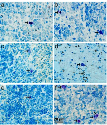
Stained with toluidine blue, MCs were identified in tissue sections from their characteristic granular, deep blue-purple metachromatic appearance against blue orthochromatic background tissue. Toluidine blue stained sections of the mesenteries and spleens from different groups at 9-10 days p.i. were shown in Figures β and γ, respectively. Stained with immunofluorescence for tryptase, MCs from their characteristic green fluorescence were identified in tissue sections of the mesenteries and spleens from different groups at 9-10 days p.i. (Figures 4 and 5, respectively). MCs were intact in uninfected mice with PBS treatment (Figures βa, γa, 4a, and 5a); MCs had mild or obvious granula release (Figures βb, γb, 4b, and 5b) in T. gondii-infected control mice. However, MCs had marked granule release in uninfected (Figures βc, γc, 4c, and 5c) and T. gondii-infected mice (Figures βd, γd, 4d, and 5d) with C48/80 treatment. MCs were intact in uninfected (Figures βe, γe, 4e, and 5e) and T. gondii-infected mice (Figures βf, γf, 4f, and 5f) with DSCG treatment, and the latter appeared morphologically indistinguishable from the uninfected controls.
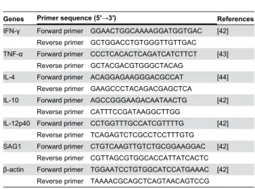
Table 1. Primer sequences of mouse target cytokines and housekeeping genes used for quantitative real-time polymerase chain reaction (qRT-PCR) assays.
Genes Primer sequence (5′→3′) References
IFN- Forward primer GGAACTGGCAAAAGGATGGTGAC [4β] Reverse primer GCTGGACCTGTGGGTTGTTGAC TNF-α Forward primer CCCTCACACTCAGATCATCTTCT [4γ]
Reverse primer GCTACGACGTGGGCTACAG
IL-4 Forward primer ACAGGAGAAGGGACGCCAT [44] Reverse primer GAAGCCCTACAGACGAGCTCA
IL-10 Forward primer AGCCGGGAAGACAATAACTG [4β] Reverse primer CATTTCCGATAAGGCTTGG
IL-1βp40 Forward primer CCTGGTTTGCCATCGTTTTG [4β] Reverse primer TCAGAGTCTCGCCTCCTTTGTG SAG1 Forward primer CTGTCAAGTTGTCTGCGGAAGGAC [4β]
Reverse primer CGTTAGCGTGGCACCATTATCACTC -actin Forward primer TGGAATCCTGTGGCATCCATGAAAC [4β]
Reverse primer TAAAACGCAGCTCAGTAACAGTCCG doi: 10.1γ71/journal.pone.0077γβ7.t001
Spleen MC densities
MC count was assessed by examining sections of spleen tissues by both metachromatic staining with toluidine blue and immunofluorescence staining of tryptase. As shown in Figure 6, there were only a low density (the number of MCs per mmβ)
positively stained MCs with undegranulation observed in the spleen tissues of uninfected mice treated with PBS, while there were significantly higher densities of MCs in T. gondii-infected control mice. In uninfected mice, C48/80 administration did not change the number of MCs; while DSCG administration increased the MC density in the spleens by γ.1 fold by toluidine blue staining (P < 0.01) and 1.8 fold by immunofluorescence staining of tryptase (P < 0.01) relative to that in uninfected mice with PBS. T. gondii infection increased the density of MCs by 4.0 fold by toluidine blue staining (P < 0.01) and 1.7 fold by immunofluorescence staining of tryptase (P < 0.01) relative to that in uninfected mice with PBS. In contrast, in T. gondii -infected mice that received C48/80, the density of MCs was no change by both staining, whereas in T. gondii-infected mice that received DSCG, the density of MCs was increased by 1γ.0 fold by toluidine blue staining (P < 0.01) and 4.6 fold by immunofluorescence staining of tryptase (P < 0.01) relative to that in uninfected mice with PBS. Compared with toluidine blue staining, there were significantly higher MC densities in spleen tissues in all the groups when using immunofluorescence staining of tryptase (P < 0.01). C48/80 treatment of the spleens degranulated MCs, which resulted in a lack of both toluidine blue staining of granule matrix proteoglycans and
immunofluorescence staining of tryptase. However, it is important to notice that not all MCs were degranulated or undegranulated by these treatments.
Severe liver, spleen, and mesentery inflammation in T. gondii-infected mice with C48/80 treatment
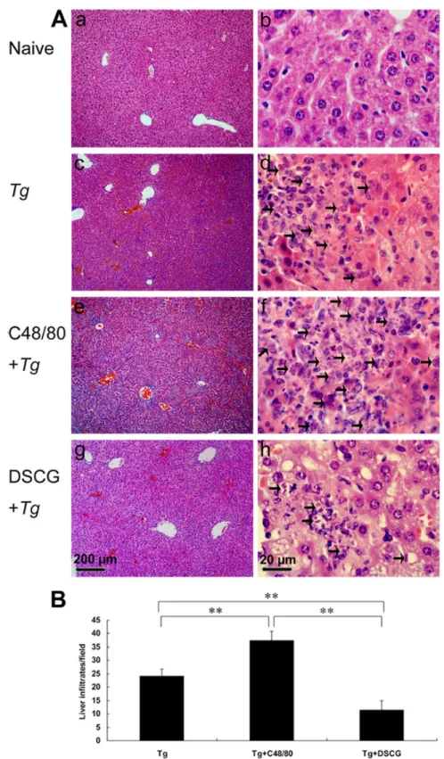
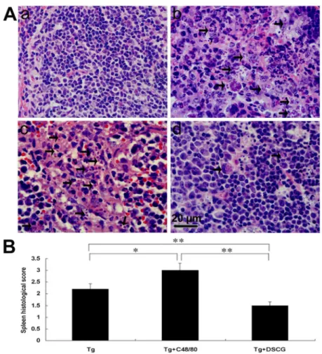
To investigate the effects of the mediators released by MCs on tissue pathological changes, the liver (Figure 7A), spleen (Figure 8A), and mesentery (Figure 9A) tissues from different groups were examined histological. Control sections of liver (Figures 7a and b), spleen (Figure 8a), and mesentery (Figure 9a) from uninfected mice treated with PBS were negative for both inflammation and necrosis foci and T. gondii staining. After primary i.p. T. gondii RH strain infection, severe damage (obvious inflammation and necrosis foci) and a great number of RH tachyzoites were observed in the liver (Figure 7c and d), spleen (Figure 8b), and mesentery (Figure 9b) tissues of infected control mice. In comparison, even severer damage (stronger inflammation and more necrosis foci) and a greater number of RH tachyzoites were observed in the liver (Figure 7e and f), spleen (Figure 8c), and mesentery (Figure 9c) tissues of
T. gondii-infected mice treated with C48/80; whereas attenuated or moderate histological evidence (mild inflammation and fewer necrosis foci) and a lower number of RH tachyzoites were observed in the liver (Figure 7g and h), spleen (Figure 8d) and mesentery (Figure 9 d) tissues of T. gondii-infected mice treated with DSCG. Treatment with C48/80 or DSCG did not change the tissue histology from Figure 1. Mice survival after infection with 102 RH tachyzoites of T. gondii.
Survival of naïve mice treated with PBS (open square, n=8); uninfected mice treated with C48/80 (dash, n=8); uninfected mice treated with DSCG (open upright triangle, n=8); T. gondii-infected control mice (filled square, n=7), T. gondii-infected mice with C48/80 treatment (asterisk, n=9), and T. gondii-infected mice with DSCG treatment (filled upright triangle, n=8). The mice were monitored for survival on a daily basis until the termination of the experiment.
doi: 10.1γ71/journal.pone.0077γβ7.g001
uninfected mice, comparing with that of uninfected mice received PBS (data not shown).
Quantitative analysis of the severity of inflammation and necrosis of liver sections (e.g. the number of inflammatory foci per field, γ slides/animal) of different groups of mice was performed (Figure 7B). A great number of inflammatory foci of neutrophil infiltrates were observed in the liver of T. gondii -infected control mice. In comparison, significantly increased inflammatory foci of neutrophil infiltrates were observed in the
T. gondii-infected mice with C48/80 treatment (P < 0.01), whereas significantly reduced inflammatory foci of neutrophil infiltrates were observed in the T. gondii-infected mice with DSCG treatment (P < 0.01). Semiquantitative histological evaluation of spleen (Figure 8B) and mesentery (Figure 9B) sections (γ slides/animal) of different groups of mice were performed. Severe pathology was shown in the spleen and mesentery tissues of T. gondii-infected mice without treatment. In comparison, even severer pathology were shown in the spleen and mesentery tissues of T. gondii-infected mice with C48/80 treatment (P < 0.05); whereas attenuated pathology
were shown in the spleen and mesentery tissues of infected mice with DSCG treatment (P < 0.01).
Increased parasite burden in T. gondii-infected mice with C48/80 treatment
To investigate whether MC activation and degranulation are important in host defense, live T. gondii tachyzoites were recovered from the peritoneal lavage fluids of infected mice with either C48/80 or DSCG treatment, or without treatment at 9-10 days p.i when mice were becoming moribund, and counted by hemocytometer (Figure 10A). Compared with T. gondii-infected control mice, there was a significant increase (β.γ-fold) in the number of T. gondii tachyzoites in the peritoneal lavage fluids of infected mice treated with C48/80 (P < 0.01), whereas there was a significant decrease (β.1-fold) in the number of T. gondii tachyzoites in that of mice treated with DSCG (P < 0.01). In addition, a significant decrease (4.8-fold) in the number of T. gondii tachyzoites from infected mice treated with DSCG in comparison with that from infected mice Figure 2. Light photomicrographs of metachromatic MCs in mesenteries by toluidine blue staining. Infected mice i.p. inoculated with 10β RH tachyzoites of T. gondii from different groups were killed at 9-10 days p.i. Metachromatic MCs were
evaluated in mesentery tissue from uninfected mouse treated with PBS (a), infected control mouse displaying mildly degranulated MCs (b), uninfected mouse treated with C48/80 (c) and infected mouse treated with C48/80 (d), both displaying degranulated MCs (arrows); uninfected mouse treated with DSCG (e) and infected mouse treated with DSCG (f), both displaying intact MCs.
doi: 10.1γ71/journal.pone.0077γβ7.g00β
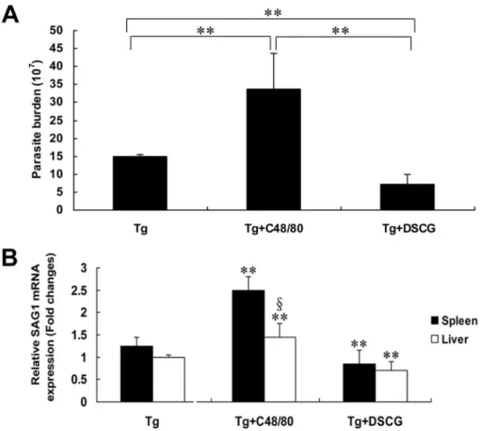
treated with C48/80 (P < 0.01). To confirm the parasite burden of T. gondii tachyzoite in tissues, qRT-PCR was performed to determine the levels of mRNA transcripts for tachyzoite SAG1-stage specific gene in both liver and spleen tissues from different groups of mice at 9-10 days p.i (Figure 10B). Compared with T. gondii-infected controls, there was a significantly increased mRNA transcripts for SAG1 in both liver (P < 0.01) and spleen (P < 0.01) of infected mice treated with C48/80, whereas there was a significantly decreased mRNA transcripts for SAG1 in both liver (P < 0.01) and spleen (P < 0.01) of infected mice treated with DSCG (P < 0.01).
Th1 and Th2 mRNA cytokine responses in the spleen and liver of different groups
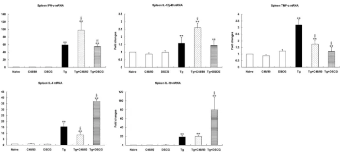
The effect of MC mediator release on Th1 and Thβ cytokine responses after T. gondii infection was evaluated by measuring IFN- , IL-1βp40, TNF-α, IL-4, and IL-10 mRNA expressions in the spleens (Figure 11) and livers (Figure 1β) of different groups. Cytokine mRNA expressions in naïve mice were not
altered by C48/80 or DSCG treatment itself. However, compared with uninfected mice treated with PBS, there were significantly increased mRNA expressions of IFN- , IL-1βp40, TNF-α, IL-4, and IL-10 in the livers and spleens of T. gondii -infected control mice at days 9-10 p.i. (P < 0.01), using qRT-PCR. Compared with T. gondii-infected controls, the Th1 cytokine (IFN- , IL-1βp40, and TNF-α) expressions were significantly increased (P < 0.01) and the Thβ cytokine (IL-10) was significantly decreased (P < 0.01) in the livers, and the expressions of IFN- (P < 0.01) and IL-1βp40 (P < 0.01) were significantly increased but TNF-α (P < 0.01) and IL-4 (P < 0.01) were significantly decreased in the spleens of infected mice treated with C48/80 at day 9-10 p.i. Whereas the expressions of Th1 cytokine [IFN- (P < 0.01) and IL-1βp40 (P < 0.05)] were significantly increased in the liver, and IFN- (P < 0.05) and TNF-α (P < 0.01) were significantly decreased in the spleens of the infected mice treated with DSCG at day 9-10 p.i. However, the Thβ cytokine (IL-4 and IL-10) expressions in both spleen and liver were significantly increased (P < 0.01) in the infected mice treated with DSCG.
Figure 3. Light photomicrographs of metachromatic MCs in spleens by toluidine blue staining. Infected mice i.p. inoculated with 10β RH tachyzoites of T. gondii from different groups were killed at 9-10 days p.i. Metachromatic MCs (arrows) were evaluated
in spleen tissue from uninfected mouse treated with PBS (a), infected control mouse displaying a degranulated MC (b), uninfected mouse treated with C48/80 (c) and infected mouse treated with C48/80 (d), both displaying degranulated MCs; uninfected mouse treated with DSCG (e) and infected mouse treated with DSCG, both displaying intact MCs (f).
doi: 10.1γ71/journal.pone.0077γβ7.g00γ
Discussion
Toxoplasmosis, an inflammatory disease, can lead to severe pathology in both humans and animals, involves leukocyte recruitment, immune responses, and inflammatory cytokine production, and immunity to infection relies on the development of a strong cell-mediated immune response, such as T cells and the production of IFN- [17,18]. It has been reported that a significant increase in the number of MCs in animals inoculated either i.p. or via the conjunctiva with T. gondii tachyzoites has been observed previously [19]. After incubation with T. gondii in vitro, peritoneal lavage MCs of Sprague-Dawley rats were found to degranulate and release LTB4, which damage T. gondii tachyzoites [β0]. Despite these observations, little is known about the relationship between MCs and pathogenesis of T. gondii infection, as well as the MCs mediators generated in response to T. gondii infection. KM mice are the most widely used outbred colony in China. Using this murine model of T. gondii infection in the present study, we show here that the activation of MCs in mice via C48/80 injection resulted in increased T. gondii load and enhanced inflammatory infiltrate, whereas the MC stabilization with DSCG diminished T. gondii
load and attenuated inflammatory reaction. Our data demonstrate that MCs play a crucial role in the course of T. gondii infection.
In this study, our data demonstrated that infection with T. gondii not only increased the number of MCs in the analyzed tissues but also induced noticeable MC degranulation at 9-10 days p.i., by both toluidine blue staining and immunofluorescence staining of tryptase. As it has been reported that immunohistochemical staining for tryptase is a highly specific and sensitive method for identifying MCs [β1], we also found MC density was significantly higher with immunofluorescence staining of tryptase compared with that of toluidine blue staining, due to the strong immunofluorescence staining of both intact and degranulated MCs. MC activation and degranulation most commonly result from multivalent antigens binding to the IgE bound to the high-affinity IgE receptor (FcεRI) on the surface, which results in noncytotoxic degranulation and the release of a variety of preformed and newly synthesized mediators [ββ]. The degranulation of MCs observed in T. gondii-infected animals is probably due to the presence of excreted-secreted antigens from T. gondii in tissues [βγ]. The C48/80 has been used to study allergies and Figure 4. Light photomicrographs of tryptase positive-MCs in mesenteries by immunofluorescence staining. Infected mice i.p. inoculated with 10β RH tachyzoites of T. gondii from different groups were killed at 9-10 days p.i. MCs were evaluated in
mesentery tissue from uninfected mouse treated with PBS (a), infected control mouse displaying a degranulated MC (arrow) (b), uninfected mouse treated with C48/80 (c) and infected mouse treated with C48/80 (d), both displaying degranulated MCs (arrows); uninfected mouse treated with DSCG (e) and infected mouse treated with DSCG (f), both displaying intact MCs.
doi: 10.1γ71/journal.pone.0077γβ7.g004
anaphylaxis, because it can vigorously activate the release of histamine via the mechanism of cellular exocytosis [β4]. In vivo studies have shown that C48/80 is a potent activator of MCs [β5], a receptor mimetic that directly activates G proteins and stimulates vigorous MC degranulation, and releasing MC mediators independently of FcεRI activation [β6]. Thus, C48/80 has been widely used to degranulate MCs in live animals. To determine whether regulation of MC activation controls acute toxoplasmosis, we injected C48/80 into T. gondii-infected mice before infection with T. gondii, and mice received daily injection of C48/80 during the experiment. Thus, MCs are repeatedly stimulated to release mediators under the conditions used in the present study. Compared with infected controls, in T. gondii-infected mice with C48/80 treatment, the presence of normal numbers of degranulated MCs containing granules at the site of infection with T. gondii correlates with the development of severer pathology, which presented as significantly more inflammation sites or higher pathological scores. Pharmacological treatment of mice with C48/80 triggers MC activation and the release of preformed mediators such as histamine, tryptase, chemokines, and interleukins that are important in the initial events of the inflammatory response [β7].
DSCG is a drug widely used in the treatment of asthmatic patients [β8], and observations from in vitro tests and animal models show that the effect of DSCG is related to MC stabilization [14]. DSCG prevents MC degranulation and acts as antiinflammatory agent [β9], and the effect of DSCG is due to its ability to stabilize the MC membrane and to prevent release of histamine and inflammatory mediators. In the current study, compared with infected controls, there were significantly increased MC numbers in the spleens, accompanied with significantly impaired pathogenesis of T. gondii infection in the analyzed tissues of the infected mice with DSCG treatment. Our data suggest that mediators released by MCs results in impairment of T. gondii clearance and reduced MC degranulation limits pathogenesis caused by T. gondii infection, which indicates that MC activation/inhibition mechanisms are potential novel targets for T. gondii infection prevention and control.
It is well known that activated MCs synthesize and release a large number of cytokines and chemokines [γ0]. To directly evaluate the in vivo role of MCs in acute murine toxoplasmosis, the effect of MC mediator release on Th1 and Thβ cytokine responses was evaluated in the spleens and livers in different Figure 5. Light photomicrographs of tryptase positive-MCs in spleens by immunofluorescence staining. Infected mice i.p. inoculated with 10β RH tachyzoites of T. gondii from different groups were killed at 9-10 days p.i. MCs were evaluated in spleen
tissue from uninfected mouse treated with PBS (a), infected control mouse displaying degranulated MCs (arrows) (b), uninfected mouse treated with C48/80 (c) and infected mouse treated with C48/80 (d), both displaying degranulated MCs (arrows); uninfected mouse treated with DSCG (e) and infected mouse treated with DSCG (f), both displaying intact MCs.
doi: 10.1γ71/journal.pone.0077γβ7.g005
groups. Importantly, increased pathogenesis of T. gondii
infection, accompanied with enhanced mRNA expressions of Th1 cytokine (IFN- , IL-1βp40, or TNF-α), and decreased Thβ cytokine (IL-4 or IL-10) in liver and spleen in C48/80-treated mice, suggesting that C48/80 promotes MC activation or degranulation and thereby affects the release of MC mediators. MC degranulation produces the initial signals responsible for regulating neutrophil and mononuclear cell recruitment in the bronchoalveolar space through release of both pro- and antiinflammatory mediators [β7]. Activation of MCs and the subsequent release of their granular constituents is a major mechanism whereby MCs participate in pathobiological processes [γ1]. These findings suggest that release of mediators after MC activation plays an important role in modulating acute inflammation during T. gondii infection. MCs likely affect pathogenesis of T. gondii infection by up-regulating the expressions of Th1 cytokine (IFN- , IL-1βp40, or TNF-α), and down-regulating the expressions of Thβ cytokine (IL-4 or IL-10), but other unmeasured mediators may also involve this process. Whereas infected mice treated with DSCG, the expressions of Th1 cytokine (IFN- or TNF-α) were significantly decreased and Thβ cytokine (IL-4 and IL-10) were significantly increased in the spleens or livers. IL-4 is the main promoter of type-β responses and is classically reported as counter-regulating type-1 immunity [γβ], and IL-10 plays a vital role in controlling the inflammatory response during acute T. gondii
infection [γγ]. In the course of toxoplasmosis in patients, the level of IL-10 is five-fold higher than that in healthy controls; however, the levels of IL-1β and TNF-α are comparable to those observed in healthy controls [γ4]. MCs and MC-derived IL-10 limit leukocyte infiltration, inflammation, and tissue
damage associated with immunological or innate responses [9]. Histamine, the main preformed mediator stored in MC granules, stimulates alveolar macrophages to release neutrophil and monocyte chemotactic factors [β7]. In the present study, the role played by MCs in neutrophil recruitment in analyzed tissues may be attributed to the consequence of a reduction in the expressions of IL-4 or IL-10 at the site of the infection in infected mice with C48/80 treatment. In addition, the results presented here may also be due to an indirect effect of the release of mediators by MCs on the production and release of cytokines and chemokines by other cells. MCs have been proposed to be an important source of TNF [γ5]. However, MCs do not contribute to the rapid appearance of TNF in the serum of LPS-treated mice [γ6]. Our data showed that MCs contribute significantly to local (the liver tissue) TNF-α production in this experimental model.
IFN- -mediated immune responses are essential for controlling tachyzoite proliferation during both acute acquired infection and reactivation of infection in the brain [γ7], and has also been demonstrated to regulate the T. gondii load and interconversion in the eye [γ8]. However, there were increased IFN- mRNA expressions in both livers and spleens in mice treated with C48/80 in this study, thus, the inability to control T. gondii replication observed in mice treated with C48/80 seems not to be a consequence of an increase in the expression of IFN- . IL-4 is protective against development of TE by preventing formation of T. gondii cysts and proliferation of tachyzoites in the brain [γ9]. In this study, there were significantly decreased levels of IL-4 and IL-10 in spleen and liver, respectively, from mice treated with C48/80. It has been reported that IL-10 limits parasite burden in murine Figure 6. The numbers of metachromatic and tryptase-positive MCs in spleen tissues from different groups expressed as MCs mm−2. There were 4 mice per group, and the data are representative of two experiments. Statistically significant differences for comparison with the uninfected mice with PBS (**, P < 0.01) and for comparison with the infected controls (§, P < 0.01).
doi: 10.1γ71/journal.pone.0077γβ7.g006
Figure 7. The liver histological analysis of T. gondii-infected mice from different groups.
Infected mice i.p. inoculated with 10β RH tachyzoites of T. gondii were killed at 9-10 days p.i. (A) Representative microscopic
pictures show sections from uninfected mouse treated with PBS (a and b), infected control mouse (c and d), infected mouse treated with C48/80 (e and f), and infected mouse treated with DSCG (g and h). Tachyzoites were indicated with arrows. H&E stain. (B) Quantitative analysis of the number of inflammatory foci per field in liver sections from different groups. There were 4 mice per group, and the data are representative of two experiments. *, P < 0.05; **, P < 0.01 (compared to control).
doi: 10.1γ71/journal.pone.0077γβ7.g007
Trypanosoma cruzi infection [40], and IL-10 mRNA levels directly correlate with parasite load in lesions tissues of post kala azar dermal leishmaniasis patients [41]. This finding suggests that mediators released by C48/80-treated MCs result in impairment of T. gondii clearance, which may be related to the decreased IL-4 or IL-10 levels; whereas infected mice treated with DSCG result in lower parasite burden, which may be related to the increased IL-4 and IL-10 levels in this model. Our data indicated that MC activation is important in the regulation of the inflammatory response to host defense against T. gondii infection, and the cellular immune response may be partially impaired in infected mice treated with C48/80, which is crucial to the destruction and elimination of T. gondii. We cannot outline the mechanism increasing the parasite burden in acute toxoplasmosis with C48/80 treatment in the current study; however, the fact that it involves MCs degranulation brings new aspect of the problem. In addition, we
found that the levels of T. gondii -specific IgG were no differences among the infected groups (data not shown), which suggested that the administration of either C48/80 or DSCG does not change the humoral immunity during acute T. gondii
infection.
In summary, this study showed that MC stimulator were able to deteriorate the pathology and increase parasite burden in T. gondii-infected mice with C48/80 treatment; whereas MC stabilizers were able to improve the pathology and decrease parasite burden in T. gondii-infected mice with DSCG treatment. Our data indicate that MCs contribute to susceptibility and systemic inflammation during acute murine T. gondii infection through the production and secretion of mediators including cytokines that play a role in the recruitment and activation of inflammatory cells in this experimental model, and these findings propose a novel mechanism that MCs play important roles for host immunity against T. gondii infection. Figure 8. The spleen histological analysis of T. gondii-infected mice from different groups.
Infected mice i.p. inoculated with 10β RH tachyzoites of T. gondii were killed at 9-10 days p.i. (A) Representative microscopic
pictures show sections from uninfected mouse treated with PBS (a), T. gondii-infected control mouse (b), T. gondii-infected mouse treated with C48/80 (c), and T. gondii-mouse treated with DSCG (d). Tachyzoites were indicated with arrows. H&E stain. (B) Histological score analysis of spleen tissues. There were 4 mice per group, and the data are representative of two experiments. *, P
< 0.05; **, P < 0.01 (compared to control).
doi: 10.1γ71/journal.pone.0077γβ7.g008
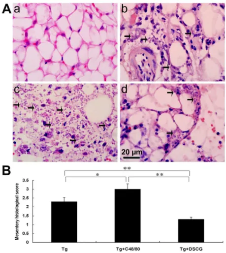
Figure 9. The mesentery histopathology of T. gondii-infected mice from different groups.
Infected mice i.p. inoculated with 10β RH tachyzoites of T. gondii were killed at 9-10 days p.i. (A) Representative microscopic
pictures show sections from uninfected mouse treated with PBS (a), T. gondii-infected control mouse (b), T. gondii-infected mouse treated with C48/80 (c), and T. gondii-infected mouse treated with DSCG (d). Tachyzoites were indicated with arrows. H&E stain. (B) Histological score analysis of mesentery tissues. There were 4 mice per group, and the data are representative of two experiments. *, P < 0.05; **, P < 0.01 (compared to control).
doi: 10.1γ71/journal.pone.0077γβ7.g009
Figure 10. Parasite burden of T. gondii RH strain tachyzoites in the peritoneal lavage fluids and tissues.
(A) Parasite burden of T. gondii RH strain tachyzoites in the peritoneal lavage fluids and (B) normalized mRNA expression levels of
T. gondii tachyzoite SAG1 gene in the spleens and livers using qRT-PCR, from different groups i.p. inoculated with 10βT. gondii RH
strain tachyzoites at 9-10 days p.i. There were 4 mice per group, and the data are representative of two experiments. Symbols indicate statistically significant differences (P < 0.01) for comparison with the uninfected controls (**) and for comparison between group means (§).
doi: 10.1γ71/journal.pone.0077γβ7.g010
Figure 11. Cytokine mRNA expressions in spleens from different groups i.p inoculated with 102 T. gondii RH strain tachyzoites at 9-10 days p.i., using qRT-PCR.
There were 4 mice per group, and the data are representative of two experiments. Symbols indicate statistically significant differences (P < 0.01) for comparison with the uninfected control mice (**) and the infected controls (§), and statistically significant differences (P < 0.05) for comparison with the infected controls (#).
doi: 10.1γ71/journal.pone.0077γβ7.g011
Figure 12. Cytokine mRNA expressions in livers from different groups i.p. inoculated with 102 T. gondii RH strain tachyzoites at 9-10 days p.i., using qRT-PCR.
There were 4 mice per group, and the data are representative of two experiments. Symbols indicate statistically significant differences (P < 0.01) for comparison with the uninfected control mice (**) and the infected controls (§), and statistically significant differences (P < 0.05) for comparison with the infected controls (#).
doi: 10.1γ71/journal.pone.0077γβ7.g01β
Author Contributions
Conceived and designed the experiments: FL BH SH LHK. Performed the experiments: BH SH YC HZ. Analyzed the data:
BH SH FL. Contributed reagents/materials/analysis tools: JS Z-RL YW. Wrote the manuscript: FL BH SH LHK.
References
1. Reid AJ, Vermont SJ, Cotton JA, Harris D, Hill-Cawthorne GA et al. (β01β) Comparative genomics of the apicomplexan parasites Toxoplasmagondii and Neosporacaninum: Coccidia differing in host range and transmission strategy. PLOS Pathog 8: e100β567. PubMed: ββ457617.
β. Cavalcanti MG, Mesquita JS, Madi K, Feijó DF, Assunção-Miranda I et al. (β011) MIF participates in Toxoplasma gondii-induced pathology following oral infection. PLOS ONE 6: eβ5β59. doi:10.1γ71/ journal.pone.00β5β59. PubMed: β1977ββ8.
γ. Gersch C, Dewald O, Zoerlein M, Michael LH, Entman ML et al. (β00β) Mast cells and macrophages in normal C57/BL/6 mice. Histochem Cell Biol 118: 41–49. PubMed: 1β1ββ446.
4. Gurish MF, Boyce JA (β006) Mast cells: ontogeny, homing, and recruitment of a unique innate effector cell. J Allergy Clin Immunol 117: 1β85–1β91. doi:10.1016/j.jaci.β006.04.017. PubMed: 16750988. 5. Matsuguchi T (β01β) Mast cells as critical effectors of host immune
defense against gram-negative bacteria. Curr Med Chem 19: 14γβ– 144β. doi:10.β174/09β98671β7998β8γ19. PubMed: ββγ60480. 6. Hepworth MR, Daniłowicz-Luebert E, Rausch S, Metz M, Klotz C et al.
(β01β) Mast cells orchestrate type β immunity to helminths through regulation of tissue-derived cytokines. Proc Natl Acad Sci U S A 109: 6644–6649. doi:10.107γ/pnas.111ββ68109. PubMed: ββ49γβ40. 7. Collington J, Williams TJ, Weller CL (β011) Mechanisms underlying the
localisation of mast cells in tissues. Trends Immunol γβ: 478–485. doi: 10.1016/j.it.β011.08.00β. PubMed: β19175ββ.
8. Lu LF, Lind EF, Gondek DC, Bennett KA, Gleeson MW et al. (β006) Mast cells are essential intermediaries in regulatory T-cell tolerance. Nature 44β: 997–100β. doi:10.10γ8/nature05010. PubMed: 169β1γ86. 9. Grimbaldeston MA, Nakae S, Kalesnikoff J, Tsai M, Galli SJ (β007)
Mast cell-derived interleukin 10 limits skin pathology in contact dermatitis and chronic irradiation with ultraviolet B. Nat Immunol 8: 1095–1104. doi:10.10γ8/ni150γ. PubMed: 1776716β.
10. Dawicki W, Marshall JS (β007) New and emerging roles for mast cells in host defence. Curr Opin Immunol 19: γ1–γ8. doi:10.1016/j.coi. β006.11.006. PubMed: 171β6541.
11. Im SJ, Ahn MH, Han IH, Song HO, Kim YS et al. (β011) Histamine and TNF-alpha release by rat peritoneal mast cells stimulated with Trichomonas vaginalis. Parasite 18: 49–55. doi:10.1051/parasite/ β011181049. PubMed: β1γ95β05.
1β. Maurer M, Lopez Kostka S, Siebenhaar F, Moelle K, Metz M et al. (β006) Skin mast cells control T cell-dependent host defense in Leishmaniamajor infections. FASEB J β0: β460–β467. doi:10.1096/fj. 06-5860com. PubMed: 1714β795.
1γ. S Ferreira GL, Mineo JR, Oliveira JG, V Ferro EA, Souza MA et al. (β004) Toxoplasmagondii and mast cell interactions in vivo and in vitro: experimental infection approaches in Calomys callosus (Rodentia, Cricetidae). Microbes Infect 6: 17β–181. doi:10.1016/j.micinf. β00γ.11.007. PubMed: 14998515. doi:10.1016/j.micinf.β00γ.11.007 PubMed: 14998515
14. Sun J, Sukhova GK, Yang M, Wolters PJ, MacFarlane LA et al. (β007) Mast cells modulate the pathogenesis of elastase-induced abdominal aortic aneurysms in mice. J Clin Invest 117: γγ59–γγ68. doi:10.117β/ JCIγ1γ11. PubMed: 179γβ568.
15. Wu B, Huang B, Chen Y, Li S, Yan J et al. (β01γ) Upregulated expression of Tim-γ involved in the process of toxoplasmic encephalitis in mouse model. Parasitol Res 11β: β511–β5β1. doi:10.1007/ s004γ6-01γ-γ416-1. PubMed: βγ595β1γ.
16. Messina A, Knight KR, Dowsing BJ, Zhang P, Phan LH et al. (β000) Localization of inducible nitric oxide synthase to mast cells during ischemia/reperfusion injury of skeletal muscle. Lab Invest 80: 4βγ–4γ1. doi:10.10γ8/labinvest.γ780047. PubMed: 10744078.
17. Subauste CS, Koniaris AH, Remington JS (1991) Murine CD8+
cytotoxic T lymphocytes lyse Toxoplasma gondii-infected cells. J Immunol 147: γ955–γ959. PubMed: 1940γ78.
18. Gazzinelli RT, Hakim FT, Hieny S, Shearer GM, Sher A (1991) Synergistic role of CD4+ and CD8+ T lymphocytes in IFN- production
and protective immunity induced by an attenuated Toxoplasmagondii vaccine. J Immunol 146: β86–β9β. PubMed: 1670604.
19. Gil CD, Mineo JR, Smith RL, Oliani SM (β00β) Mast cells in the eyes of Calomys callosus (Rodentia: Cricetidae) infected by Toxoplasma
gondii. Parasitol Res 88: 557–56β. doi: 10.1645/00ββ-γγ95(β00β)088[0557:MAONCO]β.0.CO;β. PubMed: 1β107479.
β0. Henderson WR Jr, Chi EY (1998) The importance of leukotrienes in mast cell-mediated Toxoplasma gondii cytotoxicity. J Infect Dis 177: 14γ7–144γ. doi:10.1086/5178γγ. PubMed: 959γ04γ.
β1. Huang S, Lu F, Chen Y, Huang B, Liu M (β01γ) Mast cell degranulation in human periodontitis. J Periodontol 84: β48–β55. doi:10.190β/jop. β01β.1β0066. PubMed: ββ509749.
ββ. Okayama Y, Semper A, Holgate ST, Church MK (1995) Multiple cytokine mRNA expression in human mast cells stimulated via Fc epsilon RI. Int Arch Allergy Immunol 107: 158–159. doi: 10.1159/000βγ696γ. PubMed: 754β065.
βγ. Yamamoto YI, Mineo JR, Meneghisse CS, Guimarães AC, Kawarabayashi M (1998) Detection in human sera of lgG, lgM, and IgA to excreted/secreted antigens from Toxoplasmagondii by use of dot-ELISA and immunoblot assay. Ann Trop Med Parasitol 9β: βγ–γ0. doi:10.1080/000γ49898595β8. PubMed: 9614450.
β4. Gupta S, Duraiswamy B, Satishkumar MN (β01γ) Peritoneal mast cell stabilization potential of Pothosscandens L. Indian J Pharmacol 45: 8γ–86. doi:10.410γ/0β5γ-761γ.10644β. PubMed: βγ54β88γ.
β5. Oldenburg PJ, Mustafa SJ (β005) Involvement of mast cells in adenosine-mediated bronchoconstriction and inflammation in an allergic mouse model. J Pharmacol Exp Ther γ1γ: γ19–γβ4. PubMed: 156β67β7.
β6. Metcalfe DD, Baram D, Mekori YA (1997) Mast cells. Physiol Rev 77: 10γγ–1079. PubMed: 9γ54811.
β7. Carlos D, de Souza Júnior DA, de Paula L, Jamur MC, Oliver C et al. (β007) Mast cells modulate pulmonary acute inflammation and host defense in a murine model of tuberculosis. J Infect Dis 196: 1γ61– 1γ68. doi:10.1086/5β18γ0. PubMed: 179ββ401.
β8. Miyatake A, Fujita M, Nagasaka Y, Fujita K, Tamari M et al. (β007) The new role of disodium cromoglycate in the treatment of adults with bronchial asthma. Allergol Int 56: βγ1–βγ9. doi:10.βγγβ/ allergolint.O-06-444. PubMed: 17519581.
β9. Theoharides TC, Wang L, Pang X, Letourneau R, Culm KE et al. (β000) Cloning and cellular localization of the rat mast cell 78-kDa protein phosphorylated in response to the mast cell "stabilizer" cromolyn. J Pharmacol Exp Ther β94: 810–8β1. PubMed: 109458β8. γ0. Swedenborg J, Mäyränpää MI, Kovanen PT (β011) Mast cells:
important players in the orchestrated pathogenesis of abdominal aortic aneurysms. Arterioscler Thromb Vasc Biol γ1: 7γ4–740. doi:10.1161/ ATVBAHA.110.β1γ157. PubMed: β1β05988.
γ1. Woolley DE (β00γ) The mast cell in inflammatory arthritis. N Engl J Med γ48: 1709–1711. doi:10.1056/NEJMcibr0βγβ06. PubMed: 1β711748.
γβ. Nickdel MB, Lyons RE, Roberts F, Brombacher F, Hunter CA et al. (β004) Intestinal pathology during acute toxoplasmosis is IL-4 dependent and unrelated to parasite burden. Parasite Immunol β6: 75– 8β. doi:10.1111/j.0141-98γ8.β004.00686.x. PubMed: 15ββ5β94. γγ. Lu F, Huang S, Kasper LH (β00γ) Interleukin-10 and pathogenesis of
murine ocular toxoplasmosis. Infect Immun 71: 7159–716γ. doi: 10.11β8/IAI.71.1β.7159-716γ.β00γ. PubMed: 146γ8808.
γ4. Matowicka-Karna J, Dymicka-Piekarska V, Kemona H (β009) Does Toxoplasmagondii infection affect the levels of IgE and cytokines (IL-5, IL-6, IL-10, IL-1β, and TNF-alpha)? Clin Dev Immunol β009: γ74696. γ5. Bischoff SC, Lorentz A, Schwengberg S, Weier G, Raab R et al. (1999)
Mast cells are an important cellular source of tumour necrosis factor alpha in human intestinal tissue. Gut 44: 64γ–65β. doi:10.11γ6/gut. 44.5.64γ. PubMed: 10β05β00.
γ6. Gatti S, Faggioni R, Sironi M, Erroi A, Ghezzi P (199γ) Mast cells do not contribute to the rapid appearance of TNF in the serum of LPS-treated mice: a study with mast cell-deficient mice. Int J Immunopharmacol 15: 551–555. doi:10.1016/019β-0561(9γ)90071-6. PubMed: 8γ759γ7.
γ7. Suzuki Y, Sa Q, Gehman M, Ochiai E (β011) Interferon-gamma- and perforin-mediated immune responses for resistance against Toxoplasmagondii in the brain. Expert Rev Mol Med 1γ: eγ1. doi: 10.1017/S146βγ9941100β018. PubMed: ββ005β7β.
γ8. Norose K, Mun HS, Aosai F, Chen M, Piao LX et al. (β00γ) IFN-gamma-regulated Toxoplasma gondii distribution and load in the murine eye. Invest Ophthalmol Vis Sci 44: 4γ75–4γ81. doi:10.1167/ iovs.0γ-0156. PubMed: 1450788β.
γ9. Suzuki Y, Yang Q, Yang S, Nguyen N, Lim S et al. (1996) IL-4 is protective against development of toxoplasmic encephalitis. J Immunol 157: β564–β569. PubMed: 8805658.
40. Roffê E, Rothfuchs AG, Santiago HC, Marino AP, Ribeiro-Gomes FL et al. (β01β) IL-10 limits parasite burden and protects against fatal myocarditis in a mouse model of Trypanosoma cruzi infection. J Immunol 188: 649–660. doi:10.4049/jimmunol.100γ845. PubMed: ββ156594.
41. Katara GK, Ansari NA, Verma S, Ramesh V, Salotra P (β011) Foxpγ and IL-10 expression correlates with parasite burden in lesional tissues
of post kala azar dermal leishmaniasis (PKDL) patients. PLoS Negl Trop. Drosophila Inf Serv 5: e1171.
4β. Jones LA, Roberts F, Nickdel MB, Brombacher F, McKenzie AN et al. (β010) IL-γγ receptor (T1/STβ) signalling is necessary to prevent the development of encephalitis in mice infected with Toxoplasmagondii. Eur J Immunol 40: 4β6–4γ6. doi:10.100β/eji.β009γ9705. PubMed: 1995018γ.
4γ. Zhao J, Endoh I, Hsu K, Tedla N, Endoh Y et al. (β011) S100A8 modulates mast cell function and suppresses eosinophil migration in acute asthma. Antioxid Redox Signal 14: 1589–1600. doi:10.1089/ars. β010.γ58γ. PubMed: β114β608.
44. Jash A, Kwon HK, Sahoo A, Lee CG, So JS et al. (β011) Topical application of porcine placenta extract inhibits the progression of experimental contact hypersensitivity. J Ethnopharmacol 1γγ: 654–66β. doi:10.1016/j.jep.β010.10.054. PubMed: β1040771.