The Rh system if of great clinical interest because its antibo-dies are involved in erythrocyte immune-mediated destruction; i.e., hemolytic transfusion reaction and hemolytic disease of the newborn (HDN)1. The first reports about its importance began
around the year 1600, as a possible cause of severe jaundice and newborn death referred to as “erythroblastosis fetalis”2. A
French woman gave birth to twins: one was hydropic and the other was jaundiced, later dying of kernicterus.
This wide variety of newborn signs and symptoms which ranges from light jaundice to fatal edema was not really associated with one single agent. The first correlation of an anti-erythrocyte antibody involved against the Rh antigen was reported by Levine et al. in 1939 through the investigation of a hemolytic transfusion reaction in a post-partum woman due to the transfusion of ABO red blood cells compatible with her husband, after giving birth to a stillborn3. This woman’s serum
agglutinated her husband’s red blood cells, and that of about 80% of compatible ABO donors. It was demonstrated that this new antibody was independent of the ABO, MN and P systems, suggesting the woman had probably been immunized by a fetal antigen from her father.
In the following year, Landsteiner et al.4 described an
antibody obtained through the immunization of guinea pigs and red blood cells of rhesus macaque. This serum agglutinated approximately 85% of tested human red blood cells and the corresponding determinant was called Rh factor. The study with 60 families showed that the positive Rh was inherited with dominant character.
Also in 1940, Wiener et al.5 observed the same specificity
in the serum of people who lacked the same determinant and who had received compatible ABO transfusions in the past.
The following year, Levine et al.6 found similar antibodies in
the serum of several post-partum women and these presented similar reactions to the anti-Rhesus animal serum.
Later, in 1942, Fisk et al.7 showed the difference between
the human and animal anti-Rh, and concluded that it was not the same antibody. The Rh nomenclature, however, was maintained.
Only after 1963 Levine et al.8 proposed that the
hetero-antibody of rabbits should be named anti-LW (a tribute to Landsteiner and Wiener) and the human should be named anti-D. The Rh name resulted from the LW antigen. It was only in the mid-1940s that four additional antigens (C, c, E, e) were recognized as belonging to the Rh system. Race et
*Correspondence:
Av. Lopes de Azevedo, 888 Cidade Jardim
São Paulo – SP, Brazil CEP:05603-001 Tel: 38151-390 [email protected]
AbstrACt
The Rh system is the most polymorphic and immunogenic of all blood group systems. Currently, over 49 antigens have been identified, among which the five main antigens are D, C, c, E, e. Knowledge of the Rh system’s molecular basis, since its first cloning 17 years ago, has allowed for an understanding of the mechanism of Rh-negative phenotype and of the variants of antigens RHD and RHCE. Deletions, gene rearrangements and insertions are the main mutations. In Caucasians, the primary mechanism of Rh-negative phenotype is the complete RHD gene deletion, whereas in Afrodescendant it is the presence of pseudogene RHDø and hybrid gene RHD-CE (4-7)-D. The authors analyzed the structure of the Rh complex in red cells, the molecular basis of the Rh system, mechanisms of RHD negativity and weak and partial RHD expressions.
Key Words:Rh-Hr blood-group system. Rh isoimmunization. Antigen-Antibody complex.
the
molecular
basis
of
rh
system
and
its
applications
in
obstetrics
and
transfusion
medicine
luciano marcondes machado nardozza1*, alexandre szulman2, Jose augusto barreto3, edward arauJo Junior4, antonio fernandes moron5
Study conducted at Universidade Federal de São Paulo – UNIFESP, São Paulo, SP, Brazil
1- Livre-docente - Professor associado e chefe disciplina Medicina Fetal da Universidade Federal de São Paulo – UNIFESP, São Paulo, SP 2- Mestre – Pós-graduando do departamento de Obstetrícia da Universidade Federal de São Paulo – UNIFESP, São Paulo, SP
3- Doutor em Cardiologia - Diretor da COLSAN, São Paulo, SP
al.9, having four Rh antiserums of different specificities already
defined seven alleles, whereas Wiener10, with three antiserums,
determined six.
Since his discovery, it was suspected that these antigens were transmitted in a block; one single gene encoded agglu-tinogenic numbers, or three closely attached genes encoded several products. Currently, over 49 antigens have been iden-tified through antibodies produced after blood transfusion or pregnancy. The main antigen, from a clinical point of view, is RhD, followed by Rhc.
Antigen D is the most immunogenic of the Rh system, 20 times more powerful than c. Approximately 80% of Rh negative individuals who receive Rh positive blood will produce anti-D antibodies after the first contact11 and only 7% to 8% of Rh
negative individuals will remain non-responsive12.
Antibodies against RhD are the main cause of hemolytic disease of the newborn (HDN)13. The determination of Rh
blood time is important, and should be carried out early in the pregnancy, because HDN presents with significant perinatal morbidity and mortality. RhD negative pregnant women with Rh positive fetuses could present sensitivization during labor, which answers for 14% of alloimmunization cases14.
Even after the introduction of anti-D immunoglobin, in the late 1960s, and the combination of pre- and post-partum immu-noprophylaxis in the late 1990s, the incidence of alloimmuni-zation to antigen D ranges from 0.8% to 1.5% in RhD negative pregnant women, caused by fetal-maternal hemorrhage15.
In Brazil, Rh alloimmunization is still the main cause of HDN and anti-D is still the main cause of the indication of photo-therapy or exchange transfusion in newborns. The prophylaxis with immunoglobulin anti-D, when administered with the right dosage and in early stage, could prevent sensitivization to antigen D16-17.
Therefore, detailed knowledge of the Rh system is of great importance, particularly in Obstetrics and Transfusion Medicine.
rh complex structure in red blood cells
The Rh blood group (ISBT 004) is the most complex, polymorphic and immunogenic blood group system known in humans. After the ABO, it is the most important in Transfusion Medicine18.
Five main and important antigens -- D(RH1), C(RH2), E (RH3), c(RH4) and e(RH5) – can be distinguished and are responsible for most clinically significant antibodies. With over 49 different antigens characterized, it is the largest of all blood systems.
The Rh positive and Rh negative refer to the presence or absence of antigen D, but bith express antigens C\c and E\e. C is antithetical to c whereas antigen E is antithetical to e. Each chromosome contains the C or c and E or e genes18.
The antigens are located in two proteins expressed in the membrane of erythrocytes and their immediate precursors: rhD (CD240D) and rhCE (CD240CE), which respectively carry antigens D(rh1) and C, c, E, e (rh2-rh5) in various combi-nations (ce, cE, Ce and CE). Both proteins, RHD and RHCE, are
hydrophobic and non-glycolized, each with a molecular weight of 30 to 32 KD, made up of 417 aminoacids distributed in six extracellular segments (responsible for the immune response), 12 transmembranous and seven intracellular. The N-terminal and C-terminal portions are intracellular19.
The RhD and RhCE proteins present with 92% homology and differentiate in 35/36 aminoacids (8.4% divergence), suggesting that the corresponding genes are the result of the duplication of a common ancestral gene19-20. This concept is
based on the identification of Rh-like genes in non-human primates. The differences between RHD and RHCE occur in the extracellular region, in which loops (extracellular portions) 2, 3, 4 and 6 are restricted (Figure 1).
In the second loop, allele c differentiates proteins RhD and RhCE. Proteins C/c and E/e are produced by a mechanism of alternative pre-processing messenger RNA splicing and poly-morphisms ser103-pro and pro226-ala and are responsible for the specificity of C/c and E/e, respectively21.
On the erythrocyte membrane, the Rh protein, along with the glycoprotein associated with Rh (rhAG/rH50), encoded by another gene located on chromosome 6p12-p21, form the so called “Rh complex”22. The Rh complex is firmly attached to
the cytoskeleton of the erythrocyte membrane. Other additional proteins, such as LW, glycoprotein Duffy, band 3 and integrin-associated protein (CD47) are linked to the Rh complex, but are not necessary to its expression (Figure 2).
The expression of Rh on red blood cell surface depends on the functional RhAG protein. When there is absence of RH50 protein, antigens D,E,C,e,c are not expressed, and the pheno-type is referred to as Rhnull. Because they present homology with
other proteins, proteins Rh and RhAG appear to be involved in the ammonia transportation. Additionally, it is supposed that they are also involved in the transportation of O2 and CO2
gases. Changes in the shape of red blood cells, especially in individuals with Rhnull phenotype, indicate a strong interaction
between the Rh complex and the cytoskeleton of the erythrocyte membrane22.
Molecular basis of the rh system
The RHD gene was discovered in 1992, two years after the RHCE. Currently, although over 170 RHD alleles have been described, this gene has not yet been fully characterized. Many mammals present only one Rh gene corresponding to the RHCE gene in humans.
Experiments conducted with the Southern Blot technique using a cDNA Rh probe have shown that only three species carry more than one Rh gene: chimpanzees, gorillas and humans23.
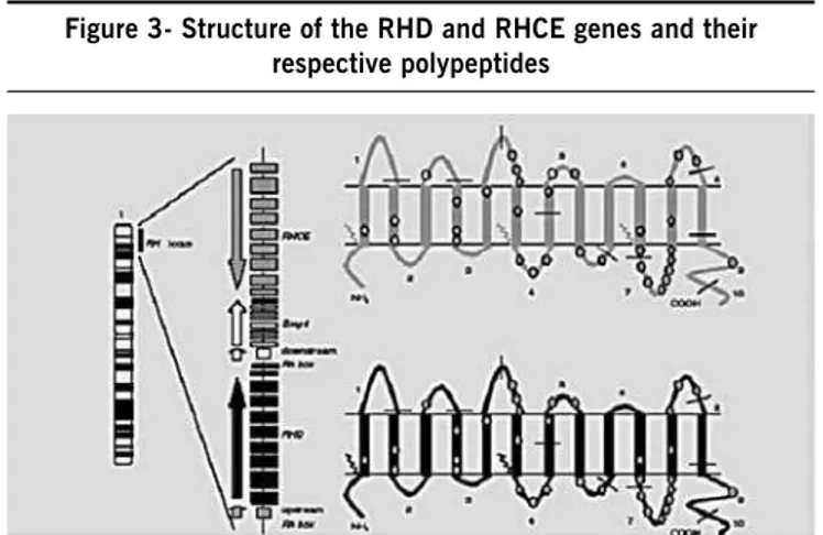
The two genes of the Rh system (RHD and RHCE) are located on the short arm of chromosome 1, locus 34-36 (Figure 3).
These are highly homologue genes (93.8%), each contai-ning 10 exons, with a total sequence of approximately 60,000 pb.
The SMP1 gene (Small Membrane Protein 1) is interposed between RHD and RHCE. SMP1 is functionally related to Rh in terms of its expression on the erythrocyte membrane. RHD is flanked by two DNA segments with the size of 9000 pb, homo-logy of 98.6% and identical orientation, called Rhesus boxes24.
rHD negativity
D negative red blood cells do not entirely express the RhD protein on the erythrocyte surface. There are basically three molecular mechanisms of RHD negativity: total deletion of RHD gene, RHD pseudogene and hybrid gene, which vary in frequency according to the race in question19. In Caucasians,
found in 15% to 17% of the population, the most common mechanism is the complete deletion of the RHD gene. The RhD negative phenotype is most commonly caused by homozygosis of a haplotype in which the RHD gene has been deleted. This deletion occurs in Rhesus boxes in a region that is identical to 1463 pb, probably caused by defective chromosome alignment during meiosis, resulting in an unequal crossing-over between Rhesus boxes. The result is characterized by the presence of a hybrid Rhesus Box24. Few cases may result from genie
rearran-gements or point mutations leading to stop codon.
The RHD gene could also not be expressed due to premature stop codon, nucleotide insertions, point mutations, or hybrid RHD/CE. Fourteen different D- alleles are described with a frequency of 1/1500 in the Caucasian population19.
The deletion of the RHD gene is observed in only 18% of the Afrodescendant population and in 60% of Asians, considering that, among the latter, the RHD gene is found in less than 1% of the population.
The most common variant in D negative Asians is the pheno-type Del RHD (K409K), i.e., 10% to 30% have the RHD gene
intact18. Various mechanisms contribute to the vast number of
non-functioning Rh alleles.
In Afrodescendants with RhD negative phenotype, the most common mechanism is the presence of the pseudogene RHD(RHDø), associated with the ce allele in the RHCE. The RHD pseudogene contains a duplicate sequence of 37 pb, compromising the the last 19 nucleotides of intron 3 and the first 18 nucleotides of exon 4, thus causing four missense mutations in exon 5 (609G>A, 654G>C, 667T>G, 674C>T) and one nonsense mutation in exon 6 (807T>G), which causes the creation of a stop codon signal(Y269X)25-29.
In this way, a non-functional protein is produced and no polypeptide D reaches the erythrocyte surface. This generates non-functional Rh protein, representing approximately 70% of Afrodescendant with an inactive gene. Currently, RHDØ detec-tion through the polymerase chain reacdetec-tion (PCR) method is considered mandatory for any antigen D prediction19.
Regarding the Rh negative phenotype due to hybrid gene, the high similarity in opposite directions and the proximity to the same chromosome favor a cis gene conversion and the formation of a hairpin loop, in which the inner part of a gene is exchanged by corresponding parts of another gene. The results of this mutation are the hybrid alleles RHD-CE-D and RHCE-D-CE.
The hybrid gene RHD-CE (4-7)D (Ces) contains both altered
genes: a hybrid gene RHD-CE-D segregated as ces allele of
the RHCE. The haplotype (C)ces has two different molecular
bases30.
- Type 1, which consists of exons 1, 2 , terminal 5´ of exons 8, 9 and 10 of the RHD gene, joined with terminal 3´ of exon 3 starting on nucleotide 455, exons 4 to 7 of the RHCE gene. - Type 2, in which the hybrid gene has a fusion of exons 1, 2, complete 3, 8, 9 and 10 of the RHD gene, joined with exons 4 to 7 of the RHCE gene.
Both encode two nucleotides 733C>G (exon 5) and 1006G>T (exon 7) carried by RHCE, generating two substitu-tions, respectively: Leu245Val in the eighth transmembranous region and Gly336Cys in the eleventh. The haplotype (C) ces encodes a partial C,c associated with a weak e and with
antigen VS.
This mechanism is the second most frequent Afrodescen-dant RhD negative phenotype, associated with phenotype VS V- with a frequency of 0.03%. In Asians, however, the hybrid gene RHD-CE (2-9) is the most common in Rh negative donors, after the Del phenotype31-32.
Weak D expression
Normally, RhD positive red blood cells have an antigenic density ranging from 15000 to 33000 antigens per cell, depen-ding on the haplotype. However, some phenotypes with density ranging from 70 to 5200 RhD antigens have been identified. These phenotypes are called weak D and are caused by the substitution of aminoacids in the transmembranous and intra-cellular portions of the RhD protein due to a single missense mutation in the RHD gene. Weak D phenotype red blood cells express an intact antigen RhD, and occur in 0.2% to 1% of caucasians33.
Currently, over 40 types of weak D have been identified on the molecular level, in among which the most frequent are weak D type 1 and 2 (70% and 18%, respectively). Due to the fact that the weak D phenotype carries the intact RhD antigen, the probability of forming anti-d alloantibody is very unlikely. However, the presence of anti-D in some weak D cases (type 1, 4.2 and 15) has been reported34-35.
The difference between weak D and partial D must not be drawn by the production of anti-D. To predict the risk of immunization in weak D individuals, the Rhesus index was proposed34. This index is based on the antigenic density of
various monoclonal antibodies, depending on the quantity of antigenic sites and on the affinity of the antibody. Theoretically, the index could range from 1 (low risk, e.g., normal RhD) to 0 (high risk, e.g., partial RhD with missing epitopes). Type 4.2 and 15 weak D present the lowest Rhesus index (0.21), with the exception of type 7 (0.03).
Partial D expression
Figure 1 – schematics of the rh proteins on the erythrocyte membrane
Legend: Adapted from Wagner EF, Flegel WA. Review: the molecular basis of the Rh blood group phenotypes. Immunoematology 2004;20:23-35.
epitopes36, involving various extracellular loops.
Partial D red blood cells are defined by the absence of one or more epitopes caused by the rearrangement of the RHD and
Figure 2 – schematics of the rh complex on the erythrocyte membrane
Legend: Adapted from Kim CLV, Colin Y, Cartron JP. Rh proteins: Key structural and functional components of the red cell membrane. Blood Reviews 2006;20:93-110.
Figure 3- structure of the rHD and rHCE genes and their respective polypeptides
Legend: Adapted from Martine GHM Tax. Rh variabiliteit vanuit multi-etnisch perspectief Con-sequenties voor RH genotypering. © 2005, Rotterdam, The Netherlands. ISBN 90-9020176-9
RHCE genes. This genetic configuration enables microconver-sion and single-direction exchanges of fragments of RHD and RHCE gene fragments or parts, leading to the formation of RHD-CE-D or RHCE-D-CE alleles, respectively37. These new
Rh aberration alleles not only produce hybrid proteins, RhD regions joined with RhCE leading to the loss of D epitopes, but also generate new antigens.
Few partial D phenotypes result from exchanges of one single aminoacid. However, in contrast with weak D, poly-morphism occurs in the extracellular segments of the RhD protein. Partial D individuals may frequently produce anti-D against those absent epitopes when exposed to the complete RhD protein.
This clarification of the molecular basis of the Rh system has enabled the development of serum-based and molecular techniques to determine these variants among the various populations. In the field of Obstetrics, it enables unveiling the Rh negative phenotype in pregnant women, thus aiding in the understanding of the correlation between phenotype, genotype and Rh alloimmunization. Although there is high concordance between the phenotyping and genotyping of the Caucasian population, false-positive results are observed in Afrodescen-dant and Asians, which could frequently occur depending on the strategy used.
Another advantage of the molecular study would be to avoid unnecessary anti-D immunoprophylaxis (3% to 5%)38 in RhD
negative pregnant women with a specific type of weak D, as well as the identification of partial D pregnant women who present a high probability of alloimmunization and require Rh immunoprophylaxis.
This reasoning can also be extended to Transfusion Medi-cine, thus preventing many complications and reactions after transfusions.
Conflict of interest: No conflicts of interest declared concer-ning the publication of this article.
r
eferences1. Simsek S, Faas BH, Bleeker PM, Overbeeke MA, Cuijpers HT, Van der Schoot CE, et al. Rapid RhD genotyping by polymerase chain reaction-based amplification of DNA. Blood. 1995;29:75-80.
2. Race RR, Sanger R. Blood groups in man. 6th ed. Oxford: Blackwell; 1975.
3. Levine P, Stetson RE. An unusual case of intra-group agglutination. J Am Med Assoc 1939;113:126-7.
4. Landsteiner K, Wiener AS. An agglutinable factor in human blood recognized by immune sera for Rhesus blood. Proc Soc Exp Biol NY. 1940;43:223. 5. Wiener AS, Peters HR. Hemolytic reactions following transfusions of blood of
the homologous group, with three cases in which the same agglutinogen was responsible. Ann Intern Med. 1940;13:2306-22.
6. Levine P, Burnham L, Katzin WM, Vogel P. The role of isoimmunization in the pathogenesis of erythroblastosis fetalis. Am J Obstet Gynecol. 1941;42:925-37.
7. Fisk RT, Foord AG. Observations on the Rh agglutinogen of human blood. Am J Clin Pathol. 1942;12:545.
8. Levine P, Celano MJ, Wallace J, Sanger R. A human ‘D-Iike’ antibody. Nature.1963;198:596-7.
9. Race RR, Taylor GL, Cappell DF, McFarlane MN. Recognition of a further common Rh genotype in man. Nature. 1944;153:52-3.
Artigo recebido: 14/04/10 Aceito para publicação: 25/08/10
11. Urbaniak SJ, Robertson AE. A successful program of immunizing Rh-negative male volunteers for anti-D production using frozen/thawed blood. Transfu-sion. 1981;21:64.
12. Issit PD. The Rh blood group system. In: Garratty G, editor. Immunobiology of transfusion medicine. New York: Marcel Dekker Inc; 1974. p.111-47. 13. Bianchi DW, Avent ND, Costa JM, Schoot CE. Noninvasive fetal Rh genotyping.
Obstet Gynecol. 2005;106:682-3.
14. Bowman JT. Five years of Rh prophylaxis. Transfusion. 2003;43:1661-6. 15. Pertl B, Pieber D, Panzitt T, Haeusler MC, Winter R, Tului L, et al. RhD
geno-typing by quantitative fluorescent polymerase chain reaction: a new approach. BJOG. 2000;107:1498-502.
16. Vicent LF, Pinto G, Serrano F. Profilaxia da isomunização RHD. Acta Méd Port. 2003;16:225-60.
17. Fung FK, Eason E, Crane J, Armson A, De La Ronde S, Farine D, et al. Maternal-Fetal Medicine Comittee, Genetics Committee. Prevention of Rh alloimunization. J.Obstet Gynaecol Can 2003;25:765-73.
18.Klein HG, Anstee DJ Mollinson´s blood transfusion in clinical medicine. 11th
ed. New York: Blackweel Science; 2005.
19. Wagner FF, Flegel WA. Review: the molecular basis of the Rh blood group phenotypes. Immunohematology 2004;20:23-36.
20. Flegel WA. The genetics of the rhesus blood group system. Dtsch Arztebl 2007;104:A 651-7.
21. Avent ND, Reid ME. The Rh blood group system: a review. Blood. 2000;95:375-87.
22. Kim CLV, Colin Y, Cartron JP. Rh proteins: key structural and functional compo-nents of the red cell membrane. Blood Rev. 2006;20:93-110.
23. Westhoff CM, Wylie DE. Investigation of the human Rh blood groups systemn nonhuman primates and other species with serologic and southern blot analysis. J Mol Evol. 1994;39:87-92.
24. Wagner FF, Flegel WA. RHD gene deletion occurred in the Rhesus box. Blood. 2000;95:3662-8.
25. Singleton BK, Green CA, Avent ND, MartinPG, Smart E, Daka A, et al. The presence of an RHD pseudogene containing a 37 base pair duplication and a non sense mutation in Africans with the Rh negative blood group phenotype. Blood. 2000;95:12-18.
26. Rodrigues A, Rios M, Pellegrino Jr, Costa FF, Castilho L. Presence of the RHD pseudogene and hybrid RHD-CE-D gene in Brazilians with the D-negative phenotype. Braz J Med Biol Res. 2002;35:767-73.
27. Westhoff CM. The structure and function of the Rh antigen complex. Semin Hematol. 2007;44:42-50.
28. Avent ND. High variability of the RH locus in different ethnic backgrounds. Transfusion. 2005;45:293-4.
29. Daniels G, Finning K, Martin P, Summers J. Fetal blood group genotyping: present and future. Ann N Y Acad Sci.2006;1075:88-95.
30.Pham BN, Peyrard T, Juszczac G, Auxerre C, Godin S, Bonin P, et al. Alloanti-c (RH4) revealing that the (C)ces haplotype encodes a partial c antigen. Transfu-sion. 2009;49:1329-34.
31. Okuda H. RHD gene is highly detectable in RHD-negative japanese donors J Clin Invest. 1997;100:373-9.
32. Gassner C, Doescher A, Drnovsek TD, Rozman P, Eicher NI, Legler TJ, et al. Presence of RHD in serologically D-,C/E individuals: a European multicenter study. Transfusion. 2005;45:527-38.
33. Muller TH, Wagner FF, Trockenbacher A, Eichner NI, Flegel WA, Schonitzer D, et al. PCR screening for common weak D types shows different distributions in three central European populations. Transfusion. 2001;41:45-52. 34. Wagner FF, Frohmajer A, Ladewig B, Eicher NI, Lonicer CB, Muller TH, et
al. Weak D alleles express distinct phenotypes. Blood. 2000;95:2699-708. 35. Roxby D, Coloma M, Flegel WA, Poole J, Martin P, Abbott R. Observation
ofan anti-D after D-positive transfusion in an individual with weak D type-1 phenotype. Vox Sang. 2003;87(Suppl 3):17-44.
36. Scott ML, Voak D, Liu W, Jones JW, Avent ND. Epitopes on Rh proteins. Vox Sang. 2000;78 (Suppl 2):117-20.
37. Flegel WA, Wagner FF. Molecular Biology of partial D and weak D: implications for blood bank practice. Clin Lab. 2002;48:53-9.