Asymptomatic Perforation of Large Bowel and
Urinary Bladder as a Complication of
Ventriculoperitoneal Shunt: Report of Two Cases
Miljan Mihajlović1, Goran Tasić2,3, Mirjana Raičević1, Milan Mrdak1, Bojana Petrović1,
Vladimir Radlović1
1University Children’s Hospital, Belgrade, Serbia;
2Clinic for Neurosurgery, Clinical Centre of Serbia, Belgrade, Serbia; 3School of Medicine, University of Belgrade, Belgrade, Serbia
INTRODUCTION
Insertion of a ventriculoperitoneal (VP) shunt represents a classical and most frequently used method in the treatment of hydrocephalus [1]. Although introduced already in 1908 and followed by numerous complications, it still remains the solution of choice in the treatment of this pathological condition [1-4].
VP shunt complications, early or late, are classified as mechanical, infective and func-tional, i.e. associated with either excessive or insufficient drainage of cerebrospinal fluid [5]. The group of rarer complications of mechanical nature, usually associated with infection and/ or poor cerebrospinal fluid drainage, includes the migration of VP shunt distal segment into the thoracic cavity, heart, large bowel, urinary bladder, scrotum, umbilicus, inguinal hernia and other body regions [6-11]. As they can remain asymptomatic for a long period of time, the penetration of a VP catheter into the visceral organs is most frequently additionally complicated and disclosed late, and are thus followed by high mortality which, according to Ghritlaharey et al. [12], rates even up to 15%.
We present two children with asymptomatic disclosed on time and adequately resolved VP shunt migration into the large bowel and uri-nary bladder.
CASE REPORTS Patient 1
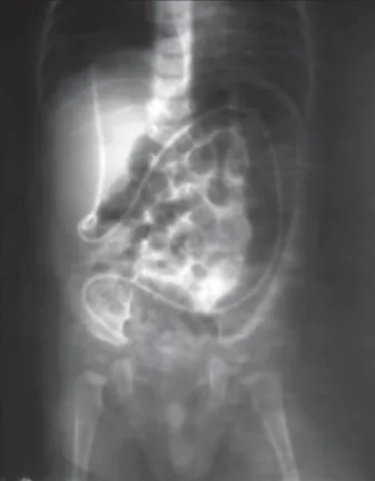
A six-month-old male neonate, with a VP shunt inserted due to congenital hydrocepha-lus with onset at age one month, presented at the hospital for an unplanned neurosurgical check-up due to a transanal protrusion of the anterior shunt noted during clothes changing of the child (Figure 1). According to parents, except for an unexplained rectal temperature of 38.5°C a week before admission, the child did not manifest any other setups. On examination neurological and general clinical findings were within normal limits. The fontanella was at the same level as the calvaria, the abdomen was soft and insensitive on palpation, with normal peristaltic sounds. Laboratory findings of liq-uor, blood count and urine were also normal. Endocranial CT imaging showed a normal shunt function, while thoracic and abdominal X-ray examination clearly showed the presence of the shunt in the large bowel lumen (Figure 2). A surgical revision of the entire VP shunt system was performed, and its peritoneal por-tion was removed and changed. There was no need to place suture at the area of colon perfo-ration. With a seven-day cessation of oral food intake, namely, the introduction of parenteral
SUMMARY
Introduction Insertion of a ventriculoperitoneal (VP) shunt, the method of choice in the treatment of hydrocephalus, is often followed by various mechanical and/or infective complications. We present two children with asymptomatic perforation of the large bowel and urinary bladder, relatively rare and potentially severe complications of this surgical procedure.
Outline ofCases In both patients a VP shunt was implanted in the first month after birth; in a boy due to congenital hydrocephalus and in a girl due to the consequences of intracranial haemorrhage. Immediately after surgery, as well as during the further course, in both children growth and development were optimal and without any signs of infection or VP shunt malfunction. In the boy at age 6 months and in the girl at age 4 years, without any signs of complications, mothers noted the prominence of the VP shunt tip from the anus in the first case and from the urethral orifice in the second one. The VP shunts were immediately changed, so that both complications were resolved without any consequences.
Conclusion Insertion of a VP shunt represents the most frequent method of choice of the surgical treat-ment of hydrocephalus, but also potentially a highly risky procedure followed by various complications about which parents should be informed when patients are children. Owing to adequate approach in the follow-up of children with implanted VP shunt, large bowel and urinary bladder perforation, examples of severe and potentially fatal complications of this surgical intervention, could be disclosed on time and adequately resolved.
Keywords: hydrocephalus; ventriculoperitoneal shunt; complications
Correspondence to: Miljan MIHAJLOVIĆ University Children’s Hospital Tiršova 10, 11000 Belgrade Serbia
nutriton and antibiotic therapy (amikacin, cephtriaxon, metronidazole), the patient completely recovered. VP shunt function, as well as control endocranial CT and abdomi-nal X-ray were completely normal. He was discharged in good general condition and without neurological sequels. Signs or any consequences of the complications were not registered during further follow-up.
Patient 2
A 4-year-old girl with inserted VP shunt at age one month due to posthaemorrhagic hydrocephalus, who came for a neurological examination due to the transurethral
promi-nence of the distal catheter of the VP shunt disclosed by chance. Neurological abnormalities, micturation problems, fever or any other upsets were not registered. A complete clinical finding also including inspection of the urethral orifice was normal. Abdominal X-ray and ultrasonographic examination of the small pelvis showed the presence of the distal portion of the VP catheter in the urinary bladder (Figures 3 and 4). A control endocranial CT examination verified the usual width of the chamber system speaking in favour of normal shunt function. Urinary analysis and uroculture confirmed urinary infection (Klebsiella spp.), while liquor was sterile. WBC count and leukocyte formula were within the referent limits, CRP was 20 mg/L and RBC sedimentation rate was 16/38. After the introduction of amikacin and cephtriaxon the peritoneal portion of the VP shunt was cystoscopically removed and a new one was percutaneously inserted. During cystoscopy in the area of the right side of the urinary bladder wall we verified the site of catheter perforation of about 3 mm in diameter sur-rounded by a field of inflammation. A Foley catheter was inserted into the urinary bladder, and antibiotic therapy was continued for further two weeks. The entire postop-erative recovery of the patient was normal, so that after a week the Foley catheter was removed. Control cystoscopy showed a preserved anatomic integrity of the urinary blad-der with a scar formation at the site of protrusion of the VP shunt distal segment. A few days after the removal of the Foley catheter the function of the detrusor and uri-nary bladder sphincter was restored. In further course the girl was in good condition, without any setups and with adequate neurological and neurosurgical findings, so that
Figure 1. Protrusion of peritoneal catheter through the anal orifice
Figure 2. Abdominal X-ray with shadow of VP catheter in the large bowel lumen
after two weeks of hospitalization she was discharged from hospital. During further follow-up, the presence of no set-ups or complications was verified.
DISCUSSION
In children with hydrocephalus, the immediate risk of sur-gical insertion of a VP shunt is relatively low; however, later complications are quite frequent and are seen in 24-47% of cases [13]. About one-fourth of complications occur at the abdominal level, most frequently involving the intestinal volvulus, peritoneal pseudocysts or extrusion and penetra-tion of the VP shunt distal part into the visceral organs [14, 15, 16]. In the literature one can sporadically find examples of VP catheter visceral perforation into the urinary bladder, vagina, gallbladder, stomach, bowels, scrotum, liver, vagina, urethra and other organs, with extrusion of its distal part through natural orifices (rectum, vagina, external urethral orifice) or the abdominal wall itself [5, 11, 15, 17, 18].
Gastrointestinal tract, and predominantly the large bowel, with an incidence of 0.1-0.7%, presents the most fre-quent area of VP shunt perforation [19]. First descriptions of large bowel perforation after the insertion of VP shut were published in 1966 by Wilson and Bertan [20]. Since then, 70 such cases have been reported in the literature, of which most occurred in children. It has been disclosed that visceral perforation of such aetiology in over 50% of cases has asymptomatic course, and that it is almost always diag-nosed only after the extrusion of the VP catheter through the natural orifices of the damaged organ, the abdominal wall itself or associated with the malfunction of the shunt [17, 18]. However, in a smaller number of children the
perforation of the visceral organs by the VP catheter is manifested by septic symptoms due to peritonitis, intra-abdominal abscess, meningitis, encephalitis, ventriculi-tis and/or brain abscesses [21, 22, 23]. Extensive clinical experience has shown that in such children underlying purulent meningitis or ventriculitis caused by Escherichia coli or some other coliform gram-negative bacteria is most probably a hidden asymptomatic bowel perforation caused by a VP catheter [18-24]. In addition, independent or com-bined with neurological indicators of shunt malfunction, intestinal or urinary perforation caused by the VP shunt is often followed by abdominal pain, vomiting diarrhoea and dysuric disorders [24, 25].
The exact basis of peritoneal catheter perforation into the lumen of body cavities has not been fully defined yet. There have been descriptions of cases of allergic and immunogenic reactions to chemical components of the VP catheter (silicon, latex), which resulted in the disrup-tion of skin continuity above the shunt, its obstrucdisrup-tion, as well as the perforation of the visceral organs [26, 27]. The formation of local inflammatory response and the resulting fibrosis, adherence and penetration of the distal portion of the catheter through the intestinal wall are presented as the stages of a possible mechanism of intestinal wall perfora-tion [18]. It has been suggested that subclinical infecperfora-tion of liquor, as well as increased protein quantity in the liquor trigger the above mechanism. Also undoubtedly, previously formed intra-abdominal adhesions can facilitate organ wall perforation by the catheter [19]. Researches conducted by certain authors have shown that in children with congeni-tal hydrocephalus and spinal dysraphism intestinal wall innervation is weak, thus leading to the increased risk of visceral organ perforation [19, 26]. Despite the publication of numerous studies, a correlation between the length of the peritoneal part of the catheter and its intra-abdominal complications has not been either proved or disapproved [12].
The operative technique of VP shunt insertion itself imposes the question of possible prevention of system migration. Having in mind the presence of the so called memory effect of peritoneal catheter twisting, as well as associated mechanical factors, such as propulsive forces, motion of extension and flexion of the child’s head, loss of subcutaneous fat tissue and positive intra-abdominal pres-sure, many authors suggest that the prevention of migra-tion may be achieved by the fixamigra-tion of shunt system using non-absorptive suture materials [28, 29, 30].
The presentation of two patients, as well as the cor-relative analysis of clinical cases and experience of other authors imposes the essential significance of adequate fol-low-up of patients with a VP shunt, so as to disclose on time and immediately remove potentially numerous complica-tions of this inevitable, but also a highly risky neurosurgical procedure. Accordingly, particularly regarding children, and above all those of the youngest age, parents should be also informed in detail. If such approach is applied, both severe and even potentially fatal complications of the VP shunt can be detected on time to be promptly followed by adequate treatment.
Insertion of a VP shunt represents an old, classical, well-checked and most frequently used method in the operative treatment of hydrocephalus, but also concurrently a poten-tially highly risky procedure followed by various complica-tions about which, especially regarding children, parents
must be informed. By cultivating such an approach in the follow-up of children with inserted VP shunt, the perfora-tion of the large bowel and urinary bladder, as well as severe and potentially fatal complications of this surgical interven-tion can be detected in time and adequately resolved.
REFERENCES
1. Lifshutz JI, Johnson WD. History of hydrocephalus and its treatments. Neurosurg Focus. 2001; 11(2):E1.
2. Melikian AG, Shakhnovich AR, Arutiunov NV. Results of endoscopic ventriculostomy of the III ventricle in the treatment of occlusive hydrocephalus. Zh Vopr Neirokhir Im N N Burdenko. 2002; 4:5-11. 3. Appelgren T, Zetterstrand S, Elfversson J, Nilsson D. Long-term
outcome after treatment of hydrocephalus in children. Pediatr Neurosurg. 2010; 46(3):221-6.
4. Kulkarni AV, Drake JM, Kestle JR, Mallucci CL, Sgouros S, Constantini S, et al. Predicting who will benefit from endoscopic third ventriculostomy compared with shunt insertion in childhood hydrocephalus using the ETV Success Score. J Neurosurg Pediatr. 2010; 6(4):310-5.
5. Surchev J, Georgiev K, Enchev Y, Avramov R. Extremely rare complications in cerebrospinal fluid shunt operations. J Neurosurg Sci. 2002; 46:100-2.
6. Akyüz M, Uçar T, Göksu E. A thoracic complication of ventriculoperitoneal shunt: symptomatic hydrothorax from intrathoracic migration of a ventriculoperitoneal shunt catheter. Br J Neurosurg. 2004; 18(2):171-3.
7. Imamura H, Nomura M. Migration of ventriculoperitoneal shunt into the heart – case report. Neurol Med Chir (Tokyo). 2002; 42:181-3.
8. Eser O, Dogru O, Aslan A, Kundak A. Umbilical perforation: an unusual complication of a ventriculoperitoneal shunt. Childs Nerv Syst. 2006; 22:1509-10.
9. Zhou F, Chen G, Yhang J. Bowel perforation secondary to ventriculoperitoneal shunt: case report and clinical analysis. J Int Med Res. 2007; 35:926-9.
10. Ramana Murthy KV, Jayaram Reddy S, Prasad DV. Perforation of the distal end of the ventriculoperitoneal shunt into the bladder with calculus formation. Pediatr Neurosurg. 2009; 45(1):53-5.
11. De Aquino HB, Carelli EF, Borges Neto AG, Pereira CU. Nonfunctional abdominal complications of the distal catheter on the treatment of hydrocephalus: an inflammatory hypothesis? Experience with six cases. Childs Nerv Syst. 2006; 22(10):1225-30.
12. Ghritlaharey RK, Budhwani KS, Shrivastava DK, Gupta G, Kushwaha AS, Chanchlani R, et al. Trans-anal protrusion of ventriculo-peritoneal shunt catheter with silent bowel perforation: report of ten cases in children. Pediatr Surg Int. 2007; 23(6):575-80. 13. Gupta DK, Dave S. Hydrocephalus. In: Gupta DK, editor. Textbook of
Neonatal Surgery. New Delhi: Modern Publishers; 2000. p.434-50. 14. Prusseit J, Simon M, von der Brelie C, Heep A, Molitor E, Volz S, et al.
Pediatr Epidemiology, prevention and management of
ventriculoperitoneal shunt infections in children. Neurosurg. 2009; 45(5):325-36.
15. Guillen A, Costa JM, Castello I, Claramunt E, Cardona E. Unusual
abdominal complication of ventriculoperitoneal shunt. Neurocirugia (Astur). 2002; 13:401-4.
16. Chowdhary S. Rare complication of intestinal volvulus with perforation of the small bowel secondary to the peritoneal end of the ventriculoperitoneal shunt (VPS) [letter]. Pediatr Surg Int. 2001; 17(2-3):248.
17. Patel CD, Matloub H. Vaginal perforation as a complication of ventriculoperitoneal shunt. J Neurosurg. 1973; 38:761-2.
18. Ozveren MF, Kazez A, Cetin H, Ziyal IM. Migration of the abdominal catheter of a VP shunt into the scrotum: case report. Neurol Med Chir (Tokyo). 1999; 39:313-5.
19. Sathyanarayana S, Wylen E, Baskaya M, Nanda A. Spontaneous bowel perforation after ventriculoperitoneal shunt surgery: case report and a review of 45 cases. Surg Neurol. 2000; 54:388-96. 20. Wilson CB, Bertran V. Perforation of the bowel complicating
peritoneal shunt for hydrocephalus. Am Surg. 1966; 32:601-3. 21. Obradović S, Stojković-Andjelković A, Vuletić B, Radovanović M.
Brain abscesses in neonates: neurosonographic diagnosis and long-term follow-up. Srp Arh Celok Lek. 2005; 133(7-8):343-7. 22. Bogdanović R, Nikolić V, Ognjanović M, Marjanović B, Sindjić M,
Djordjević M, et al. Shunt nephritis: 2 case reports and a review of the literature. Srp Arh Celok Lek. 1996; 124(1-2):29-36.
23. Sharma BS, Kak VK. Multiple subdural abscesses following colonic perforation – a rare complication of a ventriculoperitoneal shunt. Pediatr Radiol. 1988; 18(5):407-8.
24. Longstreth G, Buckwalter N. Sterile cerebrospinal fluid ascites and chronic peritonitis. N Engl J Med. 2001; 345:297-8.
25. Burnette DG Jr. Bladder perforation and urethral catheter extrusion: an unusual complication of cerebrospinal fluid-peritoneal shunting. J Urol. 1982; 127(3):543-4.
26. Berhouma M, Messerer M, Houissa S, Khaldi M. Transoral protrusion of a peritoneal catheter: a rare complication of ventriculoperitoneal shunt. Pediatr Neurosurg. 2008; 44(2):169-71.
27. Brownlee JD, Brodkey JS, Schaefer IK. Colonic perforation by ventriculoperitoneal shunt tubing: a case of suspected silicone allergy. Surg Neurol. 1998; 49(1):21-4.
28. Dominigguez CJ, Tyagi A, Hall G, Timothy J, Chumas PD. Subgaleal coiling of the proximal and distal components of a
ventriculoperitoneal shunt. An unusual complication and proposed mechanism. Childs Nerv Syst. 2000; 16:493-5.
29. Erol FS, Akgun B. Subgaleal migration of the distal catheter of a ventriculoperitoneal shunt. Acta Medica (Hradec Kralove). 2009; 52(2):77-9.
КРАТАК САДРЖАЈ
Увод Уград ња ве три ку ло пе ри то не ал ног (ВП) шан та је сте ме то да из бо ра у ле че њу обо ле лих од хи дро це фа лу са ко ја је че сто пра ће на раз ли чи тим ме ха нич ким, од но сно ин фек-тив ним ком пли ка ци ја ма. При ка зу је мо два де те та с асимп-то мат ском пер фо ра ци јом де бе лог цре ва и мо краћ не бе ши-ке, ре ла тив но рет ши-ке, али по тен ци јал но те шке ком пли ка ци је овог хи рур шког за хва та.
При каз бо ле сни ка ВП шант је код оба бо ле сни ка угра ђен то ком пр вог ме се ца по ро ђе њу: код де ча ка због раз во ја кон-ге ни тал ног хи дро це фа лу са, а код де вој чи це због по сле ди-ца ин тра кра ни јал не хе мо ра ги је. Не по сред но на кон опе ра-ци је, као и у да љем то ку, раст и раз вој оба де те та био је оп-ти ма лан, без зна ко ва ин фек ци је или ло ше функ ци је ВП шан-та. Код де ча ка је у уз ра сту од шест ме се ци, а код де вој чи це у уз ра сту од че ти ри го ди не уоче на про ми нен ци ја вр ха ВП
шан та без зна ко ва, од но сно симп то ма ком пли ка ци је, и то у пр вом слу ча ју из ану са, а у дру гом из ори фи ци ју ма уре тре. ВП шан то ви су убр зо за ме ње ни, те су обе ком пли ка ци је са-ни ра не без по сле ди ца.
За кљу чак Уград ња ВП шан та је нај че шћа ме то да из бо ра хи-рур шког ле че ња обо ле лих од хи дро це фа лу са, али и ви со ко ри зич на про це ду ра пра ће на раз ли чи тим ком пли ка ци ја ма на ко је, ка да су у пи та њу де ца, мо ра ју би ти упу ће ни ро ди те љи. За хва љу ју ћи од го ва ра ју ћем при сту пу у кли нич ком пра ће њу де це с угра ђе ним ВП шан том, пер фо ра ци ја де бе лог цре ва и мо краћ не бе ши ке, као при ме ри те шких и по тен ци јал но фа-тал них ком пли ка ци ја овог хи рур шког за хва та, мо гу се бла-го вре ме но уочи ти и аде кват но са ни ра ти.
Кључ не ре чи: хи дро це фа лус; вен три ку ло пе ри то не ал ни шант; ком пли ка ци је
пто т к
пе о
ј
де ело
е
ок ћ е
е ке
к о
ко пл к
ј
е т кулопе то е л о
т
–
п к
д
оле
к
Миљан Михајловић1, Горан Тасић2,3, Мирјана Раичевић1, Милан Мрдак1, Бојана Петровић1,
Владимир Радловић1
1Универзитетска дечја клиника, Београд, Србија;
2Клиника за неурохирургију, Клинички центар Србије, Београд, Србија; 3Медицински факултет, Универзитет у Београду, Београд, Србија