Fertility of male adult rats submitted to
forced swimming stress
Departamento de Produção e Saúde Animal, Medicina Veterinária, Universidade Estadual Paulista, Araçatuba, SP, Brasil
G.Z. Mingoti, R.N. Pereira and C.M.R. Monteiro
Abstract
We investigated whether stress interferes with fertility during adult-hood. Male Wistar rats (weighing 220 g in the beginning of the experiment) were forced to swim for 3 min in water at 32ºC daily for 15 days. Stress was assessed by the hot-plate test after the last stressing session. To assess fertility, control and stressed males (N = 15 per group) were mated with sexually mature normal females. Males were sacrificed after copulation. Stress caused by forced swimming was demonstrated by a significant increase in the latency of the pain response in the hot-plate test (14.6 ± 1.25 s for control males vs 26.0 ± 1.53 s for stressed males, P = 0.0004). No changes were observed in body weight, testicular weight, seminal vesicle weight, ventral pros-tate weight or gross histological features of the testes of stressed males. Similarly, no changes were observed in fertility rate, measured by counting live fetuses in the uterus of normal females mated with control and stressed males; no dead or incompletely developed fetuses were observed in the uterus of either group. In contrast, there was a statistically significant decrease in spermatid production demonstrated by histometric evaluation (154.96 ± 5.41 vs 127.02 ± 3.95 spermatids per tubular section for control and stressed rats, respectively, P = 0.001). These data demonstrate that 15 days of forced swimming stress applied to adult male rats did not impair fertility, but significant-ly decreased spermatid production. This suggests that the effect of stress on fertility should not be assessed before at least the time required for one cycle of spermatogenesis.
Correspondence
G.Z. Mingoti
Medicina Veterinária, UNESP Rua Clóvis Pestana, 793 16050-680 Araçatuba, SP Brasil
Fax: +55-18-622-6487 E-mail: gmingoti@fmva.unesp.br Presented at the XVII Annual Meeting of the Federação de Sociedades de Biologia Experimental, Salvador, BA, Brazil, August 28-31, 2002. Publication supported by FAPESP. R.N. Pereira was a scientific initiation student.
Received April 11, 2002 Accepted February 17, 2003
Key words
·Forced swimming stress ·Male
·Fertility ·Rat
·Spermatogonia
The testes carry out spermatogenesis and steroidogenesis in distinct compartments of the organ, i.e., seminiferous tubules and the interstitial space, respectively (1). Leydig cells, under the stimulus of luteinizing hor-mone (LH), secrete testosterone that is re-quired by Sertoli cells to support spermato-genesis during all of its phases (2).
A variety of stressors such as hyperther-mia, microorganisms, and exposure to heavy
mechan-isms leading to a reduced population of germ cells involve apoptosis, with subsequent tes-ticular atrophy (5).
Different forms of stress can produce potent analgesia. These stress-induced anti-nociceptive effects are mediated by complex endogenous antinociceptive systems (6) and the differential activation of opioid and non-opioid mechanisms of analgesia by stress appears to depend upon the characteristics of the stressor and pain test employed (7). For example, in swim stress-induced analge-sia, altering the temperature of the water influences the extent of the involvement of opioid and non-opioid mechanisms (8). A 3-min swim in 32ºC water produces a mixed opioid and non-opioid analgesia measured by the hot-plate test (7). It is well known that reproductive functions can be inhibited by opioid mechanisms because opioids strongly modulate gonadotropin and neurohypophy-seal hormone secretion during animal repro-duction (9). For example, Met-enkephalin inhibits LH secretion in males and ß-endor-phin and Leu-enkephalin can modify LH secretion (9).
The purpose of the present study was to determine whether forced swimming stress applied to adult male rats affects fertility during adulthood.
Adult male Wistar rats, weighing 220 g in the beginning of the experiment, were housed under controlled environmental conditions, with free access to laboratory chow and tap water. Animals were housed individually in metabolic cages for the determination of daily ingestion of food and water. Animals were divided into two groups of 15 each. Controls remained undisturbed in their cages and the animals of the stressed group were submitted to forced swimming for 3 min in water at 32ºC daily for 15 days. Stress was assessed by the hot-plate test after the last stressing session. In the hot-plate test, the plate temperature was 52ºC and the maximal cut-off time was 60 s. The latency time for hind paw licking after exposure to the
hot-plate surface was measured and the increase in relation to control was considered to be an index of the antinociceptive effect.
Stressed and control males were mated with sexually mature normal females who had presented at least three regular cycles, confirmed by analysis of daily vaginal smears. Females in the proestrus stage in the morn-ing were mated with males overnight (1 fe-male per 1 fe-male) and the presence of sper-matozoa in the vaginal smear on the next morning was considered to indicate the occurrence of copulation. Females were au-topsied on day 20 of pregnancy and their uteri were examined for number of live fe-tuses; no dead or incompletely developed fetuses were observed in the uterus of either group.
Males were sacrificed after copulation and the testes, seminal vesicle and ventral prostatewere removed, weighed and fixed in Bouins solution for 24 h. Six-micrometer thick paraffin sections were stained with hematoxylin and eosin for histological and histometric analysis. Spermatid production was estimated by the amount of round sper-matids in the maturation phase (stages I to VII of spermatogenesis, identified accord-ing to Ref. 10) counted in 10 seminiferous tubule cross-sections per animal.
Data are reported as means ± SEM. The statistical significance of the difference be-tween the control and stressed groups was determined by the unpaired t-test.
Probabili-ties of less than 5% (P<0.05) were consid-ered to be significant.
There was no difference between control and stressed rats in body weight (303.37 ± 15.62 vs 335.19 ± 11.53 g, respectively),
testicle weight (1.56 ± 0.07 vs 1.60 ± 0.05 g,
respectively), seminal vesicle weight (0.39 ± 0.02 vs 0.45 ± 0.03 g, respectively), ventral
prostate weight (0.32 ± 0.02 vs 0.36 ± 0.04 g,
127.02 ± 3.95 spermatids per tubular section for control and stressed rats, respectively (Table 1). No difference in food or water intake was observed between groups during the experiment (data not shown).
In the hot-plate test, a 3-min swim at 32ºC significantly prolonged the latency time for hind paw licking (14.6 ± 1.25 and 26.0 ± 1.53 s for control and stressed rats, respec-tively, P = 0.0004; Table 1).
Data for females submitted to autopsy on day 20 of pregnancy were used to estimate male fertility rates. No difference (P>0.05) was observed in the number of live fetuses per uterus of females mated with control or with stressed males (11.09 ± 1.3 vs and 10.3
± 1.41 live fetuses, respectively; Table 1). The present data demonstrate that forced swimming causes stress, as confirmed by a significant rise in latency time for the pain response in the hot-plate test. As mentioned above, others have shown thata 3-min swim in 32ºC water produces a mixed opioid/non-opioid analgesia when animals were submit-ted to the hot-plate test (7). Opiates are likely to play a role in the decreased gonadotropin secretion observed during both acute and chronic stress, having a major impact on the regulation of amplitude and frequency of the pulsatile pattern of LH secretion (11,12). Testosterone released from the Leydig cells under the influence of LH and follicle-stimu-lating hormone are the key regulators of the spermatogenic function of the testis and it is clear that both hormones are needed to ini-tiate and maintain the process of spermato-genesis (1). There is a decrease in testoster-one secretion in male rats exposed to immo-bilization-induced stress (4,10,13,14), a fact attributed to reduced LH concentration (15). Testosterone is crucial for the differentiation of round to elongated spermatids, and the specific effect of testosterone during sper-miogenesis is on the conversion of round spermatids from stage VII to stage VIII of the spermatogenic cycle (2). When intrates-ticular testosterone was reduced, few stage
VII round spermatids progressed to stage VIII (2).
On the basis of these considerations, we analyzed the effect of forced swimming stress on the number of spermatids in the seminif-erous tubules. We showed that forced swim-ming stress caused a significant decrease in the number of spermatids in seminiferous tubules. This result supports literature data showing the inhibition of spermatogenesis in response to various stressors (1,3,10). Spermatogenesis in the rat is divided into 14 stages or cell associations (16) and the com-plete sequence of these stages constitutes one cycle of the seminiferous epithelium, which lasts 48 to 53 days in the rat (16). Although the response of plasma testoster-one concentration to stressful stimuli was not measured in the present study, literature data strongly support the hypothesis that the decrease in circulating testosterone levels of stressed males can be responsible for de-pressing spermatogenesis (10,13,15). In ad-dition, stress may activate the hypothalamic-pituitary-adrenal axis (4) and the roles of corticotropin-releasing hormone, adrenocor-ticotropin (ACTH) and the adrenal gluco-corticoids in the regulation of gonadotropin-releasing hormone (GnRH) pulse generator activity have also been studied extensively
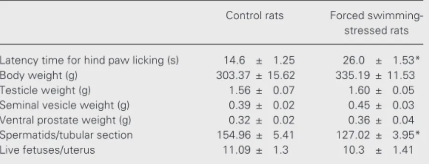
Table 1. Effect of forced swimming stress on some adult male reproductive param-eters.
Control rats Forced swimming-stressed rats
Latency time for hind paw licking (s) 14.6 ± 1.25 26.0 ± 1.53*
Body weight (g) 303.37 ± 15.62 335.19 ± 11.53
Testicle weight (g) 1.56 ± 0.07 1.60 ± 0.05
Seminal vesicle weight (g) 0.39 ± 0.02 0.45 ± 0.03 Ventral prostate weight (g) 0.32 ± 0.02 0.36 ± 0.04 Spermatids/tubular section 154.96 ± 5.41 127.02 ± 3.95*
Live fetuses/uterus 11.09 ± 1.3 10.3 ± 1.41
The forced swimming-stressed rats were submitted to 3-min daily forced swimming for 15 days. The significantly increased latency time for hind paw licking in the hot-plate test confirmed that the rats were stressed. Data are reported as means ± SEM for 15 rats.
(14). However, the exact site where ACTH, or even corticosteroids, exert their effect on the reproductive process is still not known. A wide variety of mechanisms have been proposed, including the activation of opioid pathways, a steroid-mediated decrease in the ability of the pituitary to respond to GnRH resulting in a fall in LH production, and a direct or indirect effect of ACTH and/or corticosteroids on the gonads (4,14). Previ-ous studies have demonstrated a direct effect of glucocorticoids on testicular LH recep-tors and on steroidogenesis, demonstrating the existence of regulation of testicular func-tion by the adrenals (17). As a consequence of the reduction in LH receptors, stress causes a reduction of testosterone secretion (4,14). Thus, the stressful stimulus may interfere both with endocrine and local factors, dis-turbing spermatid production.
On the other hand, the present data dem-onstrated no change in testicular weight or gross histological features after 15 days of forced swimming. Similarly, no difference was observed in the fertility of control or stressed males, as indicated by the similar number of live fetuses found in the uterus of autopsied females. The data demonstrated that 15 days of forced swimming stress ap-plied to adult male rats did not immediately impair fertility, but significantly decreased spermatid production. The normal fertility exhibited by stressed males may be explained by the fact that forced swimming lasted 15
days, a period shorter than the 48- to 53-day duration of one cycle of the seminiferous epithelium. Previous studies have shown that long-term testosterone suppression led to reduced spermatogonial and spermatocyte production, and that losses during these ear-lier stages of germ cell development led to reduction in the spermatid population (18). When intratesticular testosterone was sup-pressed for 11 weeks, the conversion of round spermatids from stage VII to stage VIII was markedly suppressed and elongated sperma-tids were not detected. However, round sper-matid conversion was not affected until 3 weeks of testosterone withdrawal, perhaps because of the persistence of androgen ac-tion for several weeks after the reducac-tion in testicular testosterone levels (18). Since stress reduces testosterone secretion (3,4,14), it is possible that impairment of male reproduc-tive function and/or spermatogenesis is due to the reduced testosterone levels. Our data suggest that short-term forced swimming stress leads to a reduction in the round sper-matid population, but the conversion of pre-existing round spermatids was apparently unaffected, as indicated by normal fertility. Thus, the present data suggest that the ef-fects of stress on fertility should be better assessed after the period of time necessary to complete an entire cycle of the seminiferous epithelium, i.e., between 48 and 53 days in the rat (16), or at least after the period of time necessary for spermatids to mature.
References
1. Ozawa N, Goda N, Makino N, Yamaguchi T, Yoshimura Y & Suematsu M (2002). Leydig cell-derived heme oxygenase-1 regu-lates apoptosis of premeiotic germ cells in response to stress.
Journal of Clinical Investigation, 109: 457-467.
2. O’Donnell L, McLachlan RI, Wreford NG & Robertson DM (1994). Testosterone promotes the conversion of round spermatids be-tween stages VII and VIII of the rat spermatogenic cycle. Endocrinol-ogy, 135: 2608-2614.
3. Almeida SA, Kempinas WG & Lamano Carvalho TL (2000). Sexual behavior and fertility of male rats submitted to prolonged immobili-zation-induced stress. Brazilian Journal of Medical and Biological
Research, 33: 1105-1109.
4. Rivier C & Rivest S (1991). Effect of stress on the activity of the hypothalamic-pituitary-gonadal axis: peripheral and central mechan-ism. Biology of Reproduction, 45: 523-532.
5. Richburg JH (2000). The relevance of spontaneous- and chemically-induced alterations in testicular germ cell apoptosis to toxicology.
Toxicology Letters, 112-113: 79-86.
6. Lewis JW, Cannon JT & Liebeskind JC (1980). Opioid and non-opioid mechanism of stress analgesia. Science, 208: 623-625. 7. Vaccarino AL, Marek P, Sternberg W & Liebeskind JC (1992). NMDA
anal-gesia in the formalin test. Pain, 50: 119-123.
8. Izumi R, Takahashi M & Kaneto H (1983). Involvement of different mechanisms, opioid and non-opioid forms, in the analgesia induced by footshock (FS) and immobilized-water immersion (IW) stress.
Japanese Journal of Pharmacology, 33: 1104-1106.
9. Parvizi N (2000). Neuroendocrine regulation of gonadotropins in the male and the female. Animal Reproduction Science, 60-61: 31-47. 10. Almeida SA, Petenusci SO, Anselmo-Franci JA, Rosa-e-Silva AAM &
Lamano Carvalho TL (1998). Chronic intermittent immobilization of male rats throughout sexual development: a stress protocol. Brazil-ian Journal of Medical and Biological Research, 31: 1443-1448. 11. Van Vugt DA, Sylvester PW, Aylsworth CF & Meites J (1982).
Counteraction of gonadal steroid inhibition of LH by naloxone. Neu-roendocrinology, 34: 274-278.
12. Kinoshita F, Nakai I, Katakami H, Kato G, Yajima H & Imura H (1980). Effect of ß endorphin on pulsatile luteinizing hormone release in conscious castrated rats. Life Sciences, 27: 843-846.
13. López-Calderón A, Ariznavarreta C, González-Quijano MI,
Tres-guerres JAF & Calderón MD (1991). Stress induced changes in testis function. Journal of Steroid Biochemistry and Molecular Biol-ogy, 40: 473-479.
14. Rivier C & Vale W (1984). Influence of CRF on reproductive func-tions in the rat. Endocrinology, 114: 914-921.
15. Demura R, Suzuki T, Nakamura S, Komatsu H, Odagiri E & Demura H (1989). Effect of immobilisation stress on testosterone and inhibin in male rats. Journal of Andrology, 10: 210-213.
16. Clermont Y, Le Blond CP & Messier B (1959). Durée du cycle d’epithelium seminal du rat. Archives d’Anatomie Microscopique et de Morphologie Experimentale, 48: 37-56.
17. Bambino TH & Hsueh AJW (1981). Direct inhibitory effect of gluco-corticoids upon testicular LH receptor and steroidogenesis in vivo
and in vitro. Endocrinology, 108: 2142-2148.
18. O’Donnell L, McLachlan RI, Wreford NG, Kretser DM & Robertson DM (1996). Testosterone withdrawal promotes stage-specific de-tachment of round spermatids from the rat seminiferous epithelium.