BIOMEDICAL SCIENCES
www.bjournal.com.br
www.bjournal.com.br
Braz J Med Biol Res, August 2012, Volume 45(8) 753-762
doi:
10.1590/S0100-879X2012007500084
Effect of skilled and unskilled training on nerve regeneration and
functional recovery
A.S. Pagnussat, S.M. Michaelsen, M. Achaval, J. Ilha, E.E.S. Hermel, F.P. Back and C.A. Netto
Institutional Sponsors
The Brazilian Journal of Medical and Biological Research is partially financed by
Faculdade de Medicina de Ribeirão Preto Campus
Ribeirão Preto
Explore High - Performance MS Orbitrap Technology In Proteomics & Metabolomics
Brazilian Journal of Medical and Biological Research (2012) 45: 753-762 ISSN 0100-879X
Effect of skilled and unskilled training on
nerve regeneration and functional recovery
A.S. Pagnussat
1, S.M. Michaelsen
2, M. Achaval
3, J. Ilha
4, E.E.S. Hermel
4,
F.P. Back
5and C.A. Netto
51Departamento de Fisioterapia, Universidade Federal de Ciências da Saúde de Porto Alegre, Porto Alegre, RS, Brasil 2Departamento de Fisioterapia, Universidade do Estado de Santa Catarina, Florianópolis, SC, Brasil 3Departamento de Ciências Morfológicas, Instituto de Ciências Básicas da Saúde,
Universidade Federal do Rio Grande do Sul, Porto Alegre, RS, Brasil
4Programa de Pós-Graduação em Neurociências, Instituto de Ciências Básicas da Saúde,
Universidade Federal do Rio Grande do Sul, Porto Alegre, RS, Brasil
5Departamento de Bioquímica, Instituto de Ciências Básicas da Saúde,
Universidade Federal do Rio Grande do Sul, Porto Alegre, RS, Brasil
Abstract
The most disabling aspect of human peripheral nerve injuries, the majority of which affect the upper limbs, is the loss of skilled hand movements. Activity-induced morphological and electrophysiological remodeling of the neuromuscular junction has
been shown to influence nerve repair and functional recovery. In the current study, we determined the effects of two different
treatments on the functional and morphological recovery after median and ulnar nerve injury. Adult Wistar male rats weighing 280 to 330 g at the time of surgery (N = 8-10 animals/group)were submitted to nerve crush and 1 week later began a 3-week course of motor rehabilitation involving either “skilled” (reaching for small food pellets) or “unskilled” (walking on a motorized treadmill) training. During this period, functional recovery was monitored weekly using staircase and cylinder tests. Histological and morphometric nerve analyses were used to assess nerve regeneration at the end of treatment. The functional evaluation
demonstrated benefits of both tasks, but found no difference between them (P > 0.05). The unskilled training, however, induced
a greater degree of nerve regeneration as evidenced by histological measurement (P < 0.05). These data provide evidence that both of the forelimb training tasks used in this study can accelerate functional recovery following brachial plexus injury.
Key words: Peripheral nerve injury; Functional recovery; Median nerve; Ulnar nerve; Nerve morphometry
Introduction
Although the majority of human peripheral nerve injuries affect the upper extremity, the sciatic nerve injury is the most widespread experimental model used for the study of nerve repair (1). Several investigators have recently defended the use of experimental brachial plexus nerve injury in studies of functional recovery (2-4).
The brachial plexus injury model has several advan-tages. The brachial plexus of rats and humans exhibits various similarities in its components and branches (5,6). Furthermore in the upper extremity of rats, the distance between the nerves and their targets is shorter than in the lower extremity, reducing the time required for studies of functional recovery (2). In general, no autonomy or contrac-tures are observed in upper extremity lesions (4,7,8).
The neuronal response following injury includes the expression of growth factors as well as other secreted mol-ecules that are involved in cell-to-cell communication (9). The mechanisms regulating the expression of the different injury-response factors, such as the neurotrophins, are not completely understood. In the central nervous system, the expression of some members of the neurotrophin family is correlated with the degree of neuronal activity (10-12). In the peripheral nervous system, moderate physical motion can exert positive effects on functional recovery after injury by means of modulation of neurotrophin expression, axonal outgrowth, and nerve maturation to promote recovery of muscle contractile properties (13,14).
There is evidence that motor activity can influence the
Correspondence: A.S. Pagnussat, Departamento de Fisioterapia, Universidade Federal de Ciências da Saúde de Porto Alegre, UFCSPA, Rua Sarmento Leite, 245, 90050-170 Porto Alegre, RS, Brasil. E-mail: [email protected]
morphological and electrophysiological remodeling of the neuromuscular junction. Although fully established that adult synapses are largely stable structures, motor nerve
endings are continuously changing and being modified in
response to functional demands (8,15,16). Adaptations of
these synapses can, therefore, influence nerve orientation
and repair. Moreover, growth factor secretion is one of the mechanisms underlying nerve regeneration (17). Neurotro-phins are unique among neurotrophic factors in their ability to act as guidance molecules for nerve growth. Strategies to enhance functional recovery after neuronal injury may depend on neurotrophin secretion to induce improvement of axonal regeneration and target reinnervation as well as to modulate abnormal plasticity of neuronal circuits (14).
Patients suffering from neuromuscular diseases undergo a great diversity in the physical therapy used for rehabilita-tion. Despite advances in the knowledge of nerve regen-eration, as well as surgical techniques, functional recovery after injury is often unsatisfactory (18). In addition, there is no consensus regarding the type and intensity of the most appropriate physical therapy (19). It is extremely important, therefore, to understand how recovery from nerve injury is
influenced by different types of exercise (20).
Rats are adept at reaching for, grasping, and bringing food to their mouths using a single paw (skilled reaching) (1). Since an attempt to identify an appropriate strategy for rehabilitation after peripheral nerve injury has unquestion-able clinical relevance, we investigated the effects of two types of physical therapies, skilled and unskilled, upon functional and morphological recovery after a brachial plexus crush lesion in adult rats.
Material and Methods
Animals
All procedures were in accordance with Brazilian Law No. 11.794/08 and decree No. 6.899/09, which regulate the
use of animals in scientific research.
Male Wistar rats (N = 42) weighing 280 to 330 g at the
time of surgery, housed in groups of four or five in Plexiglas
cages under standard conditions (12-h light/dark, 22 ± 2°C) were used. Wistar rats were obtained from the Central Ani-mal House of the Biochemistry Department, at the Institute of Basic Health Sciences from the Universidade Federal do Rio Grande do Sul, Porto Alegre, RS, Brazil. Water and standard laboratory chow were provided ad libitum except during the behavioral training and testing periods. Animals were weighed weekly. On the day before the beginning of training, animals did not receive food (in order to increase interest in new food). From then on, and after each training session, rats were provided with a measured amount of standard laboratory chow each day (12-15 g) to keep their body weight at ~80-90% of free-feeding level. The training period was completed two days prior to surgery and animals were then returned to ad libitum feeding.
Experimental design
Three weeks before surgery animals were randomly
assigned to one of the five groups described in Table 1.
During the 3 weeks (5 days/week) prior to the surgical procedure, animals were habituated to all tasks. The habitu-ation phase consisted of 3 weeks of training in the single pellet task, 2 weeks of training in the staircase test and 1 week of habituation to the electrical treadmill. A time line of experimental events is presented in Figure 1.
Surgical procedures
For the surgical procedure, all animals, except controls, were deeply anesthetized with a mixture of 90 mg/kg ket-amine and 10 mg/kg xylazine administered intraperitone-ally. Rats were immobilized on a wooden surface and a horizontal incision was made parallel to the clavicle, from the sternum to the axillary region (the forepaw chosen was that which showed higher performance in the staircase test prior to surgery). The brachial plexus was approached through this opening and the ulnar and median nerves were crushed together (1 cm distal to the sternum) with a 1-mm hemostatic forceps for 30 s (adapted from Refs. 20 and 21). The crush was performed on these two nerves because median and ulnar nerve injury affects the
capac-Table 1. Experimental groups and number of animals used for each analysis.
Group description Designation
(abbreviation)
Total number of animals
Behavioral evaluation
Morphometric analysis
Control animals (intact - unoperated) Control (C) 8 8 4
Animals submitted to all procedures (anesthesia, incision and suture), except median and ulnar nerve crush
Sham (S) 8 8 4
Animals submitted to median and ulnar nerve crush and not treated Crush (CC) 8 8 5
Animals submitted to median and ulnar nerve crush and treated with a skilled motor task
Skilled (Sk) 10 10 5
Animals submitted to median and ulnar nerve crush and treated with repetitive motor movement (unskilled task)
Unskilled (Usk)
Motor task and nerve repair 755
ity of finger flexion and grasping (2,7,22). This procedure
induces axonal interruption while preserving the connective sheaths (axonotmesis) (8).
Rehabilitation protocols
One week following median and ulnar nerve crush, ani-mals received one of the following treatments. Both protocols were performed for 3 weeks, 5 days per week.
Skilled task. This task consisted of reaching for food inside reaching boxes made of clear Plexiglas (20 x 25 x 40 cm high) (1). In the middle of the front wall, a 1.1-cm wide vertical slot allowed animals to reach for food pellets
placed on a shelf situated 4 cm above the floor. Two small
indentations (0.5 cm in diameter, 0.15 cm deep) on the up-per side of this shelf, each aligned with one side of the slit,
served to stabilize the food pellets (sweet flavored sucrose
spheres: 4.6 mm; 65 mg ± 10%; Brazilian Homeopathic Pharmacopoeia [www.anvisa.gov.br/farmacopeiabrasileira/ conteudo/3a_edicao.pdf]). The distance from the indenta-tions to the front wall was 1.5 cm.
Each animal received individual preoperative training. Once animals began to reach, the food pellets were placed in the indentation contralateral to their preferred limb to provide easier access to the food and to prevent simultaneous use of the non-preferred limb. In the preoperative training period, rats were motivated to reach during a 20-min period. The protocol was performed similarly during the postoperative training period. Reaching with the impaired forelimb was effectively enforced by the insertion of an inner chamber wall ipsilateral to this limb, and the placement of pellets, one at a time, in the wall opposite this limb. Since animals needed to cross the midline to reach the pellet, this prevented the use of the unaffected forelimb.
Unskilled task. This task consisted of walking on an adapted motorized rodent treadmill (INBRAMED TK 01, Brazil) for a 20-min (0.03 m/s - 3 initial min; 0.05 m/s - 14 min; 0.03 m/s - last 3 min) period. The grade of the treadmill remained at 0% and no aversivestimuliwere used. The low velocity was chosen to avoid possible effects of aerobic treadmill exercise.
Behavioral assessments
Behavioral assessments were performed using a skilled reaching test (staircase test) and an asymmetrical forelimb use test (cylinder test).
Skilled reaching test. Fine motor ability was assessed
by means of a staircase test, which provides a sensitive measure of the skilled reaching of both forepaws indepen-dently (23). This test has been recently demonstrated to identify the effects of axon misdirection on forelimb function (3). The boxes were made of clear Plexiglas and consisted of a chamber with a central platform. Seven steps were positioned on each side of the platform and three small food pellets were placed on each staircase (Brazilian Homeopathic Pharmacopoeia). Animals were trained for 2 weeks before the surgical procedure (2 trials/day). The rats remained in the staircase chamber for 15 min and the total number of pellets eaten on each side was recorded. Animals were tested on two different days (2 trials/day) be-fore surgery, and the reaching performance was calculated as the average of these trials. Animals were submitted to the staircase test 2 and 7 days after surgery and weekly for the 3-week treatment.
Forelimb asymmetry test. To examine the effects of median and ulnar nerve crush and treatment on spontane-ous forelimb use during exploratory activity, animals were placed individually inside a transparent cylinder (20 cm in diameter and 40 cm high) on a glass tabletop and video-recorded from below through an angled mirror for a 4-min test session. The cylindrical shape encouraged rearing and vertical exploration of the walls with the forelimbs. The number of forepaw-wall contacts used for postural support was counted and the percentage asymmetry of single-limb wall contacts [(contralateral / contralateral + ipsilateral) x 100] was calculated (24). A single cylinder test session was performed 2 days prior to surgery, 2 days after surgery, and weekly after the beginning of treatment.
Morphometric analyses
Two days after the end of the treatment period, animals were anesthetized with chloride hydrate (30%, 10 mL/kg, ip) and injected with 1000 IU heparin (Cristália, Brazil). They were then transcardially perfused through the left ventricle using a peristaltic pump (Control Company, Brazil) with 100
mL saline followed by 200 mL of a fixative solution composed
of 0.5% glutaraldehyde (Sigma, USA) and 4% paraformal-dehyde (Reagen, Brazil) in 0.1 M phosphate buffer (PB), pH 7.4, at room temperature. For nerve regeneration analysis, one short segment (~3 mm) of the ulnar and median nerves was rapidly excised 5 mm after the crush injury site. The
specimens were postfixed by immersion in the same fixa
-tive solution for 1 h. They were then postfixed in 1% OsO4
(Sigma) in PB, dehydrated in a graded alcohol series and propylene oxide (Electron Microscopy Sciences, USA), embedded in araldite (Durcupan ACM, Fluka, Switzerland), and polymerized for 48 h at 60°C.
Transverse semithin sections (1 µm) were obtained with an ultramicrotome (MT 6000-XL, RMC, USA) and stained with 1% toluidine blue (Merck, Germany) in 1% sodium tetraborate (Ecibra, Brazil). Images of the distal portions of the nerves were then digitized (initially 100X and
further amplified 200X for analysis) using a Nikon Eclipse
E-600 microscope (Japan) coupled to a Pro-Series High-Performance CCD camera and Image Pro Plus Software 6.0 (Media Cybernetics, USA). For morphological evaluation, a set of 6 images was obtained from each nerve portion: 3 random images from the periphery and 3 random images from the center of the nerve, in order to obtain a represen-tative area per nerve segment (0.010 mm2; 100% of the
area analyzed per segment) (20,25).
The morphometric measurements used to assess the dif-ferentiation of regenerating nerves included: 1) myelinated
fiber density (number of fibers/mm2); 2) average myelinated
fiber area (μm2); 3) average myelinated fiber diameter
(µm); 4) average axon diameter (µm) of the myelinated
fiber; 5) average myelin sheath thickness (µm); 6) g ratio
(the quotient axon diameter/fiber diameter, a measurement of the degree of myelination). Individual myelinated fibers
were counted and the myelinated fiber density was deter
-mined by examining the ratio of the myelinated fibers/total
area analyzed. The average myelin sheath thickness was estimated using the measurement tools of the ImagePro Plus software. The area measurements were estimated with a point-counting technique (20,26) using grids with
point density of one point per 1.87 μm2 and the following
equation: Â = Σp
.
a/p, where  is the area, Σp the sum of points, and a/p the area/point value (1.87 µm2). Toestimate the axon and fiber diameters, the area of each individual fiber was measured and the value obtained was
converted to the diameter of a circle having an equivalent
area. In each nerve segment, 10.48 μm2 was 100% of the
analyzed area.
Statistical analysis
The SPSS® 16.0 (Statistical Package for the Social
Sciences, Inc., USA) was used for data analysis. Repeated measures analysis of variance (ANOVA) was used to determine differences between groups in the behavioral evaluation. Morphological measurements were analyzed by one-way ANOVA and the Duncan multiple range test was
used when appropriate. Significance was set at P< 0.05 for
all analyses and results are reported as means ± SEM.
Results
Staircase test
Repeated measures ANOVA of the staircase test
showed time (F(1,37) = 12.02, P= 0.01) and group effects
(F(4,37) = 7.24, P < 0.01), but no significant interaction be
-tween time and group (F(4,37) = 1.71, P =0.16). As shown
in Figure 2, Duncan’s post hoc analysis revealed that nerve
lesion resulted in a significant decrease in performance of
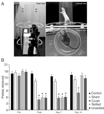
the crushed groups when compared to sham and control groups at post (evaluation performed at 24 h after crush) and day 7 (P < 0.05).At day 14, the crush group (which was not submitted to any exercise protocol) remained impaired (P < 0.05) while no difference between the sham- and control-treated groups were observed. At days 21 and 28, no differences between groups were observed (data not shown).
Cylinder test
For the forelimb asymmetry task, repeated measures ANOVA showed an effect of time (F(1,37) = 4.21, P < 0.05),
group (F(4,37) = 15.50, P < 0.01) and time-group interaction
(F(4,37) = 7.61, P < 0.01). As demonstrated in Figure 3, all
injured groups were significantly impaired immediately fol
-lowing the median and ulnar nerves crush (P < 0.05) and used the damaged limb for support less frequently than did the sham and control groups. At 14 days post-injury, both treated groups improved to sham and control levels. Untreated animals from the crush group had persistent
Figure 2. Staircase test of skilled forelimb reaching. A, Photo-graph showing the staircase apparatus. The experimental boxes were made of clear Plexiglass with a central raised platform. The narrowness of the compartment was enough to prevent the rat from turning around and to allow unilateral reaches. Pellets were provided into a removable double staircase located at the end of the box. B, Number of pellets retrieved: all damaged groups were
significantly impaired compared to control and sham animals at
Motor task and nerve repair 757
impairment (P < 0.05) at 14 days, and showed spontane-ous recovery at day 21. There were no differences between groups at days 21 and 28 (data not shown).
Histological analysis
The histological characteristics of the distal portions of the median and ulnar nerves (Figure 4) demonstrated pathological features in the crush group (Figure 4C,H), which were not seen in the control (Figure 4A,F) or sham (Figure 4B,G) groups. These consisted of enlargement of
endoneurial connective tissue between the nerve fibers, reduction of myelinated fiber diameter and myelin sheath
thickness, and presence of degeneration debris. In injured treated groups (Figure 4D,E,I,J), these pathological features were reduced and less endoneurial connective tissue and tissue debris were observed.
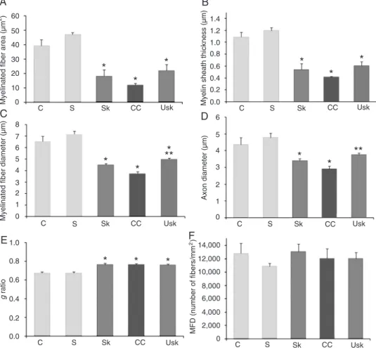
Morphometric analysis of the median nerve
In the median nerve, one-way ANOVA demonstrated an
effect of the lesion for myelinated fiber area (F (4,18) = 54.16,
P < 0.01); myelin sheath thickness (F(4,18) = 67.06, P < 0.01);
myelinated fiber diameter (F(4,18) = 54.60, P < 0.01); axon
diameter (F(4,18) = 24.80, P < 0.01), and g ratio (F(4,18) =
11.36, P < 0.01), but not for myelinated fiber density (F(4,18)
= 1.23, P=0.33). After 3 weeks of rehabilitation, Duncan
post-hoc analyses demonstrated differences between the
Figure 3.A, Photograph of the cylinder test apparatus - trans-parent cylinder (20 cm in diameter and 40 cm high). Cylinder was positioned on a glass tabletop and exploratory movements were video-recorded from below through an angled mirror. B, The forelimb asymmetry task was used to measure the number of contralateral forelimb contacts compared to ipsilateral contacts while the animal reared in a cylinder. All damaged groups showed an increase in contralateral forelimb use after median and ulnar nerve crush at post (24 h) and day 7. At day 14, both treated groups improved, with no difference between treatments. At this time, only the animals from the crush group remained impaired. Data are reported as means ± SEM. *P < 0.05 compared to the control and sham groups (repeated measures ANOVA followed by the Duncan multiple range test).
Figure 4. Morphological images of the median and ulnar nerves: digitalized images of transverse semithin sections (1 µm) ob-tained from regenerating median and ulnar nerves after 3 weeks of skilled and unskilled training. The left columns represent images from distal portions of the median nerve and the right columns represent images from distal portions of the ulnar nerve. Letters indicate distal portions from nerves of A and F, control group; B and G, sham group; C and H, crush group; D and I, skilled training group; E and J, unskilled training group. Mf =
injured (crush, skilled task and unskilled) and uninjured
groups (sham and control) in myelinated fiber area, axon
diameter and g ratio (Figure 5A,D,E, respectively). Differ-ences were found between injured and uninjured groups, as well as between crush and unskilled groups in myelin
sheath thickness (Figure 5B). A difference in myelinated fiber
diameter (Figure 5C) was demonstrated between injured and uninjured groups and between injured (crush) and in-jured treated groups (skilled and unskilled task). However,
no significant differences were observed between the two
types of treatment. In addition, no difference was observed
in myelinated fiber density between groups (Figure 5F).
Morphometric analysis of the ulnar nerve
One-way ANOVA revealed an effect of the lesion for ulnar
nerve myelinated fiber area (F(4,18) = 17.17, P < 0.01), myelin
sheath thickness (F(4,18) = 19.47, P < 0.01), myelinated fiber
diameter (F(4,18) = 15.53, P < 0.01), axon diameter (F(4,18)
= 11.15, P < 0.01), and g ratio (F(4,18) = 11.58, P < 0.01),
but no significant differences were observed in myelinated
fiber density (F(4,18) = 0.48, P =0.74).
After 3 weeks of rehabilitation, Duncan post hoc analysis revealed differences between the injured (crush, skilled and unskilled task) and uninjured groups (sham and control) in
myelinated fiber area, myelin sheath thickness and g ratio
(Figure 6A,B, and E, respectively). Differences in myelinated
fiber diameter (Figure 6C) were found between the injured
and uninjured groups, as well as between the crush and unskilled groups. Differences in axon diameter (Figure 6D) were found between the crush, control and sham groups, although no differences were apparent between the control and unskilled groups, or between the treated groups (skilled
Figure 5. Effects of treatment on the morphometric parameters of regenerating median nerve fibers.
Motor task and nerve repair 759
and unskilled task). No differences were observed in
myeli-nated fiber density between groups (Figure 6F).
Discussion
The most disabling aspect of upper extremity periph-eral nerve injury in humans is reported to be the loss of skilled hand movements (27). Rats are capable of perform-ing skilled reachperform-ing and graspperform-ing movements with their forepaws. This ability can be used to model rehabilitative treatment (28)and to assess post-injury performance (1). Previous studies, however, have used neither skill training as treatment for brachial plexus injury, nor tests of skilled forelimb function to assess recovery after peripheral nerve injury and repair. The exception is a study by Galtrey and Fawcett (3).
Although small physiological variations (such as
in-creases and dein-creases in locomotor activity for a short period) can modify the ultrastructure of the nerve terminal (16,29,30),the differences obtained with different types of physical exercises have not been examined extensively in models of brachial plexus injury.
In the present study, we investigated the hypothesis that skilled and unskilled training, beginning 1 week after the injury, would produce different effects regarding the functional recovery and morphological changes observed in the regenerating nerve. Analysis of functional activity of the forepaws using the grasping strength test revealed that median and ulnar nerve crush led to a marked reduction in grasping on the 5th day after nerve damage, and that this impairment was gradually reduced about 20 days after the lesion (8). We therefore used this chronogram for our experiments.
Our results demonstrate that both types of treatment
Figure 6. Effects of treatment on the morphometric parameters of regenerating ulnar nerve fibers.
promoted the normalization of both skilled and gross forelimb motor function after 1 week of treatment, as evaluated by the staircase and cylinder tests (Figures 2 and 3). However, the unskilled training accelerated axonal regeneration
as demonstrated by the increased fiber myelination and
tissue recovery (Figure 4). Greater myelin thickness and
myelinated fiber diameter were found in the median nerve
of the animals trained in the unskilled task compared to untreated injured animals (Figure 5). Morphological exami-nation of the ulnar nerve also revealed axons with a more mature appearance in animals of the unskilled training group compared to the crush group (as demonstrated by
analysis of myelinated fiber diameter and axon diameter of the myelinated fiber; Figure 6).
As visualized in Figure 6, sham-treated animals
dis-played a lower density of myelinated fibers compared to
control animals. This variability was present both in the ulnar and median nerve evaluations and no statistical dif-ference in this measure was observed between these two groups. These results could possibly be due to unavoid-able biological variability.
The major aim of physiotherapy after nervous system injury in humans is to avoid permanent disability as well as to restore the patient’s autonomy in activities of daily living. Experimental studies demonstrate that recovery of function does not necessarily correlate with histological and electrophysiological evidence of regeneration (6,31),
as confirmed in the present study.
Previous studies have evaluated the effects of tread-mill exercise on peripheral nerves following injury (32,33). Regeneration of neural processes is likely to be an activity-dependent process (27). The type of treatment, the intensity and duration of the protocol, and the period during which it is
applied after the injury are factors that determine beneficial
or detrimental effects on functional recovery (14). In some
cases, nerve regeneration is more directly influenced by the
quantity, rather than the type, of exercise (34). Sabatier et al. (34) demonstrated that training for longer periods at slower speeds, and training for short amounts of time (as little as
4 min) at higher speeds, increased axon profile lengths 2
weeks after nerve transection.
Physiological activities such as those used in the pres-ent study, may induce greater regeneration than more intensive training. Several studies have reported that muscle overwork is harmful to muscle reinnervation, as overexerting muscles might accelerate disease progres-sion (20,33,35,36). However, for the improvement of nerve regeneration, the intervention should be of a high enough intensity to provide a training stimulus (19).This level per-haps was not reached in those animals submitted to skill training in the current study.
Although previous studies have shown that voluntary physical activity can prime enhanced axonal regeneration after subsequent axotomy (17), in our study this bias was
avoided by submitting all animals to the same pretreat-ment protocol.
Fine dexterity is often lost or diminished after nerve injury. Goal-directed activity may promote greater use of the hand and limb (27), and morphological evaluation of animals submitted to unskilled training shows superior nerve regeneration than in those receiving skill training. The discharge of weight on a treadmill (unskilled training) de-mands more intense and global muscular activity than skilled training, such as a reaching task. The pattern of peripheral regulation of growth factors may differ according to type of training (20). Unskilled training thus may induce a greater release and expression of relevant substances and their respective receptors involved in nerve regeneration, such as brain-derived neurotrophic factor (BDNF), neurotrophins
(NT 3, 4, and 5) and specific members of the tyrosine kinase
gene family (Trk) (Refs. 13 and 36, for review).
Physical exercise after nerve injuries increases the expression of genes needed for neurite growth. Animals exercised after lesion had higher levels of BDNF, NT3, synapsin I, and GAP43 mRNAs than sedentary animals (17,37). Blocking the Trk neurotrophin receptor activity be-fore exercise inhibits the effects on synapsin I transcription and attenuates the increase in neurite growth, demonstrat-ing that neurotrophin signal transduction pathways play a critical role in exercise inducing nerve growth. Exercise can
also influence the injured neurons by stimulating sensory
afferents and exerting positive effects on the cell circuitry (17). Even during the denervation period, active exercise
stimulates muscle afferents, which may influence the axo
-tomized motoneurons by normally silent spinal synaptic connections (12).
A limitation of our study is that we used a technique that permitted visualization only of the final nerve regeneration. Further studies are necessary to determine the rate of re-generation as well as the latency to target reinnervation in animals submitted to both rehabilitation protocols.
The present study demonstrated that skilled and un-skilled forelimb training accelerates the morphological and functional recovery after brachial plexus injury. Morphologi-cal analyses revealed that the unskilled training paradigm induced a slight superior degree of tissue recovery. Further studies are needed to verify the utility of the different treat-ments for promoting neuronal reorganization and functional improvement after peripheral nervous system injury.
Acknowledgments
Motor task and nerve repair 761
References
1. Whishaw IQ, Pellis SM. The structure of skilled forelimb reaching in the rat: a proximally driven movement with a single distal rotatory component. Behav Brain Res 1990; 41: 49-59.
2. Bontioti E, Kanje M, Lundborg G, Dahlin LB. End-to-side nerve repair in the upper extremity of rat. J Peripher Nerv Syst 2005; 10: 58-68.
3. Galtrey CM, Fawcett JW. Characterization of tests of func-tional recovery after median and ulnar nerve injury and repair in the rat forelimb. J Peripher Nerv Syst 2007; 12: 11-27.
4. Santos AP, Suaid CA, Fazan VP, Barreira AA. Microscopic anatomy of brachial plexus branches in Wistar rats. Anat Rec 2007; 290: 477-485.
5. Bertelli JA, Mira JC, Gilbert A, Michot GA, Legagneux J. Anatomical basis of rat brachial plexus reconstruction. Surg
Radiol Anat 1992; 14: 85-86.
6. Nichols CM, Myckatyn TM, Rickman SR, Fox IK, Hadlock T, Mackinnon SE. Choosing the correct functional assay: a comprehensive assessment of functional tests in the rat.
Behav Brain Res 2005; 163: 143-158.
7. Bertelli JA, Mira JC. Behavioral evaluating methods in the objective clinical assessment of motor function after experi-mental brachial plexus reconstruction in the rat. J Neurosci
Methods 1993; 46: 203-208.
8. Rodrigues-Filho R, Santos AR, Bertelli JA, Calixto JB. Avul-sion injury of the rat brachial plexus triggers hyperalgesia and allodynia in the hindpaws: a new model for the study of neuropathic pain. Brain Res 2003; 982: 186-194.
9. Makwana M, Raivich G. Molecular mechanisms in success-ful peripheral regeneration. FEBS J 2005; 272: 2628-2638. 10. Adkins DL, Boychuk J, Remple MS, Kleim JA. Motor
train-ing induces experience-specific patterns of plasticity across
motor cortex and spinal cord. J Appl Physiol 2006; 101: 1776-1782.
11. Klintsova AY, Dickson E, Yoshida R, Greenough WT. Altered
expression of BDNF and its high-affinity receptor TrkB in
response to complex motor learning and moderate exercise.
Brain Res 2004; 1028: 92-104.
12. Koerber HR, Mirnics K, Lawson JJ. Synaptic plasticity in the adult spinal dorsal horn: the appearance of new functional connections following peripheral nerve regeneration. Exp
Neurol 2006; 200: 468-479.
13. Apfel SC, Wright DE, Wiideman AM, Dormia C, Snider WD, Kessler JA. Nerve growth factor regulates the expression of brain-derived neurotrophic factor mRNA in the peripheral nervous system. Mol Cell Neurosci 1996; 7: 134-142. 14. Udina E, Cobianchi S, Allodi I, Navarro X. Effects of
activity-dependent strategies on regeneration and plasticity after peripheral nerve injuries. Ann Anat 2011; 193: 347-353. 15. Deschenes MR, Maresh CM, Crivello JF, Armstrong LE,
Kraemer WJ, Covault J. The effects of exercise training of different intensities on neuromuscular junction morphology.
J Neurocytol 1993; 22: 603-615.
16. Tomas J, Santafe M, Lanuza MA, Fenoll-Brunet MR. Physi-ological activity-dependent ultrastructural plasticity in normal adult rat neuromuscular junctions. Biol Cell 1997; 89: 19-28.
17. Molteni R, Zheng JQ, Ying Z, Gomez-Pinilla F, Twiss JL.
Vol-untary exercise increases axonal regeneration from sensory neurons. Proc Natl Acad Sci U S A 2004; 101: 8473-8478. 18. Gordon T, Sulaiman O, Boyd JG. Experimental strategies to
promote functional recovery after peripheral nerve injuries.
J Peripher Nerv Syst 2003; 8: 236-250.
19. Cup EH, Pieterse AJ, Ten Broek-Pastoor JM, Munneke M, van Engelen BG, Hendricks HT, et al. Exercise therapy and other types of physical therapy for patients with neuromus-cular diseases: a systematic review. Arch Phys Med Rehabil 2007; 88: 1452-1464.
20. Ilha J, Araujo RT, Malysz T, Hermel EE, Rigon P, Xavier LL, et al. Endurance and resistance exercise training programs
elicit specific effects on sciatic nerve regeneration after
experimental traumatic lesion in rats. Neurorehabil Neural
Repair 2008; 22: 355-366.
21. Bridge PM, Ball DJ, Mackinnon SE, Nakao Y, Brandt K, Hunter DA, et al. Nerve crush injuries - a model for axonot-mesis. Exp Neurol 1994; 127: 284-290.
22. Papalia I, Tos P, Scevola A, Raimondo S, Geuna S. The ulnar test: a method for the quantitative functional assessment of posttraumatic ulnar nerve recovery in the rat. J Neurosci
Methods 2006; 154: 198-203.
23. Pagnussat AS, Michaelsen SM, Achaval M, Netto CA. Skilled forelimb reaching in Wistar rats: evaluation by means of Montoya staircase test. J Neurosci Methods 2009; 177: 115-121.
24. Schallert T. Behavioral tests for preclinical intervention as-sessment. NeuroRx 2006; 3: 497-504.
25. de Medinaceli L. Interpreting nerve morphometry data after experimental traumatic lesions. J Neurosci Methods 1995; 58: 29-37.
26. Gundersen HJ, Jensen EB. The efficiency of systematic
sampling in stereology and its prediction. J Microsc 1987; 147: 229-263.
27. Duff SV. Impact of peripheral nerve injury on sensorimotor control. J Hand Ther 2005; 18: 277-291.
28. Ploughman M, Attwood Z, White N, Dore JJ, Corbett D. En-durance exercise facilitates relearning of forelimb motor skill after focal ischemia. Eur J Neurosci 2007; 25: 3453-3460. 29. Ferre J, Brunet R, Santafe M, Mayayo E. Changes in motor
nerve terminals during bupivacaine-induced postsynaptic deprivation. J Anat 1989; 162: 225-234.
30. Tomas J, Batlle J, Fenoll MR, Santafe M, Lanuza MA. Activ-ity-dependent plastic changes in the motor nerve terminals of the adult rat. Biol Cell 1993; 79: 133-137.
31. Munro CA, Szalai JP, Mackinnon SE, Midha R. Lack of asso-ciation between outcome measures of nerve regeneration.
Muscle Nerve 1998; 21: 1095-1097.
32. Marqueste T, Alliez JR, Alluin O, Jammes Y, Decherchi P. Neuromuscular rehabilitation by treadmill running or electri-cal stimulation after peripheral nerve injury and repair. J Appl
Physiol 2004; 96: 1988-1995.
33. van Meeteren NL, Brakkee JH, Helders PJ, Gispen WH. The effect of exercise training on functional recovery after sciatic nerve crush in the rat. J Peripher Nerv Syst 1998; 3: 277-282.
35. Gutmann E, Jakoubek B. Effect of increased motor activ-ity on regeneration of the peripheral nerve in young rats.
Physiol Bohemoslov 1963; 12: 463-468.
36. van der Kooi EL, Lindeman E, Riphagen I. Strength training and aerobic exercise training for muscle disease. Cochrane
Database Syst Rev 2005; CD003907.