DOI 10.1007/s00044-017-1877-y
RESEARCH
O R I G I N A L R E S E A R C H
Antibacterial effect of chalcogenoesters on planktonic cells and
bio
fi
lms of
Streptococcus mutans
and
Streptococcus parasanguinis
Leonardo Silva de Sousa1●Agena Camara-Leimbach1●
Mayron Alves de Vasconcelos1●Francisco Vassiliepe Sousa Arruda1●
Rafael Santos da Silva2●Luciano Dornelles2●Oscar Endrigo Dorneles Rodrigues2●
Edson Holanda Teixeira2
Received: 5 December 2016 / Accepted: 11 March 2017 / Published online: 23 March 2017 © Springer Science+Business Media New York 2017
Abstract The purpose of this work was to evaluate the
antibacterial and antibiofilm activity of five
chalcogenoe-sters synthetics on oral bacteria,Streptococcus mutans, and
Streptococcus parasanguinis. Five chalcogenoesters were synthesized, purified by chromatography in silica gel and its chemical structure determined by nuclear magnetic reso-nance. Antibacterial assays were performed using the microdilution methodology. The antibiofilm activity of chalcogenoesters was determined on biofilm formation and preformed biofilms by biofilm mass quantification (by crystal violet staining) and colony forming unit enumera-tion. Moreover, biofilms were also analyzed by scanning electron microscopy. In general, it was observed that the chalcogenolesters showed antibacterial activity against
Streptococcus mutans and Streptococcus parasanguinis. Regarding biofilm formation and preformed biofilm, all compounds reduced efficiently the biofilm mass and of
viable cells of Streptococcus parasanguinis and
Strepto-coccus mutans biofilms. However, Streptococcus mutans
biofilms showed greater resistance to action of chalco-genolesters. Scanning electron microscopy examination confirmed the results, showing a reduction of the
extracellular polymeric substance and number of cells in biofilm. Our result indicated that the chalcogenoesters tested here can be considered as promising molecules for prevention and control of oral biofilms formed by
Streptococcus mutans andStreptococcus parasanguinis.
Keywords Chalcogenoesters ●Antibacterial●Biofilm●
Streptococcus
Introduction
The human oral cavity is colonized by several bacterial
species (Caufield et al. 2015). Such cavity comprises
dif-ferent habitats, including teeth, gingival sulcus, tongue, cheeks, hard and soft palates, and tonsils, which can serve
as shelter for bacterial development (Dewhirst et al.2010).
More than 700 bacterial species or phylotypes have been detected in the oral cavity and some of these have been implicated in oral diseases such as dental caries and
periodontitis (Aas et al. 2005). In fact, dental caries is
considered the most prevalent human disease, affecting 80–90% of the world population (Simon-Soro and Mira
2015).
A key factor in the development of dental caries is the natural adhesion of certain bacteria to the tooth surface and subsequent formation of a dental plaque, which is a classic
example of biofilm (Marsh2010). A biofilm is an
assem-blage of microbial cells that is associated to a surface and enclosed in a self-produced matrix of hydrated extracellular
polymeric substances (EPS) (Donlan 2002; Flemming and
Wingender2010). Bacterial cells in biofilms display a
dif-ferent phenotype from those growing planktonically, being
* Edson Holanda Teixeira [email protected]
1
Laboratório Integrado de Biomoléculas, Departamento de Patologia e Medicina Legal, Universidade Federal do Ceará, Fortaleza, CE, Brazil
2
NanoBio-LabSelen, Departamento de Química, Universidade Federal de Santa Maria, Santa Maria, RS, Brazil
resistant to several antibiotics and antimicrobial agents
(Marsh2004; Hall-Stoodley et al.2004).
Given the importance of biofilms in human health as well as in industry, many studies have been carried out to develop new strategies to control its formation on either
biotic or abiotic surfaces (Simoes et al.2010). Therefore,
the search for new compounds able in inhibiting its for-mation or controlling its growth is of great importance.
Organochalcogens are a class of organic compounds that have aroused interest by the scientific community mainly due to their several biological properties, among them antioxidant, antitumor, and antimicrobial activities (Mugesh
et al.2001; Nogueira et al.2004; Sarma and Mugesh2008;
Das et al.2008; Bhabak and Mugesh2009; de Souza et al.
2015). Chalcogenoesters (selenol, thiol, and tellurol esters)
are useful synthetic intermediates that have been employed for several chemical transformations (Boger and Mathvink
1989; Boger and Mathvink 1992; Lucas and Schiesser
1996; Keck and Grier1999; Pattenden et al.2009; Rampon
et al. 2010). Additionally, some organoselenium
com-pounds have been screened regarding their antimicrobial properties showing some prominent responses (Vargas et al.
2012).
Therefore, this study was performed to evaluate the antimicrobial and antibiofilm activities of synthetic
chal-cogenoesters on oral bacteriaStreptococcus parasanguinis
American Type Culture Collection (ATCC) 903 and
Streptococcus mutans ATCC 25175, both involved in dental caries formation.
Materials and methods
Synthesis of chalcogenoesters
The chalcogenoesters were synthesized as previously
described. S501 and S503 (Ren et al. 2010), S502
(Nar-ayanaperumal et al.2011) andS505andS506(Wang et al.
2014). The chemical structures were confirmed by1H and
13
C NMR.
Se-phenyl 4-methylbenzoselenoate (S501)
1
H NMR (CDCl3, 400 MHz),δ(ppm): 7.81 (d,J=8.31 Hz,
2H), 7.57 (m, 2H), 7.38 (m, 3H), 7.24 (d,J=8.31 Hz, 2H).
13
C NMR (CDCl3, 100 MHz),δ(ppm): 192.4, 144.8, 136.3,
136.2, 129.5, 129.2, 128.8, 127.4, 126.1, 21.6.
Se-phenyl 4-nitrobenzoselenoate (S502)
1
H NMR (CDCl3, 400 MHz),δ(ppm): 8.34 (d,J=9.04 Hz,
2H), 8.07 (d,J=9.04 Hz, 2H), 7.56 (m, 2H), 7.44 (m, 2H).
13
C NMR (CDCl3, 100 MHz),δ(ppm): 192.4, 143.1, 136.1,
131.6, 129.6, 129.6, 129.1, 128.1, 124.2.
Se-p-tolyl 4-methylbenzoselenoate (S503)
1
H NMR (CDCl3, 400 MHz),δ(ppm): 7.80 (d,J=8.07 Hz,
2H), 7.44 (d,J=8.07, 2H), 7.26–7.15 (m, 4H), 2.40 (s, 3H),
2.38(s, 3H). 13C NMR (CDCl3, 100 MHz),δ(ppm): 193.1,
144.8, 139.01, 136.3, 130.2, 129.5, 127.4, 122.3, 21.7, 21.3.
S-4-chlorophenyl 4-methylbenzothioate (S505)
1
H NMR (CDCl3, 400 MHz), δ (ppm): 7.89 (d, J=8.31
Hz), 7.41–7.38 (m, 4H), 7.28 (d,J=8.31 Hz), 2.42 (s, 3H).
13
C NMR (CDCl3, 100 MHz),δ(ppm): 172.1, 144.6, 130.3,
129.2, 126.7, 21.7.
S-4-chlorophenyl 4-nitrobenzothioate (S506)
1
H NMR (CDCl3, 200 MHz),δ(ppm): 8.34 (d,J=9.04 Hz,
2H), 8.18 (d, J=9.04 Hz, 2H), 7.50–7.38 (m, 4H). 13C
NMR (CDCl3, 50 MHz), δ (ppm): 188.4, 150.7, 140.9,
136.6, 136.1, 129.8, 128.5, 124.5, 113.8.
Microorganisms
Microorganisms used in this study were S. mutans ATCC
25175 and S. parasanguinis ATCC 903, which were
obtained from the ATCC.
Culture conditions
Bacteria were grown in Brain Heart Infusion Agar (BHI
Agar; Himedia, India) by incubation at 37 °C in 5% CO2for
24 h. After growth on solid medium, some isolated colonies were removed and inoculated into 10 ml fresh BHI and
incubated again at 37 °C in 5% CO2for 24 h under constant
agitation. Prior to use, the cell density of each inoculum was
adjusted to 2×10⁶cells/ml.
Antibacterial activity on planktonic cells
The effect of chalcogenoesters on planktonic cells was determined by the broth microdilution method described in
the guideline “Methods for Dilution Antimicrobial
Sus-ceptibility Tests for Bacteria that Grow Aerobically;
Approved Standard—Nineth Edition (CLSI document
M07-A9)”, with modifications. Briefly, in 96-well
poly-styrene plates, the compoundsS501,S502,S503,S505, and
S506 were diluted in BHI broth containing 4% dimethyl
sulfoxide in concentrations ranging from 250 to 3.9µg/ml.
The assay was performed by the addition of 100μl of
with 1% sucrose and 100μl of each chalcogenoester at different concentrations. The microplates were then
incu-bated at 37 °C in 5% CO2for 24 h. The minimum inhibitory
concentration (MIC) was determined as the lowest chalco-genoester concentration showing a complete inhibition of visible bacterial growth.
Effect of chalcogenoesters on biofilm formation
The methodology used to monitor the effects of chalco-genoesters on biofilm formation was based on the microtiter
plate test developed by Stepanovic et al. (2000) with
some modifications. Briefly, sterile flat-bottom 96-well
polystyrene plates were prepared following a similar pro-cedure to that used to determine the effects on planktonic growth. However, the effect on biofilm formation was
assessed by two distinct assays: biomass quantification by
crystal violet staining and enumeration of biofilm-entrapped viable cells.
Biofilm mass quantification
The biofilm mass was quantified by crystal violet staining. After biofilm development, the content of each well was
removed, and the wells were washed twice with 200µl of
ultrapure water to remove weakly adhered cells. Forfixation
of biofilm mass, 200µl of 99% methanol were added to
each well, and after 15 min, the methanol was removed and the plates were allowed to dry at room temperature.
After-wards, 200µl of crystal violet stain (Merck, Germany) were
added to each well and after 5 min, the excess of crystal violet was removed. The plates were then washed twice
with water and after drying, 200µl of acetic acid (33%, v/v)
were added to wells to dissolve the bound crystal violet. The absorbance of each well was then measured at 590 nm
(OD590) using a microplate reader (SpectraMax® I3).
Enumeration of biofilm-entrapped cells
In order to determine the effect of chalcogenoesters on the viability of biofilm-entrapped cells the wells were washed
twice with 200µl of ultrapure water. Afterwards, the wells
were filled with 200µl of ultrapure water and the plates were placed in an ultrasonic bath for 10 min to release the cells entrapped within the biofilm matrix. In order to quantify the viability, serial decimal dilutions from the obtained suspensions were plated on BHI agar and grown
for 24 h in 5 % CO2 at 37 °C. The number of colony
forming units (CFU) was determined and expressed as CFU
per ml (log10CFU/ml).
Effect of chalcogenoesters on pre-formed biofilms
In order to evaluate the activity of chalcogenoesters on
bacterial mature biofilms, cells suspensions (200µl of
106cells/ml) were added to each well and incubated for 24 h
at 37 °C in 5% CO2under constant agitation. After biofilm
development the wells were washed twice with 200µl of
ultrapure water and 200µl of each chalcogenoester in BHI
(at concentrations ranging from 7.8 to 200µg/ml) was
added to wells. The plates were incubated for 24 h at 37 °C
in 5% CO2 under constant agitation. After of 24 h, the
medium was aspirated, and each well was washed twice with ultrapure water. Pre-formed biofilms were then eval-uated by biomass quantification and enumeration of biofilm viable cells as previously described.
Statistical analysis
Statistical analyses were performed by GraphPad Prism® version 5.0 from Microsoft Windows®. Data from all assays were compared using one-way analysis of variance, with Bonferroni post hoc test. Data were considered
sig-nificant whenp<0.05.
Scanning electron microscopy
Bacterial biofilms were grown in the presence of the
com-pound S501 at 250µg/ml and observed by scanning
elec-tron microscopy (SEM) in a Quanta 450 FEG (Fei, USA). The biofilm formation assay was carried out in 24-well microplates. After biofilm formation, the plates were washed with sterilized water, dehydrated with alcohol (70% ethanol for 10 min, 95% ethanol for 10 min, and 100% ethanol for 20 min), and allowed to dry prior to gold
coating as described by Vasconcelos et al. (2014).
Results
Chemical synthesis
Five analogs of chalcongenesters were properly synthesized
(S501,S502, S503, S505, and S506) using the respective
methodologies described previously in the literature.
Chemical structures were elucidated using 13C and 1H
nuclear magnetic resonance (NMR). The structures are
shown below (Fig.1).
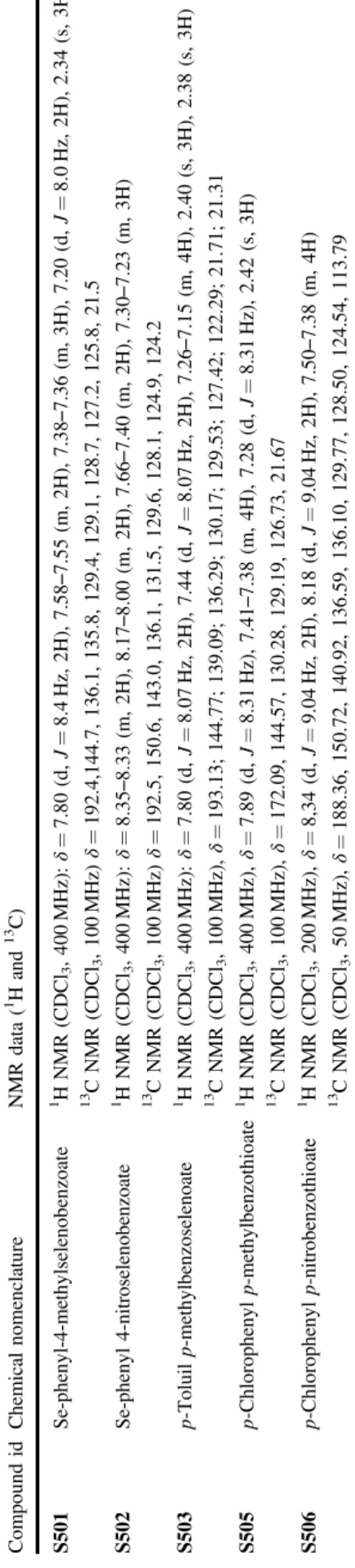
Table1shows data acquired in experiments using NMR.
Minimal inhibitory concentration and minimal bactericidal concentration
The values of MIC of each compound onS. parasanguinis
andS. mutansare described in Table2.
Effect on biofilm formation
Biofilm mass
The biofilm mass of S. parasanguinis was reduced when
biofilms were grown in the presence of S503, S505, and
S506at different concentrations and fully inhibited by the
treatment with S501 and S502 at 250 and 31.25µg/ml,
respectively (Fig.2). Regarding S. mutans strain, the
bio-film mass was reduced by the treatment withS501andS502
at most concentrations assayed, while S503, S505, and
S506were effective only at higher concentrations (Fig.2).
Viability of biofilm-entrapped cells
The number of viable cells embedded within the matrix of biofilms grown in the presence of chalcogenoesters was also determined. With few exceptions, all compounds reduced the viability of both strains in a dose-dependent manner
(Fig.3). In general,S. parasanguiniswas more susceptible
to all agents. The compoundsS501,S502,S503and S506
reduced the viability ofS. parasanguinis in all
concentra-tions tested, with reduction levels ranging from 2 to 4.5 log.
On the other hand, S505 was effective only at higher
concentrations.
Regarding S. mutans, the effect of compounds on its
viability was lower than that achieved onS. parasanguinis.
Curiously, the compounds S501, S502, S503, and S505
were less effective at higher concentrations, being the best effect achieved at lower concentration. On the other hand,
the compoundS506reduced the viability ofS. mutansin all
tested concentrations with reduction levels ranging from 1.9
to 3.7 log, which were achieved at higher concentrations
(Fig. 3).
Effect on preformed biofilms
Biofilm mass
The chalcogenoesters were also evaluated regarding their
effect on biofilm mass of S. parasanguinis and S. mutans
preformed biofilms. The results showed that all compounds
inhibited the biomass of S. parasanguinis (Fig. 4). The
compound S506 at 250µg/ml reduced S. parasanguinis
biomass at about 85%. Even in the lowest concentration
tested (7.8µg/ml), the same compound was effective on
biomass, reducing it in approximately 31%. The compounds S501,S502, andS505reduced biomass in 55, 63, and 56%, respectively.
The same results were not seen onS. mutans, since the
biomass was reduced only in higher concentrations
eval-uated (Fig. 4).
Viability of biofilm-entrapped cells
The viability of biofilm-entrapped cells of preformed
bio-films was assessed on both strains after the treatment with
chalcogenoesters. All compounds reduced the viability of
S. parasanguinis. Moreover, this effect was seen mainly in
higher concentrations (Fig. 5). The best reduction was
achieved by the treatment with S502, which reduced the
viability at about 1.9, 1.2 and 1.1 log at concentrations of
250, 125 and 62.5µg/ml, respectively.
Regarding S. mutans, the viability was reduced mainly
by the treatment withS501,S502, andS503. Interestingly,
as previously reported (in the section Effect on preformed biofilms), the best effect on preformed biofilms was also
achieved when S. mutans biofilms were treated with
chal-cogenoesters at lower concentrations (Fig. 5). The same
trend was not seen in biofilms treated withS505andS506,
since the viability was better reduced by the treatment with higher concentrations of compounds.
Analysis of the biofilms by SEM
The effect of S501 on biofilm architecture of both
strains was evaluated by SEM inspection (Fig. 6). The
images revealed morphological alterations in biofilms of
both species. Regarding control groups,S. mutansbiofilms
showed a large number of cells and EPS accumulation
(Fig. 6a). On the other hand, S. parasanguinis showed a
lower biofilm content in comparison with S. mutans
(Fig. 6b).
Biofilms formed by both strains in the presence ofS501
showed an evident decrease in the number of cells as well as
EPS content (Fig.6c and d).
Discussion
This study evaluates for the first time the effect of
five synthetic chalcogenoesters on planktonic cells and
biofilms fromS. parasanguinisandS. mutans, two
impor-tant pathogens involved in dental plaque and caries development.
S. parasanguinis plays a key role in development of cariogenic biofilms, since it express several genes related to adhesion proteins, such as Fap1 gene, which is responsible
for the production of fimbriae, which mediates microbial
adhesion to dental surfaces and BapA1 gene, which is responsible for the direct link between initial and secondary colonizers, and thus contributing to the development of
biofilms (Garnett et al. 2012; Geng et al. 2012). On the
other hand, S. mutansis widely known by its aciduric and
acidogenic properties, being considered one of the most important pathogen involved in dental caries (Liu and
Burne 2009). Oral biofilms directly contribute to
develop-ment of dental caries. Thus, the discovery of substances able in inhibiting biofilm formation by pathogens or even in destroying those already established is extremely important for human health.
The MIC found in this work indicates that each com-pound exerts a specific action against tested bacteria. This
fact can be observed by the effect of selenoestersS501and
Table 1 Chemical nomenclature and NMR data of synthesized chalcogenoesters Compound id Chemical nomenclature NMR data ( 1 H and 13 C) S501 Se-phenyl-4-methylselenobenzoate 1H NMR (CDCl 3 , 400 MHz): δ = 7.80 (d, J = 8.4 Hz, 2H), 7.58 – 7.55 (m, 2H), 7.38 – 7.36 (m, 3H), 7.20 (d, J = 8.0 Hz, 2H), 2.34 (s, 3H) 13 C NMR (CDCl 3 , 100 MHz) δ = 192.4,144.7, 136.1, 135.8, 129.4, 129.1, 128.7, 127.2, 125.8, 21.5 S502 Se-phenyl 4-nitroselenobenzoate 1H NMR (CDCl 3 , 400 MHz): δ = 8.35 – 8.33 (m, 2H), 8.17 – 8.00 (m, 2H), 7.66 – 7.40 (m, 2H), 7.30 – 7.23 (m, 3H) 13 C NMR (CDCl 3 , 100 MHz) δ = 192.5, 150.6, 143.0, 136.1, 131.5, 129.6, 128.1, 124.9, 124.2 S503 p -Toluil p -methylbenzoselenoate 1 H NMR (CDCl 3 , 400 MHz): δ = 7.80 (d, J = 8.07 Hz, 2H), 7.44 (d, J = 8.07 Hz, 2H), 7.26 – 7.15 (m, 4H), 2.40 (s, 3H), 2.38 (s, 3H) 13 C NMR (CDCl 3 , 100 MHz), δ = 193.13; 144.77; 139.09; 136.29; 130.17; 129.53; 127.42; 122.29; 21.71; 21.31 S505 p-Chlorophenyl p -methylbenzothioate 1 H NMR (CDCl 3 , 400 MHz), δ = 7.89 (d, J = 8.31 Hz), 7.41 – 7.38 (m, 4H), 7.28 (d, J = 8.31 Hz), 2.42 (s, 3H) 13 C NMR (CDCl 3 , 100 MHz), δ = 172.09, 144.57, 130.28, 129.19, 126.73, 21.67 S506 p -Chlorophenyl p -nitrobenzothioate 1 H NMR (CDCl 3 , 200 MHz), δ = 8,34 (d, J = 9.04 Hz, 2H), 8.18 (d, J = 9.04 Hz, 2H), 7.50 – 7.38 (m, 4H) 13 C NMR (CDCl 3 , 50 MHz), δ = 188.36, 150.72, 140.92, 136.59, 136.10, 129.77, 128.50, 124.54, 113.79
Table 2 Minimal inhibitory concentration of chalcogenesters determined after 24 h growth ofS. mutansandS. parasanguinis
Strain MIC (µg ml−1)
S501 S502 S503 S505 S506
Fig. 2 Biofilm mass ofS. parasanguinisATCC 903aandS. mutans ATCC 25175bafter a 24 h growth in the presence of chalcogenesters. Biomass quantification was performed by staining biofilms with
crystal violet and reading the absorbance at 590 nm.Error bars indi-cate the standard deviation (SD). *=p<0.05 compared to the control group
Fig. 3 Viability of biofilm-entrapped cells ofS. parasanguinisATCC 903aandS. mutansATCC 25175bafter a 24 h growth in the pre-sence of chalcogenoesters. The viability was determined by the
Fig. 4 Biofilm mass ofS. parasanguinisATCC 903aandS. mutans ATCC 25175bpreformed biofilms. The biofilms were treated with chalcogenoesters. Biomass quantification was performed by staining
biofilms with crystal violet and reading the absorbance at 590 nm. Error barsindicate the standard deviation (SD). *p<0.05 compared with the control group
Fig. 5 Viability of biofilm-entrapped cells ofS. parasanguinisATCC 903aandS. mutansATCC 25175bpreformed biofilms. The biofilms were treated with chalcogenoesters. The viability was determined by
S502, which were more effective on S. parasanguinis
planktonic cells. When the structures of selenoesters are
compared, it is observed that S502displays a deactivator
group of the benzene ring. Such group is probably the
responsible for better actions exerted by S502 thanS501,
since the last presents an electron donating activator group
(–CH3), which probably generates a more stable radical.
Unlike selenoesters, the thioesters analogsS505and S506
were more effective on S. mutans planktonic cells, with
MIC values of 250 and 62.5µg/ml, respectively. The
dif-ferences in actions should be explained by the presence of
polar groups in benzene rings ofS506, which are able to
deactivate and remove electrons of such rings (–Cl and
–NO2). Such characteristic probably generates unstable ions
that can act as free radicals againstS. mutansand in a less
extent onS. parasanguinis.
Radhakrishna et al. (2010) showed that the
organosele-nium3-[(phenylcarbonyl)selenyl]propanoic acid and differ-ent ester derivatives from that compound showed potdiffer-ential
antibacterial activity against Staphylococcus aureus,
Sal-monella typhimurium, Escherichia coli and Bacillus
subtilis. Moreover, some chiral chalcogenoamines showed
antibacterial activity against Bacillus cereus, Listeria
monocytogenes and different species of Paenibacillus
(Vargas et al.2012).
Concerning the effects on biofilm formation of both
strains, the results showed that all compounds were able in reducing significantly the biofilm formation. The viability
of cells embedded in EPS matrix of biofilms grown in the
presence of compounds was also evaluated. All compounds
reduced the viability of S. parasanguinis and S. aureus.
Curiously, the effect onS. mutans was seen mainly by the
treatment withS501,S502,S503, andS505, which reduced
the number of viable cells as the concentration of com-pounds decreased. Interestingly, the biofilm mass was higher in concentrations that reduced cell viability, sug-gesting that compounds initially stress bacterial cells, thus promoting high amounts of biomass. Afterwards, cells lose
their viability. The compoundS506was the most efficient
in reducing viability ofS. mutans. Probably the two polar
groups and deactivators of the benzene ring were respon-sible for this phenomenon.
The effect of compounds on preformed biofilms of both strains was lower than that seen on biofilm formation. Both biofilm mass and cell viability were higher when compared to biofilm formation assay. This is not surprisingly, since bacterial growth in biofilms promotes protection against the action of antibiotics and environmental stressors mainly because the physical barrier formed by the extracellular matrix. In addition, when microbial cells are surrounded by EPS matrix, bacterial communication mechanisms stimulate the production of enzymes and proteins important in the
physiological adaptation of biofilm (Corbin et al. 2011;
Soto2013).
Organochalcogens are lipophilic compounds that can interact with layers of polysaccharides, fatty acids, and phospholipids in bacterial membrane increasing its perme-ability and causing damage to the structures (Goldbeck et al.
2014). The mechanism of action of these compounds could
be explained by the disruption of the peptidoglycan layer, altering the cytoplasmic membrane, changing its perme-ability and releasing cellular constituents. Corroborating
with this hypothesis, Rosseti et al. (2015) showed that the
diphenyl diselenide [(PhSe)2] inhibited the growth and
biofilm formation of Candida albicans by increasing the
membrane permeability.
SEM images corroborated with results obtained in the assays for quantification of biomass and the number of
viable cells in biofilms.S. mutans biofilm has a thick EPS
matrix evolving from bacterial cells, which was clearly
reduced in the presence of S501 (Figs. 6a and b). EPS
matrix is an important component that promotes mechanical stability and protection to biofilms (Flemming and
Win-gender 2010), antimicrobial compounds able in reduce or
promote changes in biofilm matrix can be considered
pro-mising molecules. Regarding,S. parasangunis biofilm, the
SEM images showed a strong decrease in the number of
bacterial cells (Fig. 6c and d). In fact, S501 reduced
abruptly the number of viable cells of S. parasanguinis
biofilm (Fig.3a).
In general, the chalcogenoesters were more effective on
S. parasanguinis. Nevertheless,S. mutansstrain used is this study produces high amounts of biofilms (Liu and Burne
2009), which may explain its increased resistance to
chalcogenoesters.
In conclusion, this study demonstrates, for thefirst time,
the antibacterial activity of chalcogenoesters on the plank-tonic growth, biofilm formation and disruption of the pre-formed biofilm of oral bacteria. Moreover, chalcogenoesters can be considered as promising molecules with potential for the treatment of some streptococcal biofilms, which play important roles in dental caries.
Acknowledgements The authors are grateful to the Fundação Cearense de Apoio ao Desenvolvimento Científico e Tecnológico
(FUNCAP), Conselho Nacional de Desenvolvimento Científico e Tecnológico (CNPq), Coordenação de Aperfeiçoamento de Pessoal de Nível Superior (CAPES) and Central Analítica-UFC/CT-INFRA/ MCTI-SISNANO/Pró-Equipamentos CAPES. E. H. Teixeira is a member of the Brazilian Academy of Sciences.
Conflict of interest The authors declare that they have no conflict of interest.
References
Aas JA, Paster BJ, Stokes LN, Olsen I, Dewhirst FE (2005) Defining the normal bacterialflora of the oral cavity. J Clin Microbiol 43 (11):5721–5732
Bhabak KP, Mugesh G (2009) Synthesis and structure–activity correlation studies of secondary- and tertiary-amine-based glutathione peroxidase mimics. Chem Eur J 15(22):9846–9854 Boger DL, Mathvink RJ (1989) Phenyl selenoesters as effective
pre-cursors of acyl radicals for use in intermolecular alkene addition reactions. J Org Chem 54(8):1777–1779
Boger DL, Mathvink RJ (1992) Acyl radicals: intermolecular and intramolecular alkene addition reactions. J Org Chem 57 (5):1429–1443
Caufield PW, Schön CN, Saraithong P, Li Y, Argimón S (2015) Oral lactobacilli and dental caries: a model for niche adaptation in humans. J Dent Res 94(2):110–118
Corbin A, Pitts B, Parker A, Stewart PS (2011) Antimicrobial pene-tration and efficacy in an in vitro oral biofilm model. J Anti-microb Chemother 55(7):3338–3344
Das D, Roy G, Mugesh GJ (2008) Antithyroid drug carbimazole and its analogues: synthesis and inhibition of peroxidase-catalyzed iodination of l-tyrosine. Med Chem 51(22):7313–7317
de Souza D, Mariano DOC, Nedel F, Schultze E, Campos VF, Seixas F, da Silva RS, Munchen TS, Ilha V, Dornelles L, Braga AL, Rocha JBT, Collares T, Rodrigues OED (2015) New orga-nochalcogen multitarget drug: synthesis and antioxidant and antitumoral activities of chalcogenozidovudine derivatives. J Med Chem 58(8):3329–3339
Dewhirst FE, Chen T, Izard J, Paster BJ, Tanner AC, Yu WH, Lakshmanan A, Wade WG (2010) The human oral microbiome. J Bacteriol 192(19):5002–5017
Donlan RM (2002) Biofilms: microbial life on surfaces. Emerg Infect Dis 8(9):881–890
Flemming HC, Wingender J (2010) The biofilm matrix. Nat Rev Microbiol 8(9):623–633
Garnett JA, Simpson PJ, Taylor J, Benjamin SV, Tagliaferri C, Cota E, Chen YY, Wu H, Matthews S (2012) Structural insight into the role of Streptococcus parasanguinis Fap1 within oral biofilm formation. Biochem Biophys Res Commun 417(1):421–426 Geng J, Chiu CH, Tang P, Chen Y, Shieh HR, Hu S, Chen YY (2012)
Complete genome and transcriptomes of Streptococcus para-sanguinis FW213: phylogenic relations and potential virulence mechanisms. PLoS One 7:347–360
Goldbeck JC, Victoria FN, Motta A, Savegnago L, Jacob RG, Perin G, Lenardão EJ, Silva WP (2014) Bioactivity and morphological changes of bacterial cells after exposure to 3-(p-chlorophenyl)thio citronellal. Lwt- Food Sci Technol 59(2):813–819
Hall-Stoodley L, Costerton JW, Stoodley P (2004) Bacterial biofilms: from the natural environment to infectious diseases. Nat Rev Microbiol 2:95–108
Liu Y, Burne RA (2009) Multiple two-component systems of Strep-tococcus mutans regulate agmatine deiminase gene expression and stress tolerance. J Bacteriol 191(23):7363–7366
Lucas MA, Schiesser CH (1996) (Aryltelluro)formates as precursors of alkyl radicals: thermolysis and photolysis of primary and secondary alkyl (aryltelluro)formats. J Org Chem 61(17): 5754–5761
Marsh PD (2004) Dental plaque as a microbial biofilm. Caries Res 38 (3):204–211
Marsh PD (2010) Microbiology of dental plaque biofilms and their role in oral health and caries. Dent Clin N Am 54 (3):441–454
Mugesh G, Du Mont WW, Sies H (2001) Chemistry of biologically important synthetic organoselenium compounds. Chem Rev 101 (7):2125–2179
Narayanaperumal S, Alberto EE, Gul K, Kawasoko CY, Dornelles L, Rodrigues OED, Braga AL (2011) Zn in ionic liquid: an efficient reaction media for the synthesis of diorganyl chalcogenides and chalcogenoesters. Tetrahedron 67(25):4723–4730
Nogueira CW, Zeni G, Rocha JBT (2004) Organoselenium and organotellurium compounds: toxicology and pharmacology. Chem Rev 104(12):6255–6285
Pattenden G, Stoker DA, Winne JM (2009) A synthetic approach to C-nor-D-homosteroids based on a cascade of radical cyclisations from a vinylcyclopropane-substituted acyl radical precursor. Tetrahedron 65:5767–5775
Radhakrishna PM, Sharadamma KC, Vagdevi HM, Abhilekha PM, Rubeena Mubeen S, Nischal K (2010) Synthesis and antibacterial activity of novel organoselenium compounds. Int J Chem 2 (2):149–154
Rampon DS, Rodembusch FS, Goncalves PFB, Lourega RV, Merlo AA, Schneider PH (2010) An evaluation of the chalcogen atom effect on the mesomorphic and electronic properties in a new homologous series of chalcogeno esters. J Braz Chem Soc 21 (11):2100–2107
Ren K, Wang M, Liu P, Wang L (2010) Iron-catalyzed syhnthesis of selenoesters from diselenides and acyl chlorides or acid anhy-drides in the presence of magnesium dust. Synthesis (Mass) 7:1078–1082
Rosseti IB, Rocha JB, Costa MS (2015) Diphenyl diselenide (PhSe)2 inhibits biofilm formation byCandida albicans, increasing both ROS production and membrane permeability. J Trace Elem Med Biol 29:289–295
Sarma BK, Mugesh G (2008) Thiol cofactors for selenoenzymes and their synthetic mimics. Org Biomol Chem 6:965–974
Simoes M, Simoes LC, Vieira MJ (2010) A review of current and emergent biofilm control strategies. Lwt- Food Sci Technol 43 (4):573–583
Simon-Soro A, Mira A (2015) Solving the etiology of dental caries. Trends Microbiol 23(2):76–82
Soto SM (2013) Role of efflux pumps in the antibiotic resistance of bacteria embedded in a biofilm. Virulence 4(3):32–41
Stepanovic S, Vukovic D, Dakic I, Savic B, Svabic-Vlahovic M (2000) A modified microtiter-plate test for quantification of staphylococcal biofilm formation. J Microbiol Methods 40 (2):175–179
Vargas J, Narayanaperumal S, Gul K, Ravanello BB, Dornelles L, Soares LC, Alves CFS, Schneider T, Vaucher RA, Santos RCV, Rodrigues OED (2012) Synthesis of chiralβ-chalcogen amine derivatives and Gram-positive bacteria activity. Tetrahedron 68:10444–10448
Vasconcelos MA, Arruda FVS, Santos HS, Rodrigues AS, Bandeira PN, Albuquerque MRJR, Cavada BS, Teixeira EH, Henriques M, Pereira MO (2014) Effect of a casbane diterpene isolated from Croton nepetaefoliuson the prevention and control of biofilms formed by bacteria and Candida species. Ind Crops Prod 61:499–509