Nonprimary Cytomegalovirus Fetal Infection
Infeção fetal não primária por citomegalovírus
So
fi
a Rodrigues
1Daniela Gonçalves
1Ricardo Taipa
2Maria do Céu Rodrigues
11Gynecology and Obstetric Service, Centro Materno Infantil do Norte,
Centro Hospitalar do Porto, Porto, Portugal
2Pathological Anatomy Service, Centro Materno Infantil do Norte,
Centro Hospitalar do Porto, Porto, Portugal
Rev Bras Ginecol Obstet 2016;38:196–200.
Address for correspondence Sofia Pina Rodrigues, MD, Centro Hospitalar do Porto, Largo Prof. Abel Salazar, 4099-001, Porto, Portugal (e-mail: sofi[email protected]).
Keywords
►
cytomegalovirus fetal
infection
►
nonprimary infection
►
lissencephaly
Abstract
Cytomegalovirus (CMV) is the most common congenital viral infection, causing
hearing, visual and psychomotor impairment. Preexisting maternal CMV immunity
substantially reduces, but not eliminates, the risk of fetal infection and affectation. This
article is about a case of nonprimary maternal CMV infection during pregnancy, with
vertical transmission, resulting in severe fetal affectation. Preconceptional analysis
indicated maternal CMV past infection. Pregnancy progressed uneventfully until the
20th week ultrasound (US), which revealed cerebral abnormalities: thin and
hyper-echogenic cerebral cortex with prominent lateral ventricles, bilateral periventricular
hyperechogenicities, cerebellar vermis hypoplasia and absent corpus callosum. The
MRI suggested these
fi
ndings were compatible with congenital infection rather than
primary brain malformation.
The fetal karyotype was normal. The title of CMV
’
s IgG antibodies almost tripled. Since the
fi
rst semester, analysis of the polymerase chain reaction (PCR) for CMV DNA in the amniotic
fl
uid was negative. The pregnancy was terminated at 23 weeks. Neuropathological
fi
ndings
at autopsy showed severe brain lesions associated with CMV infection.
Palavras-chave
►
infeção fetal por
citomegalovírus
►
infeção não primária
►
lisencefalia
Resumo
O citomegalovírus (CMV) é a infeção viral congénita que mais comumente causa
de
fi
ciência auditiva, visual e psicomotora. A preexistência de imunidade materna reduz
substancialmente, mas não elimina, o risco de infeção e afetação fetal. Trata-se de um
caso de infeção materna não primária por CMV durante a gravidez, com transmissão
vertical, resultando em afetação fetal severa. As análises preconcepção indicavam
infecção passada por CMV. A gravidez decorreu sem intercorrências até a ecogra
fi
a
efetuada na 20ª semana, que revelou alterações cerebrais: córtex cerebral
fi
no e
hiperecogénico com ventrículos laterais proeminentes, hiperecogenecidades
perivent-riculares bilaterais, hipoplasia do vérmis cerebeloso e ausência de corpo caloso. A
ressonância magnética sugeriu que estes achados eram mais favoráveis a uma infeção
congénita do que com uma malformação cerebral primária. O cariótipo fetal era
normal. O título de anticorpos IgG para CMV havia triplicado desde a dosagem do
primeiro trimestre. O PCR para o DNA do CMV no líquido amniótico foi negativo. A
gravidez foi interrompida na 23ª semana. Os achados neuropatológicos na autópsia
mostraram lesões cerebrais severas associadas a infeção por CMV.
received
September 2, 2015
accepted
January 11, 2016
DOI http://dx.doi.org/ 10.1055/s-0036-1583170.
ISSN 0100-7203.
Copyright © 2016 by Thieme Publicações Ltda, Rio de Janeiro, Brazil
Introduction
Cytomegalovirus (CMV) is the most common congenital viral infection, with a birth prevalence of 0,2 to 2,2%,1causing hearing and visual impairment and delay of psychomotor development.1
Primary CMV infection during pregnancy has an inci-dence of 1 to 4%,2 and the global risk of transplacental infection is 40%.3,4 The risk of fetal infection increases with the gestational age1,2,5(according to a study involving 508 pregnancies, vertical CMV transmission rate by gesta-tional age at the time of maternal infection was 0% in the pregestation group, 4.6% in the periconception group, 34.8, 42 and 58.6% respectively in the first, second and third trimester groups).3 Severe disease may affect fetuses in-fected during thefirst trimester as well as the second, but rarely affects fetuses infected later on in pregnancy.3,5Also, congenital infection impairs placental development and functions, and should be considered as an underlying cause of intrauterine growth restriction (IUGR), regardless of virus transmission to the fetus.6
Ten to 15% of congenitally infected newborns are overtly symptomatic at birth and will present neurodevelopmental damage within thefirst three years.1From those, 20 to 30% die, mostly of disseminated intravascular coagulation, hepatic dysfunction, or bacterial superinfection; severe neurologic morbidity occurs in 50 to 60% of survivors.1,5,7Of the asymp-tomatic infants at birth (85 to 90%), 5 to 15% will develop long-term neurodevelopmental morbidity, such as sensorial hearing loss, delay of psychomotor development and visual impairment.1
The criteria necessary to recommend universal screening for CMV infection in pregnant women haven’t yet been met, and are not recommended by the Centers for Disease Control and Prevention or the American College of Obstetricians and Gynecologists.2,8 Prenatal diagnosis is mainly performed when US abnormalities are observed.4
Nonprimary maternal CMV infection (reactivation or reinfection with a different strain) occurs more rarely, with less probability of fetal infection (1,5% in a study with 543 cases of suspected maternal nonprimary infection)9and only 0.2 to 2% of newborns will develop a symptomatic disease,6which indicates a minor fetal affectation in these cases.
This case draws attention to the gravity of a nonprimary CMV infection.
Case Description
The patient is 37 years old, with irrelevant personal and family medical histories, and in her fourth spontaneous pregnancy. The patient has a history of two spontaneous abortions and a term vaginal delivery, which resulted in a healthy child. The spouse is healthy; the couple is not consanguineous.
Preconceptional analysis indicated maternal immunity to Rubella, Toxoplasmosis and CMV. First trimester routine analy-sis at the 11th week of pregnancy indicated CMV IgG and IgM of respectively 848.9 Ul/mL (positive threshold1) and 0.379 Ul/
mL (negative threshold<0,8). Thefirst trimester US did not
show fetal anomalies, and the combined prenatal screening for chromosomal abnormalities was negative.
At 20 weeks of gestation, US revealed cerebral abnormali-ties: thin and hyperechogenic cerebral cortex with promi-nent lateral ventricles, bilateral periventricular hyperechogenicities, cerebellar vermis hypoplasia and ab-sent corpus callosum (►Fig. 1). Amniocentesis was per-formed and the karyotype was normal (46, XY).
An extended serology study was then performed, and it showed no immunity to Herpes simplex virus type 2 and immunity to Toxoplasmosis, Herpes simplex virus type 1, Parvovirus and CMV. However, CMV IgG title increased almost three times since thefirst trimester analysis, and CMV IgM kept negative (IgG 2695.0 Ul/mL and IgM 0.333 Ul/mL) – these serologic tests were performed in the same laboratory as the
first trimester tests. IgG avidity testing was high.
When asked aboutflu–like symptoms, the patient mentions an episode during the second trimester of pregnancy; however, as it was mild, it was difficult to precise the time of it.
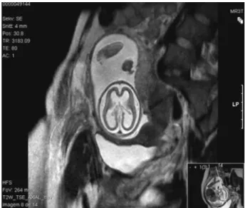
Magnetic resonance imaging (MRI) at 22 weeks of gesta-tion confirmed USfindings and raised the hypothesis of
Fig. 1 (A) Ultrasound at 20 weeks of gestation–thin and
fetal infection:“[…] enlargement of the cerebrospinalfluid circulation spaces at the expense of marked reduction in the thickness of the cerebral parenchyma of both hemispheres of lissencephalic appearance [...] suggesting serious sequel-ae of brain infection (group TORCH? other?) rather than primary malformative injury of the central nervous system” (►Fig. 2).
Outcome and Follow-up
The pregnancy was terminated at 23 weeks.
At this time, a new sample of amniotic fluid was collected to perform PCR for CMV DNA. The result was negative. Pathologic examination study of fetal and placen-tal tissues revealed cerebral ventricular dilation with
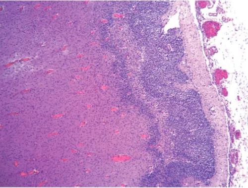
necro-hemorrhagic and calcified periventricular lesions, necro-hemorrhagic lesions also at the level of the basal ganglia, hippocampus, periventricular regions of brainstem and cerebellar hemispheres. There were areas of four-layer polymicrogyria and frequent cellular viral inclusions (cy-tomegalic cells), particularly in the germinal matrix and near the necrotic lesions. These cells tested positive for CMV immunohistochemistry (►Figs. 3,4and5). Immuno-histochemical detection of CMV DNA was found not only in the fetus brain, but also in his liver, heart and in the placenta.
Discussion
All the investigation and outcomes in this case suggest a probable CMV secondary infection, which can be due to a reactivation or reinfection with a different strain. According to the maternal serology and the morphologic US performed, the maternal CMV secondary infection must have occurred between 11 and 20 weeks of pregnancy.
The preferred approach for diagnosing fetal infection is PCR testing for DNA virus in the amnioticfluid,1,3,4which has a sensitivity of 75–100%.4Gestational age at amniocentesis and the time elapsed since the suspicion of infection are critical factors for the sensitivity of the method.1,3,4
The PCR for CMV DNA negative result was indeed a false negative. Although this method is recognized as the gold standard for prenatal diagnosis of fetal CMV infection, the gestational age and the time lag between maternal infec-tion and amniocentesis is a critical factor influencing sensitivity. To achieve the highest sensitivity, amniocente-sis should be performed after 21 weeks of gestation and at least 6 to 7 weeks after the onset of maternal infection reflecting the time it takes for placental infection, trans-mission to the fetus, viral replication in the fetal kidney, and excretion into amnioticfluid.1,3,8In spite of having fulfilled thefirst prerequisite, the time lag might not have been achieved. Nevertheless, we were reaching the legal limit for terminating the pregnancy, and even without a prenatal diagnosis, the USfindings foretold a poor outcome for the offspring.
The idea that nonprimary CMV maternal infection will not result in severe fetal sequelae and congenital disease has been proved different in several published cases.5,10,11The preexisting maternal CMV seropositivity substantially re-duces, but not eliminates, the risk of fetal infection and affectation; therefore, the use of prenatal invasive methods for the detection of fetal infection in nonprimary maternal infections may be justified.
Neurons migrate from the areas where they are born to the areas where they will settle into their proper neural circuits. Neuronal migration starts in the second month of gestation and is controlled by a complex assortment of chemical guides and signals. When these signals are absent or incorrect, neurons do not end up where they belong, resulting in a primary malformative injury called lissence-phaly. The lack of gyral development after a defective neuro-nal migration in the developing brain can be associated with
Fig. 2 Magnetic resonance imaging at 22 weeks of gestation with
lissencephalic appearance.
Fig. 3 Macroscopicalfindings–coronal section at the level of the
the agenesis of the corpus callosum or cerebellar hypoplasia, and is also due to some genetic syndromes.12
Neuropathological studies of brains infected prenatally by CMV show evidence of lissencephaly, polymicrogyria, cerebellar hypoplasia and ventricular dilation in conse-quence of loss of volume (atrophy),7,12findings suggestive of the severity and risk of neurological and motor sequelae. Still, a normal US examination cannot exclude neonatal or long-term morbidity.1,13Mild ventriculomegaly, cerebel-lar vermis hypoplasia and lissencephaly can also be asso-ciated with extra central nervous system abnormalities, chromosomal aberration and other fetal infections like rubella and toxoplasmosis.13In this case, no other abnor-malities were found beyond the fetal central nervous system; the karyotype was normal and the other fetal infections were excluded.
There is insufficient evidence to support the use of passive immunization to prevent congenital infection,2and the only known means of reducing the risk of congenital CMV is by reducing exposure to the virus through hygiene measures.2,5 Therapies for treatment of congenital CMV are in the experimental stage. In an uncontrolled study published in 2005, administration of CMV-specific hyperimmune globu-lin to pregnant women with primary CMV infection signifi -cantly reduced the rate of intrauterine transmission, from 40 to 16%.14 A 2014 randomized placebo-controlled phase 2 trial of virus-specific hyperimmune globulin for the preven-tion of congenital CMV infecpreven-tion, involving 124 women with primary CMV infection from 5 to 26 weeks of gestation, showed that the treatment with hyperimmune globulin did not significantly modify the course of primary CMV infection during pregnancy.15Other phase 3 studies are under way to further our understanding of the efficacy and safety of
Fig. 4 Histopathologicalfindings showing four-layer polymicrogyria (HE). Magnification, 50x.
Fig. 5 (A) Histopathologicalfindings–neurons of germinal matrix with
hyperimmune globulin administration as a means of pre-venting congenital CMV infection.
Although it is still not recommended, our institution offers pregnant women systematic screening for CMV infection, as well as education about hygienic measures. However, we do not provide any therapy for pregnant women with suspicious or confirmed CMV primary or nonprimary infections.
Nevertheless, the US and MRIfindings in this case sug-gested the presence of severe advanced disease and high risk of long-term neurodevelopmental impairment, so the option of pregnancy termination was obviously raised.
This article is about a rare case of nonprimary maternal CMV infection during pregnancy, with vertical transmission, resulting in severe fetal affectation. The preexisting maternal CMV seropositivity substantially reduces, but not eliminates, the risk of fetal infection and affectation. This article high-lights the importance of clinical suspicion and the difficulty of prenatal confirmation of a CMV infection.
References
1 Yinon Y, Farine D, Yudin MH, et al; Fetal Medicine Committee, Society of Obstetricians and Gynaecologists of Canada. Cytomegalovirus infection in pregnancy. J Obstet Gynaecol Can 2010;32(4):348–354
2 Johnson J, Anderson B. Screening, prevention, and treatment of congenital cytomegalovirus. Obstet Gynecol Clin North Am 2014; 41(4):593–599
3 Feldman B, Yinon Y, Tepperberg Oikawa M, Yoeli R, Schiff E, Lipitz S. Pregestational, periconceptional, and gestational primary maternal cytomegalovirus infection: prenatal diagnosis in 508 pregnancies. Am J Obstet Gynecol 2011;205(4):342.e1–342.e6
4 Picone O, Vauloup-Fellous C, Cordier AG, et al. A series of 238 cytomegalovirus primary infections during pregnancy: descrip-tion and outcome. Prenat Diagn 2013;33(8):751–758
5 Benoist G, Leruez-Ville M, Magny JF, Jacquemard F, Salomon LJ, Ville Y. Management of pregnancies with confirmed cytomeg-alovirus fetal infection. Fetal Diagn Ther 2013;33(4): 203–214
6 Pereira L, Petitt M, Fong A, et al. Intrauterine growth restriction caused by underlying congenital cytomegalovirus infection. J Infect Dis 2014;209(10):1573–1584
7 Ornoy A, Diav-Citrin O. Fetal effects of primary and secondary cytomegalovirus infection in pregnancy. Reprod Toxicol 2006; 21(4):399–409
8 Walker SP, Palma-Dias R, Wood EM, Shekleton P, Giles ML. Cytomegalovirus in pregnancy: to screen or not to screen. BMC Pregnancy Childbirth 2013;13:96
9 Yoeli-Ullman R, Lipitz S, Schiff E, Feldman B. 216: Prenatal diagnosis of fetal cytomegalovirus infection in 543 cases of suspected maternal non-primary infection. Am J Obstet Gynecol 2007;197(6, Suppl)S71
10 Boppana SB, Fowler KB, Britt WJ, Stagno S, Pass RF. Symptomatic congenital cytomegalovirus infection in infants born to mothers with preexisting immunity to cytomegalovirus. Pediatrics 1999; 104(1 Pt 1):55–60
11 Stagno S, Pass RF, Dworsky ME, et al. Congenital cytomegalovirus infection: The relative importance of primary and recurrent maternal infection. N Engl J Med 1982;306(16):945–949
12 Cheeran MC, Lokensgard JR, Schleiss MR. Neuropathogenesis of congenital cytomegalovirus infection: disease mechanisms and prospects for intervention. Clin Microbiol Rev 2009;22(1): 99–126
13 Picone O, Teissier N, Cordier AG, et al. Detailed in utero ultrasound description of 30 cases of congenital cytomegalovirus infection. Prenat Diagn 2014;34(6):518–524
14 Nigro G, Adler SP, La Torre R, Best AM; Congenital Cytomegalovi-rus Collaborating Group. Passive immunization during pregnancy for congenital cytomegalovirus infection. N Engl J Med 2005; 353(13):1350–1362