61
Radiol Bras. 2015 Jan/Fev;48(1):59–64Letters to the Editor
this latter presentation following sclerotomal distribution. Long bones of the lower limbs are most frequently affected(1–5). The disease rarely affects the spine, skull and the face. The diagnosis is essentially clinical and radiological. Laboratory tests results are usually normal and histological findings are nonspecific.
The classical radiological presentation is that of sclerosis in only one side of the cortical bone, with linear and segmental dis-tribution, resembling melted candle wax dropping along the bone axis and projecting over the medullary space. Such an alteration may distally extend to finger bones.
Other observed presentations are similar to osteoma, striated osteopathy, osteopoikilosis, and myositis ossificans, with calcifi-cations in adjacent soft tissues (1).
Computed tomography (CT) shows in more detail the scle-rotic alterations as well as reduction of the medullary space. At magnetic resonance imaging (MRI) such alterations present with low signal intensity at T1- and T2-weighted sequences, a finding that is compatible with cortical bone. The involvement of soft tis-sues may also be observed, with variable calcification degrees at CT; and MRI shows images with heterogeneous signal intensi-ties corresponding to mineralization, areas of fat and fibrovascu-lar tissue(1,2).
Thus, melorheostosis is highlighted as a relevant differen-tial diagnosis among bone diseases, particularly because of the characteristic radiographic findings of this disease.
REFERENCES
1. Suresh S, Muthukumar T, Saifuddin A. Classical and unusual imaging appearances of melorheostosis. Clin Radiol. 2010;65:593–600. 2. Nuño C, Heili S, Alonso J, et al. Melorreostosis: presentación de un caso
y revisión de la literatura. Rev Esp Enferm Metab Oseas. 2001;10:50–5. 3. Salman Monte TC, Rotés Sala D, Blanch Rubió J, et al. Melorheostosis,
a case report. Reumatol Clin. 2011;7:346–8.
4. Mariaud-Schmidt RP, Bitar WE, Pérez-Lamero F, et al. Melorheostosis: unusual presentation in a girl. Clin Imaging. 2002;26:58–62. 5. Gagliardi GG, Mahan KT. Melorheostosis: a literature review and case
report with surgical considerations. J Foot Ankle Surg. 2010;49:80–5.
Paulo Marcus Vianna Franca1, Cid Sérgio Ferrreira1, Reginaldo Figueiredo1, João Paulo Kawaoka Matushita1 1. Universidade Federal de Minas Gerais (UFMG), Belo Horizonte, MG, Brasil. Mailing Address: Dr. Paulo Marcus Vianna Franca. Rua Itaí, 570, ap. 102, Santa Efigênia. Belo Horizonte, MG, Brazil, 30260-290. E-mail: [email protected].
http://dx.doi.org/10.1590/0100-3984.2013.0019
Pre- and postnatal findings of a dicephalus tetrabrachius-dipus conjoined twins with a diaphragmatic hernia
Dear. Editor,
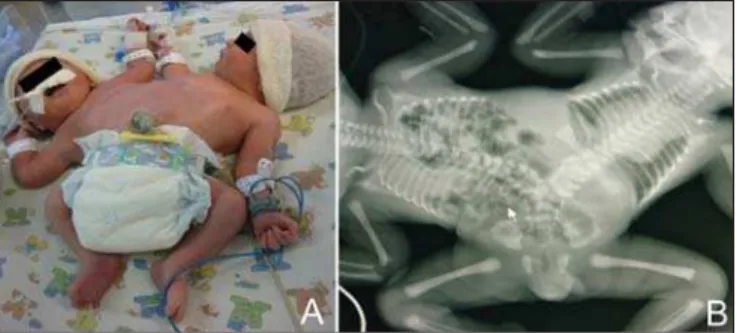
A 17-year-old primigravida attended the service at the 31st gestational week for evaluation of monochorionic, monoamniotic twin gestation. First trimester sonographic images were not avail-able. Morphological ultrasonography (US) demonstrated the fe-tuses joined at the level of their abdomen and pelvis, and presence of a diaphragmatic hernia in the second twin. The woman denied previous history of health problems or use of medicines and illicit drugs. Her 25-year-old husband was healthy, with negative history of consanguinity. No family history of genetic diseases and mal-formations was reported. Fetal magnetic resonance imaging (MRI) revealed a dicephalus tetrabrachius-dipus conjoined twin. The fetus at right presented with a left diaphragmatic hernia containing stomach, small bowel and colon. The twins shared a single liver and a urinary bladder. Two kidneys connected each other at the level of their lower poles, and two vertebral spines were fused at the level of the sacrum (Figure 1). Echocardiography was normal.
The conjoined twin was born by Cesarean section at the 35th gestational week, weighting 3,765 grams. Upper eyelid coloboma was found in the twin with diaphragmatic hernia. Radiographic evaluation demonstrated vertebral spines fusion at the level of the lumbar region, besides the presence of bowel loops in the tho-racic cavity of the twin at right (Figure 2). Surgery for the dia-phragmatic hernia could not be performed. The conjoined twin died at the 17th day of life.
Imperfect twinning occurs in approximately one per 250,000 live births(1,2) and is classified according to the fusion site added by the term pagus(3). “Parapagus” twins (meaning “extensive lat-eral fusion”) correspond to 28% of cases of conjoined twins(4). The subtype dicephalus tetrabrachius-dipus, as observed in the present case, is considered rare (4/10,000,000 births)(5).
US has shown to be the best method for initial evaluation of the gestation, and can identify imperfect twinning as early as at the 12th gestational week(1). However, US is subjected to limitations such as maternal biotype and presence of either oligohydramnios
Figure 2. Postnatal image of the dicephalus tetrabrachius-dipus twin (A). Radio-graphic evaluation showing vertebral spines fusion at the L4 level and a single pelvis. A diaphragmatic hernia is observed in the fetus at right (there is evidence of the presence of bowel loops within the thoracic cavity), without identification of the heart and airways (B).
Figure 1. Fetal MRI T2-weighted image showing the dicephalus tetrabrachius-dipus conjoined twin. The fetus at right presents with a left diaphragmatic hernia. Mediastinal structures (heart and large vessels) and pulmonary hypoplasia (A) are identified. Hepatic fusion is also visualized (B).
62
Radiol Bras. 2015 Jan/Fev;48(1):59–64 Letters to the EditorCongenital abnormalities not related to the fusion site are observed in 10% to 20% of cases of conjoined twins. Diaphrag-matic hernia such as the one observed in the present case is one of the described findings(7). Upper eyelid coloboma that was also identified in the present case is considered to be a rare abnormal-ity(8).
Thus, the correct determination of the type of imperfect twin-ning as well as of the fusion extent may be useful in the evalua-tion of the condievalua-tion severity and in the postnatal surgical plan-ning. Determining the severity of the condition is of paramount importance considering that the Brazilian laws allows for gesta-tion terminagesta-tion in cases where the extrauterine life is not pos-sible(3).
REFERENCES
1. McHugh K, Kiely EM, Spitz L. Imaging of conjoined twins. Pediatr Radiol. 2006;36:899–910.
2. Denardin D, Telles JA, Betat RS, et al. Imperfect twinning: a clinical and ethical dilemma. Rev Paul Pediatr. 2013;31:384–91.
3. Nomura RM, Brizot ML, Liao AW, et al. Conjoined twins and legal au-thorization for abortion. Rev Assoc Med Bras. 2011;57:205–10.
4. Spencer R. Theoretical and analytical embryology of conjoined twins: part I: embryogenesis. Clin Anat. 2000;13:36–53.
5. Martínez-Frías ML, Bermejo E, Mendioroz J, et al. Epidemiological and clinical analysis of a consecutive series of conjoined twins in Spain. J Pediatr Surg. 2009;44:811–20.
6. Hibbeln JF, Shors SM, Byrd SE. MRI: is there a role in obstetrics? Clin Obstet Gynecol. 2012;55:352–66.
7. Mackenzie TC, Crombleholme TM, Johnson MP, et al. The natural history of prenatally diagnosed conjoined twins. J Pediatr Surg. 2002;37: 303–9.
8. Mansour AM, Mansour N, Rosenberg HS. Ocular findings in conjoined (Siamese) twins. J Pediatr Ophthalmol Strabismus. 1991;28:261–4.
http://dx.doi.org/10.1590/0100-3984.2013.0021
Rafael Fabiano Machado Rosa1, Luciano Vieira Targa2, Stephan Philip Leonhardt Altmayer1, Karen Lizeth Puma Lliguin1, Daniela Denardin2, André Campos da Cunha2
1. Universidade Federal de Ciências da Saúde de Porto Alegre (UFCSPA), Porto Alegre, RS, Brasil. 2. Hospital Materno Infantil Presidente Vargas (HMIPV), Porto Alegre, RS, Brasil. Mailing Address: Dr. Rafael Fabiano Machado Rosa. Rua Sarmento Leite, 245/403, Centro. Porto Alegre, RS, Brazil, 90050-170. E-mail: [email protected].
Epidural cavernous hemangioma of the spine: magnetic resonance imaging findings
Dear Editor,
A previously healthy male, 52-year-old patient complaining of progressive lower limbs paraparesis for four months and recent onset of urinary incontinence, with no history of local trauma. At admission, the patient was afebrile and, at physical examination presented with spastic paraparesis with sensitive level at D8. Labo-ratory tests (blood count and biochemical blood tests) did not dem-onstrate any significant alteration.
Magnetic resonance imaging (MRI) demonstrated the pres-ence of an elongated lesion with regular contour, intermediate signal intensity on T1-weighted and marked hypersignal on T2-weighted sequences, with intense and homogeneous contrast
en-hancement, located in the epidural space, extending from D5 to D7. Such a lesion determined a significant narrowing of the rachid-ian canal and medullary compression on the corresponding seg-ments, with consequential medullary hypersignal on T2-weighted and STIR sequences compatible with compressive myelopathy (Figure 1).
The patient was submitted to surgical procedure where a wine-colored lesion compressing the dural sac was observed. The lesion was completely resected with no significant complication. Anatomopathological analysis demonstrated proliferation of middle-sized vessels filled with blood, with no atypias, compatible with cavernous hemangioma.
Hemangiomas are benign proliferative vascular lesions. Ac-cording to the predominant type of vascular canal, hemangiomas are classified as follows: venous, arteriovenous, capillary and
cav-Figure 1. Sagittal MRI T1-weighted (A), T2-weighted (B), STIR (C) and T1-weighted sequences with fat saturation following intravenous contrast injection (D) dem-onstrating an elongated, expansile well-delimited lesion with regular contour, located in the epidural space of the posterior region of the dorsal spine, extending from D5 to D7. The lesion presents intermediate signal on T1-, marked hypersignal on T2-weighted and STIR sequences, with intense and homogeneous contrast enhance-ment, suggestive of hemangioma. Such a tumor causes remarkable narrowing of the rachidian canal, determining high signal intensity in the spinal cord (better visualized on STIR sequences) due to compressive myelopathy.