ARTICLE
Stimulated jitter with concentric needle in 42
myasthenia gravis patients
Jitter estimulado obtido com agulha concêntrica em 42 pacientes com miastenia gravis
João Aris Kouyoumdjian1, Erik Stålberg2
Neuromuscular junction (NMJ) disorders, such as myas-thenia gravis (MG), can be evaluated by single-iber electro-myography (SFEMG), which is a technique developed in the early 1960s by Erik Stålberg and Jan Ekstedt in Sweden1-3 for
measuring neuromuscular jitter parameters. his parameter is the most useful one, together with impulse blocking, for electrodiagnosis of these conditions.
MG is an autoimmune disorder of the NMJ usually asso-ciated with antibodies to the postsynaptic acetylcholine re-ceptor (AChR), however it can sometimes be associated with those to muscle-speciic tyrosine kinase (MuSK), another protein at the NMJ.
he SFEMG jitter represents the variation in time inter-vals between pairs of single iber action potentials (SFAPs) in the voluntarily activated technique or the time measured be-tween stimulation pulse and SFAPs in the stimulated tech-nique. With high jitter values, the neuromuscular transmis-sion is so disturbed that occatransmis-sional impulse blocking occurs. he sensitivity of SFEMG has been found to be 88% in ocular MG and 95 to 100% in generalized MG4. Similarly,
Benatar5, in a systematic review, conirmed high speciicity of
SFEMG for the diagnosis of generalized MG. For ocular MG, the sensitivity was ranging from as low as 66% to as high as 98%. Mercelis and Merckaert6 found a speciicity of 97% and
1MD, PhD; Faculdade de Medicina de São José do Rio Preto (FAMERP); Associate Professor, Department of Neurological Sciences, Neuromuscular
Investigation Laboratory, São Paulo SP, Brazil;
2MD, PhD; Department of Clinical Neurophysiology, Emeritus Professor, Institute of Neurosciences, Uppsala University, Uppsala, Sweden.
Correspondence: João Aris Kouyoumdjian; Rua Luiz Antônio da Silveira 1.661; 15025-020 São José do Rio Preto SP - Brasil; E-mail: [email protected]
Support: Fundação de Amparo à Pesquisa do Estado de São Paulo (FAPESP).
Conflict of interest: There is no conlict of interest to declare.
Received 13 September 2012; Received in inal form 19 October 2012; Accepted 26 October 2012.
ABSTRACT
Objective: To estimate jitter parameters in myasthenia gravis in stimulated frontalis and extensor digitorum muscles using the concentric needle electrode. Methods: Forty-two conirmed myasthenia gravis patients, being 22 males (aged 45.6±17.2 years-old) were studied. Jitter was expressed as the mean consecutive difference (MCD). Results: MCD in extensor digitorum was 61.6 µs (abnormal in 85.7%) and in fron-talis 57.3 µs (abnormal in 88.1%). Outliers represented 90.5% for extensor digitorum and 88.1% for frontalis. At least one jitter parameter was abnormal in 90.5% of the combined studies. Acetylcholine receptor antibody was abnormal in 85.7% of the cases. Conclusions: Stimulated jitter recordings measured from muscles using concentric needle electrode can be used for myasthenia gravis diagnosis with high sensitivity. Extensive normative studies are still lacking and, therefore, borderline indings should be judged with great caution.
Key words: jitter, myasthenia gravis, concentric needle electrode, Extensor Digitorum, Frontalis, single-iber electromyography.
RESUMO
Objetivo: Mensurar os valores do jitter em pacientes com miastenia gravis nos músculos frontalis e extensor digitorum pela téc-nica estimulada, utilizando-se eletrodo de agulha concêntrica. Métodos: Foram estudados 42 pacientes, sendo 22 homens (idade 45,6±17,2 anos), com miastenia gravis confirmada. O jitter foi expresso como a média das diferenças consecutivas (MDC). Resultados: A MDC para o extensor digitorum foi 61,6 µs (anormal em 85,7%) e para o frontalis 57,3 µs (anormal em 88,1%). Outliers representa-ram 90,5% para o extensor digitorum e 88,1% para o frontalis. Pelo menos um parâmetro do jitter foi anormal em 90,5% dos estudos combinados. Anticorpo receptor de acetilcolina estava anormal em 85,7% dos casos. Conclusões:Jitter estimulado mensurado por meio de eletrodo de agulha concêntrica pode ser utilizado para diagnóstico de miastenia gravis com elevada sensibilidade. Estudos normativos mais amplos ainda são necessários e, portanto, valores limítrofes devem ser avaliados com cautela.
238 Arq Neuropsiquiatr 2013;71(4):237-243
a sensitivity of 80% for ocular MG in stimulated-SFEMG for
orbicularis oculi (OOc).
Disposable concentric needle electrodes (CNE) have been tested for measurements of jitter7,8, due to the
in-creasing concern for the transmission of infections. Some papers have presented normative data and diagnostic value of the test in MG7-13. Farrugia et al.14 found no
dif-ferences in mean jitter values for extensor digitorum (ED) and OOc muscles in 24 MG patients using both SFE and CNE. Papathanasiou and Zamba-Papanicolaou15 found no
significant differences between mean jitter measured by disposable or reusable SFE in 18 MG patients in the OOc muscle stimulation technique.
In order to improve recording selectivity and suc-cessfully use a CNE for jitter measurement, the low-fre-quency filter should typically be raised from 500 Hz to 1 or 2 kHz to suppress the activity from distant muscle fi-bers7,8. A filter setting with a 1 kHz high-pass filter,
rath-er than highrath-er, has been suggested for optimal quality16.
This setting seems to provide good balance between the desired effect of low frequency suppression with a rea-sonably preserved original signal shape and an accept-able signal-to-noise ratio.
As the signals obtained with CNE recording do not al-ways represent a single-iber action potential, but rather a summation of many, the term jitter recording with CNE from apparent single iber action potential (ASFAPs) is preferable, rather than SFEMG with CNE.
Jitter studies for MG electrodiagnosis using CNE are still incipient and relatively few of them have been done. In our previous report17, we have used electrical stimulation for
jit-ter measurements in 20 MG patients. he aim of this study was to evaluate in a larger cohort whether stimulated CNE in the EDand frontalis (FR) muscles in MG patients could give similar results as described with the SFE.
MATERIAL AND METHODS
Patients
Forty-two MG patients were studied between August 2009 and December 2011 for jitter measurement using CNE. All of them had confirmed MG based on the clini-cal picture ( fluctuating weakness), unequivoclini-cal cliniclini-cal response to edrophonium and/or pyridostigmine, positive acetylcholine receptor antibody (AChRAb) titer, and/or decrement of at least 10% on slow ( from 2 to 3 Hz) re-petitive nerve stimulation studies (RNS) from the first to fourth responses. None of them had previous jitter mea-surements. All patients stopped pyridostigmine at least 24 hours before testing. Cholinesterase inhibitors may mask abnormal jitter when the abnormality of neuromuscular transmission is mild18.
he MG Group consisted of 22 men and 20 women with a mean age of 45.6±17.2 years-old (range, from 21 to 82). he mean time of MG symptoms was 75.6±72.6 months (range, from 2 to 288). he mean time since diagnosis was 55.5±61.7 months (range, from 1 to 246). he mean age of debut was 39.3 years-old (13 to 80).
Disease severity was determined at each clinic visit ac-cording to the MG Foundation of America (MGFA) clinical classiication from I, ocular weakness, to II-V, generalized weakness19. In the worst period, it comprised: I (6 patients),
IIb (5), IIIa (5), IIIb (4), IVa (11), IVb (2), and V (9). he classi-ication proile at the time of CNE jitter measurement was I (8 patients), IIa (8), IIb (1), IIIa (10), IIIb (3), and IVa (3); nine patients were asymptomatic but eight of those were still taking pyridostigmine. Worst MGFA was considered for ocular (6 cases, 14.3%) and generalized (36 cases, 85.7%) distinctions; in all but one ocular case the time of symptoms was more than 24 months.
CNE jitter recordings and antibody measurements, either to AChRAb or MuSK, were studied at the same day. he most pronounced RNS decrements were taken from the iles at any time from the MG diagnosis and included several nerve — muscle settings either distal, proximal, or facial.
Reference jitter parameters for CNE in the stimulated ED muscle were taken from our previously published data20:
upper limit of normal (ULN), 97.5% (non-Gaussian), for mean consecutive diference (MCD)=22.6 µs and for out-liers=30 µs. he results for the stimulated FR muscle were also taken from our previously published data21 as follows:
ULN, mean +2 SD (Gaussian), for MCD=21.5 µs and 97.5% (non-Gaussian) for outliers=30 µs.
Jitter recording
A KeypointNet electromyograph (Medtronic Skovlunde, Denmark) with built-in jitter software was used for recording and analyzing all patients, using a peak detection algorithm for time measurements. he recordings were performed us-ing a CNE with a diameter of 0.30 mm and a recordus-ing area of 0.019 mm2 (this CNE is the smallest “facial needle” from
Medtronic/Alpine bioMed, Denmark).
Stimulation technique for Frontalis muscle
raised, ever so little. In case of submaximal stimula-tion, the jitter decreased, otherwise it was unchanged. To be accepted for measurements, the ASFAPs should have a fast rising phase without notches or shoulders, be constant at consecutive discharges, i.e., have parallel rising phases seen on superimpositions of the signals separated by more than 150 µs, and have a well-defined peak.
Jitter was expressed as the mean of MCD values. On average, 31.6 potentials were analyzed per patient. For each jitter analysis, a minimum of 50, and ideally 100, consecutive traces was recorded. The filter settings were from 1 to 10 kHz. The Keypoint software also calculated the mean peak latency.
Stimulation technique for extensor digitorum
muscle
In this muscle, small nerve branches cannot be reached outside it, therefore intramuscular axonal stim-ulation must be used. Stimstim-ulation was made with a dis-posable monopolar needle electrode, 15x0.35 mm, 28 G (Medtronic, Denmark) inserted near the motor end-plate zone, between the proximal and middle thirds of ED. A disposable scalp needle electrode, 10x0.30 mm, 30 G (Medtronic, Denmark), was used as the anode and was inserted subcutaneously about 2 cm away from the cathode. Stimulation parameters were 10 Hz using rect-angular pulses of 0.04 ms duration. In general, this could be achieved at about 2 to 6 mA. As described, special and time-consuming adjustments must be done to assure that the spike under study be supramaximally stimulated. Jitter was expressed as the mean of MCD. On average, 28.8 po-tentials were analyzed per patient. Other parameters were similar to the FR muscle stimulation.
Abnormal jitter parameters
Stimulation jitter was considered abnormal, after techni-cal one due to submaximal stimulation was excluded, if: the mean MCD was above the ULN; and equal or more than 10% of individual jitter values for the ASFAPs for each study were above the ULN for outliers.
Ethics
he study was approved by the Ethics Committee, and in-formed consent was obtained from each subject.
RESULTS
Patients
The AChRAb was positive in 36 cases (85.7%). The worst MGFA classification in the six negative cases was: I (two), IIb (one), IIIa (two), and IVa (one). The mean abnormal AChRAb value was 11.62±8.14 nmol/L (range from 0.18 to
31.3). Antibodies to MuSK were negative in all 6 AChRAb negative MG patients. Antibodies to striated muscle tis-sue were found in 3 out of 41 MG patients (7.3%); in one of them, a thymoma was found. In the retrospective analysis, the following number of RNS studies were: ulnar-Abductor Digiti Minimi in 37/42, median-Abductor Pollicis Brevis in 24/42, accessory-Trapezius in 10/42, facial-OOc in 7/42, fa-cial-Nasalis in 7/42, facial-Orbicularis Oris in 4/42,
radial-Anconeus in 3/20, and fibular-Extensor Digitorum Brevis in 2/42 patients. Abnormal decrement was found in 37 from 42 patients (88.1%) in any time of the disease. Nine pa-tients were asymptomatic at the time of jitter and anti-body studies, however only one was completely drug free (remission). Thymectomy was performed in 16 from 42 pa-tients (38.1%), and thymoma was found in five of them. Thirty-eight patients (90.5%) were on pyridostigmine from 60 to 420 mg a day. Twenty patients (47.6%) were using prednisone; 12 from 5 to 40 mg every other day, and 8 from 2.5 to 60 mg every day. Five participants (11.9%) were tak-ing azathioprine from 100 to 150 mg every day.
Jitter parameters in extensor digitorum
he total number of ASFAPs analyzed was 1,209, varying from 13 to 35 for each patient except in one case in whom only ive ASFAPs with very high jitter were obtained. he mean MCD was 61.6±43.0 µs ( from 12.4 to 221). Abnormality was found in 85.7%, and 36 from 42 patients had a MCD greater than 22.6 µs.
Using the criterion for outliers, abnormality was found in 90.5% of the patients, meaning that 38 from 42 patients had at least 10% of ASFAPs with a MCD greater than 30 µs. In 4 of 42 patients, both MCD and outliers still remained nor-mal, therefore the abnormality in any was 90.5%. he mean latency between stimulus and each spike was 6.9±3.0 ms (1.34 to 19.5).
here were no hematomas in the patients studied; the test duration was about 40 to 60 minutes, and it was report-ed to be more painful than in FR, since more nereport-edle inser-tions and position correcinser-tions (stimulation and recording) were necessary.
Jitter parameters in frontalis
he total number of ASFAPs analyzed was 1,327, vary-ing from 25 to 46 for each patient. he mean MCD was 57.3±27.3 µs ( from 12.3 to 124). Abnormality was found in 88.1% of the patients, meaning that 37 from 42 patients reached a MCD greater than 21.5 µs.
240 Arq Neuropsiquiatr 2013;71(4):237-243
Some recordings from ED and FR, with abnormal jitter and impulse blocking, are shown on Figure. Detailed results from jitter parameters, AChRAb, RNS, and MGFA-w class are provided in Table 1.
Comparisons of jitter abnormalities between ED and FR are shown in Table 2. When ED or FR were together con-sidered, abnormality was found in 90.5% either as MCD or outliers.
Jitter parameters in ocular versus generalized cases
In spite of the small cohort, comparison between ocular (worst MGFA class I, 6 patients) and generalized MG (worst MGFA class II to V, 36 patients) was made for the same pa-rameters (Table 3). Follow-up for ocular form (2–93 months) revealed that three patients (50%) considered themselves asymptomatic (all taking pyridostigmine), and the remain-ing three (50%) were unchanged. Follow-up for general-ized forms ( from 2 to 288 months) revealed that 6 patients (16.6%) considered themselves asymptomatic (all but one
taking pyridostigmine), 4 (11.1%) were unchanged, 1 (2.8%) was worsened, and the remaining 25 (69.4%) were improved.
DISCUSSION
We studied a small and heterogeneous MG cohort re-garding age, MGFA class, time of disease, thymus pathology, use of acetylcholinesterase inhibitors, and ongoing immu-nosuppressive treatment. Patients selected for jitter analysis and AChRAb titer determination only included conirmed MG cases under treatment for months or years from diag-nosis. Nine patients were asymptomatic but only one was completely free from medication. his study was designed to study the general feasibility of stimulation CNE jitter analysis in MG, and the results do not represent sensitivity and speci-icity of the technique.
Jitter was found abnormal in 90.5% of the subjects in either or both ED and FR in all patients regardless ocular or generalized, similar to the 90% in our previous article17, Figure. Stimulation concentric needle jitter in myasthenia gravis cases. (A and B): highly abnormal jitter (57.6 and 143 µs) with impulse blocking on B. Notice fast rising phase spikes without notches or shoulders and well-deined peak without shape changes at consecutive discharges. (C and D): false jitter (27.9 and 50.2 µs). Note summation with notches, shoulders, and nonparallel rising phases, not suitable for analysis. For all spikes shown in the igure, the stimulation strength was checked to be supramaximal.
MG 23 - Frontalis MG 35 - Frontalis MG 27 - ED
A B C D
57.6 µs 143 µs 27.9 µs 50.2 µs
0.5 mV/D 0.5 ms/D
5 mV/D 0.5 ms/D
0.2 mV/D 0.2 ms/D
2 mV/D 0.5 ms/D 0.5 mV/D 0.5 ms/D 0.3 mV/D 0.5 ms/D
0.3 mV/D 0.8 ms/D
and also close to results described by others18 that found
abnormality with voluntary SFEMG in either or both ED or FR in 92%.
he analysis of the four patients with normal jitter pa-rameters showed one case with both RNS and AChRAb nor-mal, one with AChRAb abnormal and RNS nornor-mal, one with both RNS and AChRAb abnormal, and one with RNS abnor-mal and AChRAb norabnor-mal.
he AChRAb titer was abnormal in 66.7% for the ocular MG (mean value of 3.49 nmol/L) and in 88.8% for the gener-alized MG (mean value of 12.63 nmol/L). In a previous simi-lar study with 20 MG patients17, the present authors found
75.0 and 87.5%, respectively. In larger published reports, the AChR antibody titer was increased in 55% of those with ocular MG and in 80% of those with generalized MG18.
RNS was abnormal in 66.7% for ocular MG and in 94.4% for generalized MG cases. An abnormal decrement was found in muscles described in 75% of patients with generalized MG, and in 50% of those with ocular MG18. The
higher percentage of RNS abnormality in this and previ-ously study17 compared to others18 gives an impression of
the general severity of our patients; also, it should be em-phasized that RNS was collected from the files and some-times were done more than three some-times over the disease duration (months or years).
Patients with ocular MG had abnormal mean MCD in 66.7% for ED and 83.3% for FR. Outliers were 83.3% in both ED and FR. In our previous study17 with 20 MG cases, we found
75% for both parameters and muscles. Sanders and Howard4
found MCD abnormality in 63% ED and in 88% if any of ED and facial (FR, OOc or orbicularis oris) muscles were tested close to our present results.
Patients with generalized MG had abnormal mean MCD in 88.9%, both ED or FR. Outliers were 91.7% for ED and 88.9% for FR. In our previous study17 with 20 MG cases, we found
93.7% when both ED and FR were considered. Others have found abnormality ranging from 81.4 to 95% in ED18. Again,
the results found here are quite similar to other previously described studies.
Impulse blocking was found in 25.8% of ED and 25.3% of FR in generalized MG and in 16% for ED and in 27.1% for FR in ocular MG. Overall, impulse blocking was found in 24.3% for ED and 25.6% for FR.
Impulse blocking in MG cases after therapy (similar to this study) was found in 5.2% (ED) and 20.6% (FR) for ocular and in 9.9 to 38.3% (ED) and 20.6 to 47.6% (FR) for general-ized forms18. In our previous study17 with 20 MG cases, it was
found 21.6% (ED) and 23.3% (FR) for ocular form, and 31% (ED) and 38.3% (FR) for generalized ones. We found much more impulse blocking in ED for ocular form both in this and in the previous paper. his could indicate that patients may have had unsuspected generalized symptoms during the dis-ease evolution (months or years).
Tae.f!"#$tersf%&
s'(a)ssss"extensor digitorum (ED)
and frontalis (FR) techniques. Muscle speciic tyrosine kinase antibody (MuSK) was negative in all AChRAb negative cases.
C*+,
MCD Outliers AChRAb RNS-w MGFA-w
ED FR ED* FR* nmol/L ab ≥10% Class
1 24.4 23.6 16.7 23.3 <0.10 n I
2 30.7 112 50 96.7 16.5 ab IIb
3 29.2 50.1 36.7 96.7 15.5 ab V 4 21.5 85.6 10 93.3 7.35 ab IVb 5 48.8 38.5 64.3 60.7 6.32 ab I 6 104 124 72.2 100 15.7 ab IIIa
7 29.6 114 40 100 11.61 ab IIb
8 23.6 14.4 10 0 <0.10 ab IIIa 9 78.9 75.1 90.6 90.6 6.48 ab IVa 10 35.6 62.7 55.2 94.1 3.82 ab IIIb 11 90.2 65.2 96 97.1 4.98 ab IIIb 12 64.9 80.6 76.7 85.3 3.44 ab I
13 17.6 16.7 8.8 9.1 1.53 n I
14 79.2 68.6 89.7 87.5 18.5 ab IIb 15 12.4 12.3 0 0 <0.10 n IIb 16 79.8 74.2 96.8 88.2 15.8 ab V 17 221 54.6 100 83.9 16.6 ab IVa 18 39 57.7 66.7 81.8 15.4 n IIIb 19 55.2 58.3 78.6 79.4 3.1 ab IIIa 20 144 77.6 84.6 93.9 22.3 ab IVa 21 79.3 91.2 91.2 97.8 2.22 ab V
22 157 88 100 96.9 10.8 ab V
23 121 57.9 93.7 83.3 0.18 ab V 24 34.4 23.3 48.6 18.2 1.47 ab IVa
25 45 54.6 63.6 67.5 2.7 ab I
26 22 15.5 3.3 3.3 12.5 ab IVa
27 40.6 45.5 67.6 87.1 6.8 ab V 28 40.7 64.3 63.6 83.3 26.0 ab IIIa 29 95.2 46.5 100 63.3 28.0 ab IVa 30 75.4 50.1 96.7 63.3 31.3 ab IVb 31 31.6 38.8 45.5 63.3 16 ab IIb 32 57.9 33.2 60 46.7 <0.10 ab IIIa 33 28.1 29.8 26.7 26.7 19.5 ab V
34 17 15.7 0 3 <0.10 ab IVa
35 22.3 77.9 23.3 77.8 <0.10 ab I 36 74.6 54 81.2 78.8 12.7 ab IVa
37 70.2 70.8 90 90.9 17.9 n V
38 60.4 74.8 70 85.2 2.91 ab IVa 39 43.7 43.7 65.6 59.4 20.6 ab IIIb
40 116 59.9 100 90 8.54 ab IVa
41 56.4 55.3 75 76 6.56 ab V
42 67.6 45.1 86.7 66.7 6.59 ab IVa Limit 22.6 µs 21.5 µs 30 µs 30 µs
Ab 85.7% 88.1% 90.5% 88.1% 85.7% 88.0%
242 Arq Neuropsiquiatr 2013;71(4):237-243
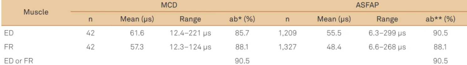
Table 2. Comparison of concentric needle electrodes jitter parameters in stimulated extensor digitorum (ED) and frontalis (FR) in 42 myasthenia gravis patients.
Muscle MCD ASFAP
n Mean (µs) Range ab* (%) n Mean (µs) Range ab** (%)
ED 42 61.6 12.4–221 µs 85.7 1,209 55.5 6.3–299 µs 90.5
FR 42 57.3 12.3–124 µs 88.1 1,327 48.4 6.6–268 µs 88.1
ED or FR 90.5 90.5
MCD: mean consecutive difference; ASFAP: apparent single iber action potentials; n: number; ab: abnormal; *>22.6 µs (ED) and 21.5 µs (FR); **outliers, at least 10% >30 µs (ED and FR).
Table 3. Some parameters comparing generalized to ocular myasthenia gravis.
Parameters Generalized Abnormal (%) Ocular Abnormal (%)
n 36 6 –
Age at debut 43.8 (21–82) 56.7 (27–78) –
Symptoms duration 82.4 months (2–288) 34.3 months (2–93) –
Mean MCD ED* 65.6 µs (12.4–221) 88.9 37.2 µs (17.6–64.9) 66.7
Mean MCD FR* 58.7 µs (12.3–124) 88.9 48.7 µs (16.7–80.6) 83.3
Outliers ED* 91.7 – 83.3
Outliers FR 88.9 – 83.3
Some blocking ED* 25.8 – 16.0
Some blocking FR* 25.3 – 27.1
AChRAb nmol/L* 12.63 (0.18–31.3) 88.8 3.49 (1.53–6.32) 66.7
RNS (1-4)** – 94.4 – 66.7
Thymectomy** 16/36 (44.4%) 0/36 (0%) –
MCD: mean consecutive difference; ED: extensor digitorum; FR: frontalis; AChRAb: acetylcholine receptor antibody; RNS: repetitive nerve conduction studies; *present; **any time.
In voluntary jitter, the percentage of recordings with blocking may be higher compared to stimulation jitter since the fact that the block in one of two ibers may be counted as a blocking pair.
In the present study, abnormal percentage of outliers was greater (ED) or equal (FR) than abnormal mean MCD, simi-lar to our previous study17. he MCD value is usually a more
sensitive index of abnormality than the number of outliers for voluntary SFEMG18. his disparity may be because the
jit-ter in voluntary recording is obtained with two motor end plates. If one has low normal jitter, and the other barely above normal values for individual neuromuscular junctions, the combined jitter of the two would be normal.
Measuring jitter and impulse blocking is technically eas-ier in FR than in ED, and this is due to small motor units in FR. Spikes are more separated and easier to record without riding, notches, and shoulders in FR.
Stimulation technique is quite dependent on the physi-cian’s ability to avoid insuicient stimulation that will give a false large jitter. herefore, in order to be accepted for analy-sis, the spike under study must fulill the ASFAP criteria and be checked for supramaximal stimulation.
Individual spikes obtained with CNE are not always ob-tained from single-muscle ibers16,22,but represent
summa-tion of more than one SFAP. For the stimulasumma-tion technique, the risk of summation is even worse, since many axons are often stimulated.
One must be careful to obtain spikes with a fast rising phase without notches or shoulders, parallel rising phases on consecutive discharges with spikes separated by more than 150 µs. he peak should be well-deined without shape changes on consecutive discharges. he recordings should show clear spikes for reliable increased jitter and impulse blocking analysis.
In CNE jitter studies, we can get SFAPs and ASFAPs (summated), and therefore separate normative data should be collected for this kind of needle. We have done it in some previous reports for ED and OOc muscles, both for voluntary and stimulated techniques and FR stimulated10,20-24. A few studies that have compared jitter
values from SFEMG and from CNE in healthy controls and patients with MG reported a good correlation be-tween results6-8.
After some previous reports on stimulation jitter analy-sis with CNE in healthy subjects20,21,24 and patients with MG17,
we have noticed that in FR the individual single iber signals may be shorter and deinitely more separated in time from each other compared to ED. his technique can be applied in pediatric patients25 after collection of reference values for
younger groups. Large limb muscles like ED are less recom-mended for stimulation studies.
1. Ekstedt J. Human single muscle iber action potentials. Acta Neurol Scand 1964;61:1-96.
2. Stålberg E, Trontelj JV. Single iber electromyography. Studies in healthy and diseased muscle. 2nd ed. New York: Raven Press; 1994. pp. 45-82.
3. Sanders DB, Stålberg E. AAEM minimonograph #25: single-iber electromyography. Muscle Nerve 1996;19:1069-1083.
4. Sanders DB, Howard JF. AAEE Minimonograph #25: Single-iber electromyography in myasthenia gravis. Muscle Nerve 1986; 9:809-819.
5. Benatar M. A systematic review of diagnostic studies in myasthenia gravis. Neuromuscul Disord 2006;16:459-467.
6. Mercelis R, Merckaert V. Diagnostic utility of stimulated single-iber electromyography of the Orbicularis Oculi muscle in patients with suspected ocular myasthenia. Muscle Nerve 2011;43:168-170.
7. Benatar M, Hammad M, Doss-Riney H. Concentric-needle single-iber electromyography for the diagnosis of myasthenia gravis. Muscle Nerve 2006;34:163-168.
8. Sarrigiannis PG, Kennett RP, Read S, Farrugia ME. Single-iber EMG with a concentric needle electrode: validation in myasthenia gravis. Muscle Nerve 2006;33:61-65.
9. Ertaş M, Baslo MB, Yildiz N, Yazici J, Öge AE. Concentric needle electrode for neuromuscular jitter analysis. Muscle Nerve 2000;23:715-719.
10. Kouyoumdjian JA, Stålberg E. Concentric needle single iber electromyography: normative jitter values on voluntary activated Extensor Digitorum. Arq Neuropsiquiatr 2007;65:446-449.
11. Kokubun N, Sonoo M, Imai T, et al. Reference values for voluntary and stimulated single-iber EMG using concentric needle electrodes: A multicentre prospective study. Clin Neurophysiol 2012;123:613-620.
12. Delgado-Delos Santos MMS, Rosales RL. Stimulated single iber electromyography using a concentric needle electrode among normal ilipino subjects. Clin Neurophysiol 2012;123:e51.
13. Lozano AMR, Corredor FO. Single iber electromyography with a concentric needle electrode and surface stimulus in the orbicularis oculi muscle. Clin Neurophysiol 2012;123:e17-e68.
14. Farrugia ME, Weir AI, Cleary M, Cooper S, Metclafe R, Mallik A. Concentric and single iber needle electrodes yield comparable jitter results in Myasthenia Gravis. Muscle Nerve 2009;39:579-585.
15. Papathanasiou ES, Zamba-Papanicolaou E. A comparison between disposable and reusable single iber needle electrodes in relation to stimulated single iber studies. Clin Neurophysiol 2012;123:1437-1439.
16. Stålberg E, Sanders DB. Jitter recordings with concentric needle electrodes. Muscle Nerve 2009;40:331-339.
17. Kouyoumdjian JA, Fanani ACS, Stålberg, EV. Concentric needle jitter on stimulated frontalis and extensor digitorum in 20 myasthenia gravis patients. Muscle Nerve 2011;44:912-918.
18. Stålberg EV, Trontelj JV, Sanders DB. Myasthenia gravis and other disorders of neuromuscular transmission. In: Single Fiber EMG. Edshagen Publishing House, Fiskebäckskil, Sweden. 3rd ed; 2010, p. 218-255.
19. Jaretzki A 3rd, Barohn RJ, Ernstoff RM, et al. Myasthenia gravis. Recommendations for clinical research standards. Task force of the medical scientiic advisory board of the Myasthenia Gravis Foundation of America. Neurology 2000;55:16-23.
20. Kouyoumdjian JA, Stålberg E. Concentric needle single iber electromyography: comparative jitter on voluntary-activated and stimulated Extensor Digitorum. Clin Neurophysiol 2008;119:1614-1618.
21. Kouyoumdjian JA, Stålberg EV. Concentric needle jitter in stimulated frontalis in 20 healthy subjects. Muscle Nerve 2012;45:276-278.
22. Stålberg E, Daube JR. Electromyographic methods. In: Stålberg E (Eds). Clinical neurophysiology of disorders of muscle and neuromuscular junction, including fatigue. Amsterdam: Elsevier; 2003. pp. 147-185.
23. Kouyoumdjian JA, Stålberg E. Reference jitter values for concentric needle electrodes in voluntarily activated Extensor Digitorum and Orbicularis Oculi muscles.Muscle Nerve 2008;37:694-699.
24. Kouyoumdjian JA, Stålberg E. Concentric needle jitter on stimulated Orbicularis Oculi in 50 healthy subjects. Clin Neurophysiol 2011;122:617-622.
25. Tidswell T, Pitt MC. A new analytical method to diagnose congenital myasthenia with stimulated single-iber electromyography. Muscle Nerve 2007;35:107-110.
References
sensitivity and specificity in suspected cases. We found a very high rate of abnormality for MCD (85.7% for ED and 88.1% for FR), outliers (90.5% for ED and 88.1% for FR), and blocking (24.3% for ED and 25.6% for FR) in this small cohort of 42 proven MG cases. This is similar to what has been reported in larger series with SFEMG. In spite of