AR
TIGO ORIGINAL / ORIGINAL AR
TICLE
PREVALENCE OF CELIAC DISEASE IN
SIBLINGS OF IRANIAN PATIENTS WITH
CELIAC DISEASE
Bashir
CHOMEILI
1, Majid
AMINZADEH
2, Amir Kamal
HARDANI
1,
Payam
FATHIZADEH
3, Pooya
CHOMEILI
1and Azarakhsh
AZARAN
3ABSTRACT – Context - Celiac disease, one of the best-known autoimmune human leukocyte antigen-dependent disorders, has a relatively increased prevalence in irst-degree relatives. Objective - To determine the prevalence of celiac disease in siblings of patients with conirmed celiac disease. Methods - Siblings of conirmed celiac disease patients in our center were identiied and enrolled in this study. Their serum immunoglobulin A and tissue transglutaminase antibody-enzyme-linked immunosorbent assay (anti-tissue transglutaminase, immunoglobulin A, and immunoglobulin G) were measured and multiple endoscopic duodenal biopsy specimens were obtained with parental consensus. Celiac disease was conirmed by observation of characteristic histological changes. Results - A total of 49 children (male, 29; female, 20; age, 2–16 years) with conirmed celiac disease in a pediatric gastroenterology ward were studied from 1999 to 2006. We found 30 siblings (female, 16) all shared in both parents. The only measurement available was for immunoglobulin A tissue transglutaminase antibody. A duodenal biopsy was performed in all 30 siblings. Clinical indings such as abdominal pain, fatigue, growth retardation and diarrhea were found in 53.3% of the completely studied siblings, and positive serology without histological changes was identiied in four cases. Both serology and biopsy (conirmed new cases) were positive in 2 of the 30 siblings. Conclusion - High prevalence of celiac disease among siblings of patients with conirmed celiac disease necessitates serologic screening (and conirmatory biopsy if indicated) in families having celiac disease. It is advantageous to diagnose the disease as soon as possible because early diagnosis and diet intervention may prevent serious complications such as growth retardation, short stature, chronic diarrhea, and malignancy.
HEADINGS - Celiac disease, epidemiology. Siblings. Iran.
INTRODUCTION
Celiac disease (CD), a permanent sensitivity to gliadin, is a common chronic and autoimmune disorder with different frequencies in different geographical areas(7, 10, 14, 16, 20). The prevalence of CD exhibits an iceberg effect; the number of asymptomatic cases with positive serology and biopsy is 5- to 7-fold higher than typical symptomatic individuals exhibiting signs and symptoms including abdominal pain, growth retardation, short stature, chronic diarrhea, iron deiciency anemia that is refractive to treatment and intestinal lymphoma(6, 12, 21, 29). Serologic tests such as the anti-endomysium IgA antibody test (EMA), the anti-tissue transglutaminase immunoglobulin (Ig) A antibody test (anti-tTG Ab) and HLA DQ2 or DQ8 genotype testing are useful for evaluation of asymptomatic subjects as well as patients with diabetes mellitus, thyroiditis, Down syndrome, Turner syndrome, William syndrome, IgA deiciency and irst-degree relatives of patients with CD(1, 4, 5, 17).
Institution: Diabetes Research Center, Department of Pediatrics, Ahvaz Jundishapur University of Medical Sciences, Ahvaz, Iran. All the authors declare that there is no conflict of interest concerning this research.
Source of Funding: Research deputy, Ahvaz Jundishapur University of Medical Sciences.
1 Pediatric Gastroenterology Unit; 2 Pediatric Endocrinology Unit, Diabetes Research Center; 3 Pathology Lab, Apadana private Hospital – Ahvaz, Iran.
Correspondence: Dr. Majid Aminzadeh – Diabetes Research Center – Ahvaz Jundishapur University of Medical Sciences, Golestan, Ahvaz, I. R. Iran. E-mail: [email protected]
METHODS
Siblings of conirmed CD patients living in Khuzestan province in southwestern Iran, who were diagnosed by or referred to the authors between 1999 and 2006, were enrolled in this study. Criteria for diagnosis of CD were as follows: clinical indings including growth retardation, short stature, abdominal pain and/or distension, proximal muscle atrophy, chronic diarrhea and fatigue, along with serologic tests and
conirmation by duodenal biopsy results. A comprehensive discussion was conducted with the parents on the subject of the disease and the importance and necessity of screening in high risk but apparently healthy irst-degree relatives with a particular emphasis on siblings. Signed written consent was obtained from the parents.
The study was approved (Approval No. p/8/20/437) by the ethical committee of Ahvaz Jundishapur University of Medical Sciences (AJUMS), Iran. In addition to a physical
Case No. Sex Age/Y Sign & symptom Anti- tTG
Ab level* 1st DUO.Biopsy 2ndDUO.Biopsy
Final diagnosis and action
1 M 6 None 8.2* Neg ND AFU
2 F 5 None 2.9 Neg ND AFU
3 F 8 CAP, SS 1222 CD CD CD, Tx
4 F 4 D 508 CD CD CD, Tx
5 M 14 P, A 18.9 Neg ND AFU
6 M 13 None 6.8 Neg ND AFU
7 M 3 None 6.2 Neg ND AFU
8 F 5 PD 3.5 Neg ND AFU
9 M 2 None 2.4 Neg ND AFU
10 M 3 None 3 Neg ND AFU
11 M 3 None 5.7 NA ND AFU
12 F 6 F 2.4 Neg ND AFU
13 M 12 F,CAP, A 113 S refused AFU
14 F 3 None 2.4 Neg ND AFU
15 M 7 None 5.3 Neg ND AFU
16 M 5 PD, D, P 26.1 Neg ND AFU
17 M 3 None 5 NA ND AFU
18 F 6 PD 18.9 S refused AFU
19 M 5 SS,P 6.6 Neg ND AFU
20 F 12 None 2.1 Neg ND AFU
21 M 7 None 3.7 NA ND AFU
22 F 2 None 2.6 NA ND AFU
23 F 13 C 2.6 Neg ND AFU
24 F 4 None 3.6 Neg ND AFU
25 M 5 I,F,SS 1.6 Neg ND AFU
26 M 6 None 8.7 Neg ND AFU
27 M 10 I, D, CAP,PD 82.6 Neg ND AFU
28 M 7 None 2.3 NA ND AFU
29 M 3 None 5.8 Neg ND AFU
30 M 5 None 15.2 Neg ND AFU
31 F 2 CAP 2.5 Neg ND AFU
32 M 16 None 1.1 NA ND AFU
33 F 14 A 7.8 Neg ND AFU
34 M 14 CAP, A, F >600 S Neg AFU
35 M 7 None 4.4 NA ND AFU
36 F 10 CAP 24.9 Neg ND AFU
37 M 3 None 2 NA ND AFU
38 F 5 None 8.2 S Neg AFU
39 M 5 None 1.8 NA ND AFU
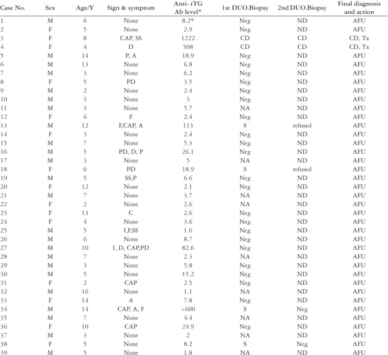
TABLE 1. Details of the studied siblings of known cases of celiac disease
Abbreviations: DUO: duodenal; CAP: chronic abdominal pain; A: anorexia; F: fatigability; PD: pallor & dizziness; D: diarrhea; P: poor weight gain; SS: short stature; I: impatience; C: constipa-tion; CD: celiac disease; Tx: treatment; S: suspicious; NA: did not attend; AFU: advised follow up; N: normal; Neg: negative; ND: not done
examination, a history of possible signs of CD was obtained and auxiliary data were collected from all individuals. A full laboratory assessment (cell blood count, erythrocyte sedimentation rate, blood urea nitrogen (BUN), creatinine, urinalysis, and stool examination for parasites, leukocytes, occult blood, etc.) was performed in all patients to rule out other possible systemic or gastrointestinal diseases. Serologic screening was performed by assessment of serum IgA (to identify possible selective IgA-deicient subjects) and anti-tTG (IgA and IgG). We used the Celi-check kit (Germany), ELISA method, and ELISA reader (ELx800, BIO-TEK Instruments, Inc., USA). Blood samples were obtained following overnight fasting and kept at -20ºC until the laboratory procedures were performed. As recommended by the manufacturer, we established our own normal range based upon our technique, control, equipment, and patient population according to our own established procedure. Our laboratory setting processed a cutoff point of 20 U/mL. Additional results (≥20 U/mL) were considered as positive for anti-tTG. HLA typing facilities were not available for selecting siblings for duodenal biopsy, so endoscopy and duodenal biopsy were performed using an Olympus endoscope on the same day that the serology test was performed to avoid observer bias for all of the 30 siblings in the study. A duodenal biopsy specimen with a villus/crypt (v/c) ratio higher than 3 (without iniltration of chronic inlammatory cells) was considered as normal, whereas a decrease of v/c along with iniltration of the lamina propria with several degrees of chronic inlammatory cells indicated a diagnosis of varying degrees of atrophy as follows: 2.5 to 1.5, mild villus atrophy; 1 to 0.5, moderate (partial) villus atrophy; and less than 0.5, severe (subtotal/ total) villus atrophy. The sample was reported as suspicious if, in addition to intraepithelial iniltration, assessment of the v/c was not possible because of inappropriate orientation or absence of muscularis mucosa.
RESULTS
The study originally included 49 conirmed CD patients since 1999 to 2006 (29 males, 20 females; mean age, 7.5 ± 2.5 years). We were not able to locate 7 families as a result of a change in address or loss of follow-up documentation. Two families chose to not participate and in 7 other families the index case was an only child. As a result, 39 children from 33 remaining families were enrolled in this study. Nine children did not permit an endoscopic biopsy and were excluded. This left 30 children (16 males, 14 females) participate in the study. Details of the siblings who participated are provided in Table 1. One or multiple clinical signs or symptoms were found in 53.3% (7 males, 9 females) of the 30 siblings (Table 2). Abnormal tTG was identiied in 44% (4 males, 3 females) of the 16 siblings with clinical signs and symptoms (Table 3) and in 33% of the 30 studied siblings. Two girls (6.6%) had characteristic intestinal changes compatible with diagnosis of CD (presence of both intraepithelial iniltration and villus atrophy). Serology also was positive in both. Four biopsy samples (from two males and two females) were suspicious,
and after recommendation for a second biopsy, two individuals refused whereas the other two were found to have normal histology in a second biopsy.
DISCUSSION
Several studies have performed screening of CD in high-risk populations. People at higher risk than normal individuals tend to have Down syndrome, Turner syndrome, selective IgA deiciency or other autoimmune diseases such as diabetes mellitus and dermatitis herpetiformis. First-degree relatives of subjects with CD are also at a higher risk for CD(2, 13). The frequency of CD in the general population depends upon the geographical area (environmental factors) and ethnicity (genetic factors)(4, 27). For instance, incidence of CD has been reported as high as 0.01%–0.5% and 1.2% of the total population in Sweden and England, respectively. Rates are higher in Arabian countries, with 0.5%–1.0% of the total population having CD(11). There have been on reports on the frequency of CD in Iran. This difference in prevalence suggests the presence of different risks in high-risk groups as well. Some other factors such as the higher incidence of consanguineous marriage in Middle Eastern countries may increase these frequencies signiicantly. HLA typing is mainly used for diagnosing CD in cases with doubtful clinical,
TABLE 2. Frequency and sex distribution of clinical signs or symptoms in 16 of 30 siblings of the studied patients with celiac disease
Signs & symptoms % Female Male
Abdominal pain 37.5 3 3
Anorexia 25 1 3
Fatigability 25 1 3
Pallor and dizziness 25 2 2
Diarrhea 18.75 1 2
Poor weight gain 18.75 - 3
Short stature 18.75 1 2
Impatience 12.5 - 2
Constipation 6.25 1
-Note: There was more than one sign or symptom in some patients
TABLE 3. Results of serology and biopsy in 9 (of 16 clinically involved) siblings
M = Male; F = Female;
anti-tTG antibody ≥20 IU considered +
Sex Age Anti-tTG Biopsy Comment
F 8 + deinite treatment
F 4 + deinite treatment
M 14 - suspicious 2nd biopsy, did not attend
F 10 + suspicious 2nd biopsy, did not attend
M 12 + suspicious 2nd biopsy, negative
F 5 - suspicious 2nd biopsy, negative
M 5 + negative follow-up
M 10 + negative follow-up
serologic, and histological indings. This can be performed by several methods, but it is costly and time consuming(31). Recently, a new method involving HLA typing of six single nucleotide polymorphism (SNPs) has been found to be eficient and cost-effective. The increased sensitivity and speciicity of this method increases the chances of identifying CD in high-risk populations(18).
Because all serologic tests are based upon assessments of serum IgA (using mucosal antibody against gliadin) and one of the high-risk groups has IgA deiciency, each serology test should be accompanied with a measurement of native IgA at the same time. Sensitivity and speciicity of IgA-Ab to tTG has been reported to be suficiently high to be considered as a diagnostic test in about 80% of situations where obtaining a biopsy is not possible(8, 28). The speciicity of the anti-tTG Ab is not signiicantly different from that of the anti-endomysium antibody, but we prefer it because it requires fewer operators. The gold standard for diagnosis is histopathology of biopsy specimens, accompanied by clinical and histological response to a gliadin-free diet and then recurrence of histological indings after the gliadin re-challenge test, which is particularly important for children below 2 years of age(9, 23, 24). This method of conirmation is dificult for everyone. Therefore, in this study, as in many others, only one typical histological inding has been considered for conirmation of the presence of the disease. Therefore, in two newly diagnosed cases of CD, we considered the clinical response (observed following administration of a gluten-free diet) as the inal conirmation of the diagnosis. Biopsy is recommended even when negative results are obtained in serologic screening when suspicious clinical signs or symptoms are evident(3, 25). A similar study was performed using HLA typing and small bowel histology in Asian irst-degree relatives of children with CD by Srivastava et al.(26). This study identiied a 4.4% prevalence of histologically conirmed CD. Our conirmed prevalence was 6.6%. Unfortunately, the genetic test and HLA typing were not available at the time our study was performed. On the basis of the rules and limitations described above, with respect to the routine screening methods for CD, it was necessary to screen all siblings with anti-tTG Ab and perform endoscopy with duodenal biopsy at the same time. These analyses were performed in a double-blinded fashion. We do not recommend intestinal biopsy to screen CD in all cases when HLA typing and serologic testing are available. A better option would be to use the low chart prepared by Srivastava et al.(26).
This study performs both serology and biopsy screening. These data can be used also for assessing the value of serologic screening in high-risk individuals. Seven patients (23.3%) were found to have positive tTG, but only 2 of 7 (6.6% of all enrolled 30 cases) were conirmed by biopsy. However, 2 of the patients with suspicious biopsies refused follow-up when a second biopsy was recommended. Suspicious biopsies were repeated immediately to rule out the possibility of inappropriate sampling (method or place) or preparation.
Our results are comparable with those of other Western studies that show a 2%–6% prevalence of clinical CD and up to 10% intestinal involvement without clinical signs in irst-degree relatives of individuals with CD(5, 7, 21). On the other hand, compatibility of anti-tTG antibody and biopsy has been reported to be 80%–95% in some studies(5, 22, 29), but was found to be 58.8% in our study (this includes the refusal of follow-up in two patients with suspicious irst biopsies). Some studies have reported that a subject with a negative biopsy but a positive anti tTG Ab (similar to case numbers 27 and 34 in our study— (Table 1)) could be classiied as a CD patient in the future. Follow-up of such patients and duodenal biopsy is recommended. We advised the members of our study group to schedule follow-up visits in the future. We were unable to ind any other study performed in Iran that was similar to the present study. The low positive predictive value of anti-tTG Ab for true histologic CD (discrepancy between serologic and histological disease) in the present study (two of seven) in comparison with other studies may be rationalized as follows: (i) as the frequency of disease in a high-risk population increases, it may lead to a higher frequency of autoimmunity alone (positive serology without intestinal disease), which would be indicated by an increased prevalence of islet cell auto antibodies in irst-degree relatives of diabetic patients relative to the normal population. (ii) Because CD can develop in elderly individuals, there is a possibility that some of the serologically positive cases progress to true disease in the future. Long-term follow-up and biopsy over subsequent years will identify additional histologically positive cases in these families. This could be considered also for siblings with negative anti tTG antibody. (iii) We might have lost two cases of true CD because two individuals with suspicious biopsies refused a second biopsy. (iv) Autoimmunity alone is a very mild form of disease on one side of the wide spectrum of this pathology. (v) Except for two of the Ab-positive subjects (cases no. 16 and 36), all others had very high levels of serum Ab, so inappropriate techniques or cutoffs could not be the cause of this discrepancy. Finally, the unavailability of genetic testing and HLA typing, the loss of follow-up because of relocation of families, unavailability of other families, and refusal to participate in re-testing or to provide biopsies must be considered as limitations of this study.
CONCLUSION
The high prevalence (6.6%) of CD in siblings of patients with CD identiied in this study and other studies conirm the necessity and importance of instituting a screening program for irst-degree relatives of children with CD to identify the disease and administer a strict gluten-free diet to prevent serious complications.
ACKNOWLEDGEMENTS
Chomeili B, Aminzadeh M, Hardani AK, Fathzadeh P, Chomeili P, Azaran A. Prevalência de doença celíaca em ilhos de pais iranianos com doença celíaca. Arq Gastroenterol. 2011;48(2):131-5.
RESUMO – Contexto - A doença celíaca, uma das mais conhecidas enfermidades autoimunes humanas, leucocitária antígeno-dependente, tem prevalência
relativamente maior em parentes de primeiro grau. Objetivo - Determinar a prevalência de doença celíaca em irmãos de pacientes conirmadamente celíacos, ilhos dos mesmos pais. Métodos - Os irmãos de pacientes com doença celíaca conirmada no Department of Pediatrics, Ahvaz Jundishapur University of Medical Sciences, em Ahvaz, Iran, foram identiicados e incluídos no estudo. A imunoglobulina A sérica e o anticorpo transglutaminase tecidual por ensaio imunoenzimático (anti-transglutaminase tecidual, imunoglobulina A e imunoglobulina G) foram medidos e múltiplas biopsias endoscópicas duodenais foram obtidas com o consenso dos pais. A doença celíaca foi conirmada pela observação das características histológicas.
Resultados - Um total de 49 crianças (29 do sexo masculino; 20 do sexo feminino; de 2 a 16 anos) com diagnóstico conirmado de doença celíaca em uma enfermaria de gastroenterologia pediátrica foi estudado de 1999 a 2006. Encontraram-se 30 irmãos (16 do sexo feminino) e todos compartilhavam os mesmos pais dos pacientes. A única medida disponível foi do anticorpo tecidual imunoglobulina A transglutaminase. A biopsia duodenal foi realizada em todos os 30 irmãos. As manifestações clínicas como dor abdominal, fadiga, retardo do crescimento e diarréia foram encontradas em 53,3% dos irmãos estudados completamente, e a sorologia positiva sem alterações histológicas foi identiicada em quatro casos. Ambas, sorologia e biopsia (novos casos conirmados) foram positivas em 2 dos 30 irmãos. Conclusão - A prevalência de doença celíaca entre irmãos de pais conirmadamente celíacos exige triagem sorológica e biopsia de conirmação, se indicada, em familiares com doença celíaca. Diagnosticar a doença o mais rápido possível traz vantagens, pois o diagnóstico precoce e a intervenção dietética podem prevenir complicações graves, como retardo do crescimento, baixa estatura, diarreia crônica e malignidade.
DESCRITORES – Doença celíaca, epidemiologia. Irmãos. Irã.
REFERENCES
1. Blackwell PJ, Hill PG, Holmes GK. Autoantibodies to human tissue transglutaminase: superior predictors of coeliac disease. Scand J Gastroenterol. 2002;37:1282-5. 2. Book L, Zone JJ, Neuhausen SL. Prevalence of celiac disease among relatives of
sib pairs with celiac disease in U. S. families. Am J Gastroenterol. 2003;98:377-81. 3. Branski D, Fasano A, Troncone R. Latest developments in the pathogenesis
and treatment of celiac disease. J Pediatr. 2006;149:295-300.
4. Carnicer J, Farré C, Varea V, Vilar P, Moreno J, Artigas J. Prevalence of coelic disease in Down’s syndrome. Eur J Gastroenterol Hepatol. 2001;13:263-7. 5. Cataldo F, Marino V. Increased prevalence of autoimmune disease in
irst-degree relatives of patients with celiac disease. J Pediatr Gastroenterol Nutr. 2003;36:470-3.
6. Catassi C, Bearzi I, Holmes GK. Association of celiac disease and intestinal lymphomas and other cancers. Gastroenterology. 2005;128:S79-86.
7. Ertekin V, Selimoğlu MA, Kardaş F, Aktaş E. Prevalence of celiac disease in Turkish children. J Clin Gastroenterol. 2005;39:689-91.
8. Farrell RJ, Kelly CP. Celiac sprue. N Engl J Med. 2002;346:180-8.
9. Fasano A, Catassi C. Current approaches to diagnosis and treatment of celiac disease: an evolving spectrum. Gastroenterology. 2001;120:636-51.
10. Fasano A, Berti I, Gerarduzzi T, Not T, Colletti RB, Drago S, Elitsur Y, Green PH, Guandalini S, Hill ID, Pietzak M, Ventura A, Thorpe M, Kryszak D, Fornaroli F, Wasserman SS, Murray JA, Horvath K. Prevalence of celiac disease in at-risk and not-at-risk groups in the United States: a large multicenter study. Arch Intern Med. 2003;163:286-92.
11. Fasano A, Catassi C. Coeliac disease in children. Best Pract Res Clin Gastroenterol. 2005;19:467-78.
12. Green PH, Fleischauer AT, Bhagat G, Goyal R, Jabri B, Neugut AI. Risk of malignancy in patients with celiac disease. Am J Med. 2003;115:191-5. 13. Gudjonsdottir AH, Nilsson S, EK J, Kristiansson B, Ascher H. The risk of celiac
disease in 107 families with at least two affected siblings. J Pediatr Gastroenterol Nutr. 2004;38:338-42.
14. Hill I, Fasano A, Schwartz R, Counts D, Glock M, Horvath K. The prevalence of celiac disease in at-risk groups of children in the United States. J Pediatr. 2000;136:86-90.
15. Hill ID, Dirks MH, Liptak GS, Colletti RB, Fasano A, Guandalini S, Hoffenberg EJ, Horvath K, Murray JA, Pivor M, Seidman EG; North American Society for Pediatric Gastroenterology, Hepatology and Nutrition. Guideline for the diagnosis and treatment of celiac disease in children: recommendation of the North American Society for Pediatric Gastroenterology, Hepatology and Nutrition. J Pediatr Gastroenterol Nutr. 2005;40:1-19.
16. Hoffenberg EJ, MacKenzie T, Barriga KJ, Eisenbarth GS, Bao F, Haas JE, Erlich H, Bugawan Tl T, Sokol RJ, Taki I, Norris JM, Rewers M. A prospective
study of the incidence of childhood celiac disease. J Pediatr. 2003;143:308-14. 17. Kagnoff MF. Celiac disease: pathogenesis of a model immunogenetic disease.
J Clin Invest. 2007;117:41-9.
18. Koskinen L, Romanos J, Kaukinen K, Mustalahti K, Korponay-Szabo I, Barisani D, Bardella MT, Ziberna F, Vatta S, Széles G, Pocsai Z, Karell K, Haimila K, Adány R, Not T, Ventura A, Mäki M, Partanen J, Wijmenga C, Saavalainen P. Cost-effective HLA typing with tagging SNPs predicts celiac disease risk haplotypes in the Finnish, Hungarian, and Italian populations. Immunogenetics. 2009;61:247-56.
19. Liu E, Li M, Bao F, Miao D, Rewers MJ, Eisenbarth GS, Hoffenberg EJ. Need for quantitative assessment of transglutaminase autoantibodies for celiac disease in screening-identiied children. J Pediatr. 2005;146:494-9.
20. Mäki M, Mustalahti K, Kokkonen J, Kulmala P, Haapalahti M, Karttunen T, Ilonen J, Laurila K, Dahlbom I, Hansson T, Höpl P, Knip M. Prevalence of celiac disease among children in Finland. N Engl J Med. 2003;348:2517-24. 21. McOmber ME, Shulman RJ. Recurrent abdominal pain and irritable bowel
syndrome in children. Curr Opin Pediatr. 2007;19:581-5.
22. Mearin ML. Celiac disease among children and adolescents. Curr Probl Pediatr Adolesc Health Care. 2007;37:86-105.
23. Norris JM, Barriga K, Hoffenberg EJ, Taki I, Miao D, Haas JE, Emery LM, Sokol RJ, Erlich HA, Eisenbarth GS, Rewers M. Risk of celiac disease autoimmunity and timing of gluten introduction in the diet of infants at increased risk of disease. JAMA. 2005;293:2343-51.
24. Revised criteria for diagnosis of coeliac disease. Report of Working Group of European Society of Paediatric Gastroenterology and Nutrition. Arch Dis Child. 1990;65:909-11.
25. Shamir R. Advances in celiac disease. Gastroenterol Clin North Am. 2003;32:931-47. 26. Srivastava A, Yachha SK, Mathias A, Parveen F, Poddar U, Agrawal S.
Prevalence, human leukocyte antigen typing and strategy for screening among Asian irst-degree relatives of children with celiac disease. J Gastroenterol Hepatol. 2010;25:319-24.
27. Sulkanen S, Halttunen T, Laurila K, Kolho KL, Korponay-Szabó IR, Sarnesto A, Savilahti E, Collin P, Mäki M. Tissue transglutaminase enzyme-linked immunosorbent assay in detecting celiac disease. Gastroenterology. 1998;115:1322-28. 28. Troncone R, Maurano F, Rossi M, Micillo M, Greco L, Auricchio R, Salerno G, Salvatore F, Sacchetti L. IgA antibodies to tissue transglutaminase: an effective diagnostic test for celiac disease. J Pediatr. 1999;134:166-71.
29. van Rijn JC, Grote FK, Oostdijk W, Wit JM. Short stature and the probability of coeliac disease, in the absence of gastrointestinal symptoms. Arch Dis Child. 2004;89:882-3.