Hydrogen-bonding one-dimensional arrangement in the crystal
of
N
-phenyl-2-hydroxyacetamide
Genivaldo Julio Perpétuo
a, Jan Janczak
b,*aDepartamento de Física, Instituto de Ciências Exatas e Biológicas, Universidade Federal de Ouro Preto, 35400-000 Ouro Preto, MG, Brazil bInstitute of Low Temperature and Structure Research, Polish Academy of Sciences, P.O. Box 1410, 50-950 Wrocław, Poland
a r t i c l e
i n f o
Article history:
Received 23 February 2009
Received in revised form 30 April 2009 Accepted 4 May 2009
Available online 10 May 2009
Keywords:
N-phenyl-2-hydroxyacetamide Crystal structure
Hydrogen bonds DFT calculations
a b s t r a c t
TheN-phenyl-2-hydroxyacetamide was obtained in crystalline form by a direct reaction of aniline with glycolic acid. The whole molecule in the crystal is non-planar, but the planar 2-hydroxyacetamide group is inclined by 7.8(1)°to the plane of the phenyl ring that is cis located to the carbonyl oxygen atom. The
hydroxyl group is in trans position to the carbonyl oxygen atom. The molecules are linked by the NAH O and OAH O hydrogen bonds into one-dimensional chains along thea-axis. Additionally, the CAH p interactions interconnect the chains stabilizing the crystal structure. The X-ray geometry of theN -phe-nyl-2-hydroxyacetamide molecule has been compared with that obtained by theab-initiomolecular orbi-tal calculated results that represents the geometry in the gas-phase.
Ó2009 Elsevier B.V. All rights reserved.
1. Introduction
Crystal engineering has significant applications in the field of materials science, molecular biology and pharmaceutical science
[1–3]. The directional interactions resulting from the multiple hydrogen bonds with the neighbours are the main key for organi-sation of molecules in solids[4,5]. Many different self-complemen-tary hydrogen bonding groups can be used to control association in supramolecular chemistry to produce different and programmed arrangement, such as chains, sheets, ribbons, tapes, rosettes, etc.
[6–9].
Besides the hydrogen bonds as directional non-covalent inter-actions responsible for organisation and design of the supramolec-ular architectures in solids, the
p
p
and CAH
p
interactions are the molecular forces, whose nature is still a matter of discussion[10–13]. In general, the such
p
p
interaction integrates two aro-matic rings arranged in a face-to-face orientation with a distance of 3.2–3.8 Å between the planes of the rings[14–16].Continuing our investigations of the crystals with NAH N, NAH O and OAH O hydrogen bonds and
p
p
or CAHp
inter-actions[17–19], in this paper we present the crystal structure of N-phenyl-2-hydroxyacetamide obtained by a direct reaction of aniline with glycolic acid. A search of the Cambridge Structural Database (Version 5.30, November, 2008) for structures containing N-substituted 2-hydroxyacetamide derivatives yielded only one compound of this class, i.e.N-(10-ethyl-20-hydroxyethyl)-2-hydrox-yacetamide[20]. Thus the present compound is the second of this type, but the first ofN-aryl substituted 2-hydroxyacetamide that has been structurally characterised. The X-ray geometry ofN -phe-nyl-2-hydroxyacetamide molecule is compared to that obtained by anab-initiomolecular orbital calculated result that represents the geometry in the gas-phase.
2. Experimental
2.1. Preparation of N-phenyl-2-hydroxyacetamide
Aniline and glycolic acid (Aldrich, both reagents of purity of 99%) were added to hot water in a molar proportion of 1:1. When the solution became homogenous it was cooled slowly and kept at room temperature. After several days, transparent yellowish crys-tals were formed. Anal. Calculated for C8H9NO2: C, 63.56%; H, 6.00%; N, 9.27% and O, 21.17%. Found: C. 63.64%, H, 5.97%; N, 9.12% and O, 22.27%.
2.2. X-ray data collection
X-ray intensity data for the crystal were collected using graph-ite monochromatic MoK
a
radiation and a four-circlej
geometry KUMA KM-4 diffractometer with a two-dimensional area CCD detector. Thex
-scan technique with Dx
= 1.0° for each image was used for data collection. The 760 images for six different runs covering over 95% of the Ewald sphere were performed. The unit cell parameters were refined by the least-squares methods on the basis of 565 reflections with intensity (I(hkl)) greater than0022-2860/$ - see front matterÓ2009 Elsevier B.V. All rights reserved. doi:10.1016/j.molstruc.2009.05.009
* Corresponding author. Fax: +48 71 344 1029.
E-mail address:[email protected](J. Janczak).
Contents lists available atScienceDirect
Journal of Molecular Structure
8
r
(I). One image was used as a standard after every 40 images for monitoring the crystal stability and data collection. No correction on the relative intensity variations was necessary. Data collection were made using the CrysAlis CCD program[21]. Integration, scal-ing of the reflections, correction for Lorenz and polarisation effects and absorption corrections were performed using the CrysAlis Red program [21]. The structure was solved by the direct methods using SHELXS-97 and refined using SHELXL-97 programs [22]. The hydrogen atoms of the phenyl ring were located in their geo-metrical positions with theUiso(H) = 1.2Ueq(C) of the carbon atom directly joined the H atoms). The other H atoms (CH2, NH and OH) were constrained: CAH = 0.97 Å with Uiso(H) = 1.5Ueq(C); NAH = 0.86 Å withUiso(H) = 1.2Ueq(N) and OAH = 0.83 Å withU i-so(H) = 1.5Ueq(O). The final difference Fourier map showed no peaks of chemical significance. The largest peaks on the finalDq
map were +0.135 and0.136 eÅ3. The details of the data collec-tion parameters, crystallographic data and final agreement param-eters are collected inTable 1. Visualisation of the structure was made with the Diamond 3.0 program[23].2.3. Quantum calculations
Ab-initiomolecular orbital calculations full geometry optimisa-tion were performed with the Gaussian98 program package[24]. All calculations were performed by the density functional three-parameter hybrid (B3LYP) methods[25,26]with the 6-31+Gbasis set starting from the X-ray geometry. As a convergence criterions the threshold limits of 0.00025 and 0.0012 a.u. were applied for the maximum force and the displacement, respectively.
3. Results and discussion
During reaction of aniline with glycolic acid in hot water solu-tion, one proton of the NH2group of aniline molecule is transferred to the hydroxyl group of COOH of glycolic acid and finally the water molecule is eliminated. Elimination of water results in the formation of the NAC bonds between the reagents as illustrate thescheme 1.
The asymmetric unit of the crystal is illustrated inFig. 1. The six-membered ring is planar, but shows slight distortions from the ideal hexagonal symmetry. The distortions result from the sub-stitution effect of the 2-hydroxyacetamide group joined to the ring and the internal C6AC1AC2 and C3AC4AC5 angles to be smaller than 120°. These distortions are in agreement with those found in similar substituted benzene derivatives [27–32]. The non-H atoms of 2-hydroxyacetamide group are planar. This plane is in-clined to the plane of the phenyl ring by 7.8(1)°. The phenyl ring is cis oriented to the carbonyl oxygen atom, whereas the hydroxyl group is in trans position the carbonyl oxygen (Fig. 1). The CAC bond lengths of the phenyl ring are in the range of 1.363(2)– 1.385(2) Å. Both the CAC bonds involving the C1 atom are longer than the remaining four CAC bonds within the phenyl ring. The elongation of the C1AC2 and C1AC6 bonds is caused by the elec-tron withdraw effect of the 2-hydroxyacetamide substituent that disturbs the delocalisation of the
p
electron in the ring.The N1AC1 bond with a distance of 1.413(2) Å between the N-amide and the C atoms of the phenyl ring is longer than the N1AC8 bond (1.339(2) Å) in the 2-hydroxyacetate group (Table 2). This can be explained by the partial delocalisation of the
p
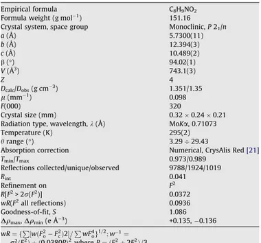
electron of the double C8AO2 bond over the N1AC8 bond, since the fragment is planar. In addition the lone-pair of electron on the N1 atom is local-ised on the p orbital, which is perpendicular to the plane of 2-hydroxyacetamide group similarly as the p orbitals of C and O atoms that form the double CAO bond. Thus the partial delcocalisation of Table 1Crystallographic data forN-phenyl-2-hydroxyacetamide. Empirical formula C8H9NO2
Formula weight (g mol1) 151.16
Crystal system, space group Monoclinic,P21/n
a(Å) 5.7300(11)
b(Å) 12.394(3)
c(Å) 10.489(2)
b(°) 94.02(1)
V(Å3) 743.1(3)
Z 4
Dcalc/Dobs(g cm3) 1.351/1.35
l(mm1) 0.098
F(000) 320
Crystal size (mm) 0.320.240.21 Radiation type, wavelength,k(Å) MoKa, 0.71073 Temperature (K) 295(2)
hrange (°) 3.2929.43
Absorption correction Numerical, CrysAlis Red[21] Tmin/Tmax 0.973/0.989
Reflections collected/unique/observed 9788/1924/1019
Rint 0.041
Refinement on F2 R[F2> 2r(F2)] 0.0372
wR(F2all reflections) 0.0936 Goodness-of-fit,S 1.086
Dqmax,Dqmin(e Å3) +0.135, 0.136
wR¼ fP½wðF2
oF2cÞ2=PwF4og1=2;w1¼
r2ðF2
oÞ þ ð0:0380PÞ2whereP¼ ðF2oþ2F2cÞ=3.
N H H
CH2OH
O
O H
N H
O
CH2OH
+ + H O2
Scheme 1.
H5
O2 H6
C5
H7B C6
H4 C4
C8 C1
C7 N1
C3
C2 H3
H1
H7A H2
O1
H1O
Fig. 1.A view ofN-phenyl-2-hydroxyacetamide molecule showing displacement ellipsoids at the 50% probability level and H atoms as a sphere of arbitrary radii.
the
p
electrons of the double C8AO2 bond over these three atoms (N, C and O) is possible due to the symmetry of the p orbitals. The inter-action of thep
-electrons of the phenyl ring and the lone-pair of elec-tron on the O2 atom makes the respective angles (C6AC1AN1, C1AN1AC8 and N1AC8AO2) significantly greater than 120°, as ex-pected for thesp2hybridisation, since according to the valence-shellelectron pair repulsion model, VSEPR[33], the lone-pair of electrons
occupies a wider region as the bonding pair. Additionally, both an-gles, N1AC8AC7 and C8AC7AO1, are significantly smaller than 120°due to the attractive interaction between the lone-pair of elec-tron on O1 atom with the H1 atom of amide group into the intramo-lecular hydrogen bond N1AH1 O1 with a D A distance of 2.658(2) Å.
Ab-initiofull-optimised molecular orbital calculation performed for an isolated molecule of N-phenyl-2-hydroxyacetamide shows quite similar correlation between the bond lengths and angles as found in the crystal (Fig. 2,Table 2). However, the conformation of the optimised molecule is slightly different than that in the crys-tal. For example the planar 2-hydroxyacetamide group is inclined by 4.3°to the plane of the phenyl ring. The inclination is smaller than in the crystal due to the intramolecular interaction between the carbonyl oxygen O2 atom and the hydrogen H6 atom in orto position in relation to the 2-hydroxyacetamide substituent with O2 H6 distance of 2.280 Å. The conformation of the molecule, in which the phenyl ring and the hydroxyl group are, respectively, in cis and trans position to the carbonyl oxygen atom is the most stable conformation of the N-phenyl-2-hydroxyacetamide mole-cule within the other possible conformers (cis–trans conformation I, seeScheme 2), according to our conformational analysis. The MO calculations on I–IV conformers were performed with constrained torsion angles C1AN1AC8AO2 and O2AC8AC7AO1 to the values 0 or 180°defining the possible conformers (Scheme 2). After optimi-sation routines, the cis/cis conformer (II), in which the phenyl ring and the hydroxyl group are in cis position in relation to carbonyl oxygen, and the trans/cis conformer (III), in which the phenyl ring and the hydroxyl group are, respectively, in trans and in cis posi-tion in relaposi-tion to carbonyl oxygen, have higher single-point en-ergy by7.2 and24.8 kcal/mol, respectively, in comparison to the lowest energy conformation cis–trans (I). The trans–trans former IV is rather unstable, since the optimisation does not con-verge. The energy differences between the conformers are about 25% (7.2 kcal/mol) and 75% (24.8 kcal/mol) of the strong hydrogen bond energy [34,35]. Concluding, the geometry of N -phenyl-2-hydroxyacetamide molecule resulting from X-ray study is mainly determined by the electronic structure, while the greater rotation of the 2-hydroxyacetamide group in relation to the phenyl rings is caused by the intermolecular hydrogen bonding system and the crystal packing forces.
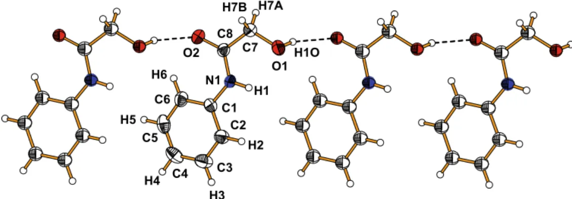
In the crystal the molecules related by a translation along the a-axis interact via the OAH O hydrogen bonds between the hydroxyl group (donor) and the carbonyl oxygen (acceptor) form-ing one-dimensional chains along [100] direction (Fig. 3). Two neighbouring chains are interconnected by the NAH O hydrogen bonds between the imine (NH) groups and the hydroxyl group (Table 3) into a double chains extending along thea-axis (Fig. 4). The double chains interact with each other by a weak CAH
p
interaction of a distance of 3.52 Å formed between the donorCH2and centroid of the
p
-aromatic ring. The CAHp
interactions between the CAH and the aromatic ring are much weaker (E= 24 kcal/mol)[36] than the NAH O or OAH O hydrogen bonds (E= 2535 kcal/mol)[34,35]. This type of intermolecular interactions have been recently recognised as equally important Table 2Bond lengths (Å) and angles (°) forN-phenyl-2-hydroxyacetamide.
C1AC6 1.382(2)
C2AC3 1.373(2)
C5AC6 1.382(2)
O2AC8 1.226(2)
C1AC2 1.385(2)
C3AC4 1.375(2)
O1AC7 1.407(2)
C7AC8 1.506(2)
C1AN1 1.413(2)
C4AC5 1.363(2)
N1AC8 1.339(2)
C6AC1AN1 123.31(12)
C8AN1AC1 128.85(11)
O2AC8AN1 125.27(13)
N1AC8AC7 116.01(11)
C3AC2AC1 120.15(14)
C3AC4AC5 118.99(14)
C5AC6AC1 119.82(14)
C2AC1AN1 117.68(11)
O1AC7AC8 113.60(11)
O2AC8AC7 118.72(12)
C6AC1AC2 119.00(13)
C2AC3AC4 120.89(15)
C4AC5AC6 121.14(14)
Fig. 2.Results of the optimized molecular orbital calculations (B3LYP/6-31+G) for isolatedN-phenyl-2-hydroxyacetamide molecule (Å,°).
N H
O H
OH
H
N H
O OH
H
H N
H
O
H OH H
N H
O O H
HH
(I) (II) (III) (IV)
factors determining the arrangement of molecules in crystals and have found a broad application in crystal engineering for design of new functional materials[37–41]. The closest contact between the hydrogen atom of CH2of 2-hydroxyacetate group and the
p
-aromatic phenyl ring points to the presence of week CAHp
inter-actions that stabilise the three-dimensional structure. The reaction of glycolic acid with amine group of aniline with elimination of water molecule yielding the N-phenyl-2-hydroxyacetamide can be useful in crystal engineering as a method for obtaining crystals of different compounds using different aromatic or aliphatic amines, ArANH2or RANH2.4. Supplementary material
Additional material comprising full details of the X-ray data col-lection and final refinement parameters including anisotropic ther-mal parameters and full list of the bond lengths and angles have been deposited with the Cambridge Crystallographic Data Center in the CIF format as supplementary publications no. CCDC 721009. Copies of the data can be obtained free of charge on the application to CCDC, 12 Union Road, Cambridge, CB21EZ, UK, (Fax: +44 1223 336 033; [email protected]).
References
[1] A. Nangia, Curr. Opin. Solid State Mater. Sci. 5 (2001) 115. [2] L. Addadi, M. Geva, CrystEngComm 5 (2003) 140.
[3] O. Almarsson, M. Zaworotko, Chem. Commun. 17 (2004) 1889.
[4] A.D. Burrows, C.W. Chann, M.M. Chowdhry, J.E. McGrady, D.M.P. Mingos, Chem. Soc. Rev. 24 (1995) 329.
[5] B. Moulton, M. Zaworotko, Chem. Rev. 101 (2002) 1629. [6] C.R. Desiraju, Acc. Chem. Res. 35 (2002) 565.
[7] J.C. MacDonald, G.M. Whitesides, Chem. Rev. 94 (1994) 2383. [8] M.J. Kirsch, J.M. Lehn, Struct. Bonding 96 (2000) 3. [9] T. Steiner, Angew. Chem. In. Ed. Engl. 34 (1995) 2311. [10] C.A. Hunter, J.K.M. Sanders, J. Am. Chem. Soc. 112 (1990) 5525. [11] C. Janiak, J. Chem. Soc. Dalton Trans. (2000) 3885.
[12] F. Cozzi, R. Annunziata, M. Benagalia, M. Cinguini, R. Raimoudi, Y.K. Baldridge, J.S. Siegel, Org. Biomol. Chem. 1 (2003) 157.
[13] Y. Kobayashi, K. Saigo, J. Am. Chem. Soc. 127 (2005) 15–54. [14] C.A. Hunter, Chem. Soc. Rev. 23 (1994) 101.
[15] C.A. Hunter, K.R. Lawson, J. Perkins, C.J. Urch, J. Chem. Soc. Perkin Trans. 2 (2001) 651.
[16] S.L. Cockroft, C.A. Hunter, K.R. Lawson, J. Perkins, C.J. Urch, J. Am. Chem. Soc. 127 (2005) 8594.
[17] G.J. Perpétuo, J. Janczak, J. Mol. Struct. 891 (2008) 429. [18] J. Janczak, Acta Cryst. C64 (2008) o159.
[19] J. Janczak, R. Kubiak, J. Mol. Struct. 920 (2009) 75.
[20] L.S.Z. Rivera, L. Carrillo, T. Mancilla, Heteroat. Chem. 10 (1999) 153. [21] CrysAlis CCD and CrysAlis Red, Version 171.32.8, Oxford Diffraction Poland,
Wrocław, Poland, 2006.
[22] G.M. Sheldrick,SHELXS97,SHELXL97, Programs for Crystal Stuctures Solution and Refinement, University of Göttingen, Göttingen, Germany, 1997. [23] K. Brandenburg, H. Putz, DIAMOND Version 3.0, Crystal Impact GbR, Bonn,
Germany, 2006.
[24] J.M. Frisch, G.W. Trucks, H.B. Schlegel, P.M.W. Gill, B.G. Johnson, M.A. Robb, J. Cheeseman, T. Keith, G.A. Petersson, J.A. Montgomery, K. Raghavachari, M.A. Al-Laham, V.G. Zakrzewski, J.V. Ortiz, J.B. Foresman, J. Cislowski, B.B. Stefanov, A. Nanayakkara, M. Challacombe, C.Y. Peng, P.Y. Ayala, W. Chen, W.M. Wong, J.L. Andres, E.S. Replogle, R. Gomperts, R.L. Martin, D.J. Fox, J.S. Binkley, D.J. Defrees, J. Baker, B.B. Stewart, M. Head-Gordon, C. Gonzales, J.A. Pople, Gaussian98, Revision A.3, Gaussian, Inc., Pittsburgh PA, 1998.
[25] A.D. Becke, J. Chem. Phys. 104 (1996) 1040. [26] C. Lee, W. Yang, R.G. Parr, Phys. Rev. B37 (1988) 785. [27] J.R. Lechat, Acta Cryst. B35 (1979) 183.
[28] H.J. Wasserman, R.R. Ryan, S.P. Layne, Acta Cryst. C41 (1985) 783.
[29] S.W. Johnson, J. Eckert, M. Barthes, P.K. McMillan, M. Muller, J. Phys. Chem. 99 (1995) 16253.
[30] J.W. Banks, A.S. Balsanov, J.A.K. Howard, O. O’Hagan, H.S. Rzepa, S. Santamaria, J. Chemm. Soc. Perkin Trans. 2 (1999) 2409.
[31] K.F. Bowes, C. Glidewell, J.N. Low, J.M.S. Skalde, J.I. Wardell, Acta Cryst. C59 (2003) o1.
H5
H4 C5
C4 H6
C6
C3
H3 C1
C2 O2
H2 N1
C8 H7A
H1 H1O C7
O1 H7B
Fig. 3.View of the OAH O hydrogen bonded chain ofN-phenyl-2-hydroxyacetamide molecules.
Table 3
Hydrogen-bond geometry (Å,°).
DAH A DAH H A D A DAH A
O1AH1O O2i 0.83 1.99 2.777(2) 160
N1AH1 O1 0.86 2.20 2.658(2) 113
N1AH1 O1ii 0.86 2.32 3.032(2) 141
C7AH1 p(C1AC6)iii 0.97 2.77 3.523(3) 139 Symmetry code: i= 1 +x,y,z;ii=x+ 2,y+ 1,z;iii= 1x, 1y,z.
O1ii H1 N1
O2i
H1O
O1 O2
a
b
c
Fig. 4.View of the crystal packing ofN-phenyl-2-hydroxyacetamide. Dashed lines represent the NAH O and OAH O hydrogen bonding as well as the CAH p
interactions that stabilise the 3D structure.
[32] G.Y. Mori, T. Chiba, T. Odani, A. Matsumoto, Cryst. Growth Des. 7 (2007) 1356. [33] R.J. Gillespie, Chem. Soc. Rev. 21 (1992) 59.
[34] J. Emsley, Chem. Soc. Rev. 9 (1980) 91.
[35] G.A. Jeffrey, An Introduction to Hydrogen Bonding (Topics in Physical Chemistry), Oxford University Press, USA, 1997.
[36] S. Tsuzuki, K. Honda, T. Uchimaru, M. Mikai, K. Tanabe, J. Am. Chem. Soc. 122 (2000) 3746.
[37] H. Okawa, K. Uyeda, S. Kida, Inorg. Chem. 21 (1982) 1594.
[38] K. Kobayashi, Y. Asakawa, Y. Kikuchi, H. Tai, Y. Aoyama, J. Am. Chem. Soc. 115 (1993) 2648.
[39] M. Nishio, M. Hirota, Y. Umezawa, The CH/pinteraction: evidence, nature and consequence, Viley-VCH, 1998.
[40] G.R. Desiraju, T. Steiner, The Weak Hydrogen Bond in Structural Chemistry and Biology, Oxford University Press, Oxford, 1999.