I
Diogo Alexandre Fialho de Almeida
Mestrado em Engenharia Física
The role of electron transfer in DNA
building blocks: evaluation of strand
breaks and their implications
Dissertação para obtenção do Grau de Doutor em Engenharia Física
Orientador: Prof. Doutor Paulo Manuel Assis Loureiro
Limão-Vieira, Professor Associado com Agregação, Faculdade de
Ciências e Tecnologia da Universidade Nova de Lisboa
Júri:
Presidente: Prof. Doutor Fernando Pina Arguente(s): Prof. Doutor Pedro Tavares
Prof. Doutor Oddur Ingólfsson
Vogais: Prof. Doutor António Varandas Prof. Doutor Gustavo García Prof. Doutor Antonio Aguillar Prof. Doutor Paulo Limão-Vieira
Doutor Filipe Ferreira da Silva
III
© Diogo Alexandre Fialho de Almeida; FCT/UNL;
UNLA Faculdade de
Ciências e
Tecnologiae a Universidade
Nova de Lisboa têm o direito, perpétuo e sem limites
geográfico
s,
de arquivar e publicar esta dissertação através
de exemplares impressos reproduzidos em papel ou de
forma digital, ou por qual
queroutro meio conheci- do ou
que venha a ser inventado,
e de a divulgar
atravésde
repositórios científicos e de admitir a sua cópia e
distribuição com objectivos educacional ou de investiga-
ção não comerciais
,
desde que seja dado crédito ao autor e
IV
Acknowledgements:
Prof. Dr. Paulo Limão-Vieira for his constant supervision, encouragement and availability throughout the course of this work, as well as for the opportunity to visit other international groups and attend several scientific meetings, from which I have learned a great deal.
Prof. Dr. Gustavo García for his invaluable support and expertise, Dr. Samuel Eden for his always friendly and available help and Dr. Filipe Ferreira da Silva for his candid and reassuring support through several stages of this thesis.
All of the remaining members of the Molecular Physics and applications research group, particularly Drª Susana Sério, Juscelino Ferreira. Prof. João Lourenço for his support and soothing advisement. Ana Cruz for the always available help and patience.
FCT/MCTES for the SFRH/BD/61645/2009 scholarship and to the several international collaboration networks EIPAM, ECCL, NANO-IBCT that over the years provided me with financial support for the several STSM's. To the Department of Physics of the New University of Lisbon and CEFITEC for providing me with the necessary working conditions.
Martina Fuss and Ana Sanz for their hospitality at the IFF, CSIC, Madrid, and Prof. Robert McCullough for his hospitality at the Centre for Plasma Physics at Queen's University Belfast.
On a more personal note, Rodrigo Antunes, Mauro Guerra, Rui Pinto, and Diana Guimarães for their much-valued and deeply thankful friendship and companionship throughout the last decade, as well as for the several unique episodes that will forever be remembered. Thumbs up, guys!
Cláudio Ribeiro and Julia Blanco for their very special and stimulating companionship and support. Shine on, you crazy diamonds.
Rita Ralha, Isa Matos (and the rest of the Matos family), Gonçalo Martins and Margarida Coelho for their much-appreciated friendship and patience, despite everything... José Samões and Rui Pedro for their availability and presence.
My family, in particular to Carla Simões, and other dear friends.
VI
Abstract:
Radiation-induced damage to biological systems, both direct and indirect processes, has increasingly come under scrutiny by the international scientific community due to recent findings that electrons are a very effective agent in damaging DNA/RNA. Indeed, much remains to be discovered regarding the exact physico-chemical processes that occur in the nascent stages of DNA/RNA damage by incident radiation. However, it is also known that electrons do not exist freely in the physiological medium, but rather solvated and/or pre-solvated states. This leads to the need for new techniques that can better explore the damaging role of “bound” electrons to DNA/RNA.
The work presented in this thesis consists on the study of electron transfer in collisions of atomic species with molecules of biological relevance. In order to study these processes, two experimental setups were used. One setup consists of a crossed beam experiment where a neutral potassium beam is created and made to collide with an effusive molecular target beam. The anionic products that stem from electron transfer in potassium atom to the molecular target collisions are then extracted and time-of-flight (TOF) mass analysed. In the second setup a beam of anionic species is formed and made to collide with a molecular target. Collisions with three different anionic beams were performed (H-, O- and OH-), as well as with different simple organic molecules, by measuring the positive and negative ion fragmentation patterns with a quadrupole mass spectrometer (QMS). A comparison between these two collisional systems can greatly help to understand the underlying mechanisms of the electron transfer processes. Finally, studies of potassium collisions with sugar surrogates D-Ribose and THF were performed. These studies show very different fragmentation patterns from DEA, although in the case of THF, it is suggested that the initially accessed states are the same as in DEA.
With these studies was also possible to show for the first time collision induced site and bond selectivity breaking, where the electron is transferred into a given state of the acceptor molecule and the resulting fragmentation pathways are exclusive to the initial anionic state. Furthermore, the role of the potassium cation post collisionwas explored and indeed its presence is suggested to induce at least partial suppression of auto-detachment. The implications that ensue from this degradation are analysed in the light of the obtained fragmentation patterns.
VIII
Resumo:
Processos directos e indirectos de dano em sistemas biológicos causados por radiação incidente têm vindo gradualmente a ser apontados pela comunidade científica internacional como agentes altamente eficientes no que diz respeito à criação de quebras simples e duplas na estrutura do ADN/ARN. Em particular, electrões criados como produto secundário destes processos têm vindo a revelar-se especialmente eficazes nos porcessos de decomposição a nível molecular. No entanto, é sabido que no meio fisiológico, não existem electrões livres, mas sim tipicamente em estados (pré-)solvatados. Assim, o uso de novas técnicas no estudo da interacção de electrões provenientes de uma espécie dadora com o meio fisiológico, mais particularmente com o ADN/ARN, tem-se vindo a revelar de uma importância crítica nos últimos anos em especial no conhecimento dos mecanismos a nível molecular.
O trabalho apresentado nesta tese consiste no estudo de processos de transferência de electrão em colisões de espécies atómicas com moléculas de interesse biológico. De forma a estudar experimentalmente estes processos, foram usados dois sistemas experimentais. Um dos sistemas usa uma geometria de feixes cruzados onde um feixe de átomos neutros de potássio colide com um feixe efusivo da molécula alvo. Os produtos aniónicos que resultam da transferência de electrão que ocorre durante a colisão são então extraídos e analisados recorrendo a um espectrómetro de massa do tipo tempo de voo (TOF). No segundo sistema experimental é criado um feixe de espécies aniónicas e os produtos iónicos resultantes (aniónicos e catiónicos) são extraídos e analisados por um espectrómetro de massa tipo quadrupólo. A comparação destes dois sistemas de colisão pode dar respostas muito relevantes quanto aos mecanismos moleculares subjacentes ao processo de transferência de electrão.
No decorrer do trabalho apresentado nesta tese, deu-se especial ênfase ao estudo de processos dissociativos de selectividade quanto à posição e ligação química. Estes mecanismos foram pela primeira vez estudados por colisões átomo-molécula. Adicionalmente, este mesmo conjunto de medidas permitiu investigar o papel do potássio catiónico após este ter cedido o seu electrão no decorrer da colisão. De facto, a presença deste catião altera significativamente os decaímentos de fragmentação devido à sua capacidade de supressão (total ou parcial) de processos de auto-ionização do anião molecular. De seguida, procedeu-se ao estudo de colisões de espécie aniónicas com moléculas de interesse biológico. Por fim, as medidas relativas aos substitutos da unidade de açúcar do ADN/ARN, D-ribose e THF, mostram perfis de fragmentação significativamente diferentes quando comparados com os de captura electrónica dissociativa. As implicações que podem decorrer da degradação destes compostos foram analisadas à luz dos padrões de fragmentação obtidos.
IX
Contents
Acknowledgements: ... IV
Abstract: ... VI
Resumo: ... VIII
Contents ... IX
Figures ... XI
Tables... XIV
Acronyms and symbols ... XV
1. Introduction ... 1
1.1. Radiation damage to biological tissue: ... 1
1.2. Indirect DNA damage by electrons (dissociative electron attachment): ... 2
1.3. DNA damage by electron transfer: ... 5
2. Theory of electron transfer in Atom-molecule Collisions ... 9
2.1. Atom-Atom collisions: ... 9
2.2. Atom-molecule collisions: ... 15
3. Experimental setups ... 19
3.1 Neutral atom collisions experimental setup: ... 19
3.1.1. Projectile beam: ... 21
3.1.2. Langmuir-Taylor surface detector: ... 22
3.1.3. Molecular target oven: ... 23
3.1.4. TOF mass spectrometer: ... 24
3.2. Anion-molecule collision experimental setup: ... 28
3.2.1. Anion formation source: ... 28
3.2.2 Anion beam mass selection and focusing:... 28
3.2.3 Interaction region and quadrupole mass spectrometer: ... 29
3.2.4 Vacuum system: ... 29
4. Site and bond selectivity in atom-molecule collisions: H
-abstraction and
de-methylation of pyrimidine bases ... 30
N-site de-methylation in pyrimidine bases as studied by low energy electrons and ab initio calculations ... 31
4.1.1. Introduction: ... 31
4.1.2. Experimental setup: ... 33
4.1.3. Theoretical methods: ... 34
4.1.4. Results and discussion: ... 37
4.1.4. Conclusions: ... 45
Selective bond cleavage in potassium collisions with pyrimidine bases of DNA ... 50
4.2.1. Introduction: ... 50
4.2.2. Experimental setup: ... 52
4.2.3. Discussion: ... 53
5. Anion collisions with simple organic molecules: negative and positive ion
formation. ... 58
Mass spectrometry of anions and cations produced in 1-4 keV H–, O–, and OH– collisions with nitromethane, water, ethanol, and methanol ... 59
5.1.1. Introduction ... 59
5.1.2. Experimental set-up ... 62
X
6. Electron transfer to DNA/RNA sugar unit surrogates: THF vs D-Ribose ... 80
Dynamic of negative ions in potassium-D-Ribose collisions ... 81
6.1.1. Introduction: ... 81
6.1.2. Experimental details: ... 83
6.1.3. Results and Discussion: ... 84
6.1.4. Conclusions: ... 90
New fragmentation pathways in K-THF collisions as studied by electron transfer experiments: negative ion formation ... 95
6.2.1. Introduction: ... 95
6.2.2. Experiment:... 97
6.2.3. Results and Discussion: ... 97
6.1.4. Conclusions: ... 101
7. Conclusions ... 108
7.1. Concluding remarks: ... 108
7.2. Future work: ... 109
XI
Figures
Figure 1.1. Chronological schematic of DNA damage by incident radiation ... 1 Figure 1.2. Secondary electron energy distribution for ejected electrons in 20 keV electron collisions
with water. The triangles represent solely second generation electrons and dots represent all generations. Taken from ref. [7]. ... 2 Figure 1.3. Thermalization of electrons after being created by interaction of ionizing radiation with the
phyisiological environment.[XX] ... 3 Figure 2.1 Adiabatic and diabatic Potential Energy Surfaces for a general atom-atom collision. Dashed red line represents the diabatic ionic curve, dashed blue line is the diabatic covalent curve. The full lines represent the resulting adiabatic curves. Adapted from [24]...14 Figure 2.2. Schematic of atom-atom scattering with b representing the impact parameter, the shaded
area representing the repulsive part of the nuclear potential. When the electron is not transferred at the first crossing, then the trajectory is named covalent. When the electron is transferred at the first crossing, the trajectory is named ionic. Adapted from[23]. ... 15 Figure 3. 2. Charge exchange beam formation. Taken from[24]...22 Figure 3.3. Picture of the solid sample oven. a) Molecular target oven; b) heating lamp; c) Stainless
steel support and alignment system; d) radiation deflector. Taken from[24]. ... 23 Figure 3. 4. Photograph of the extraction system. a) Extraction region; b) Acceleration region; c)
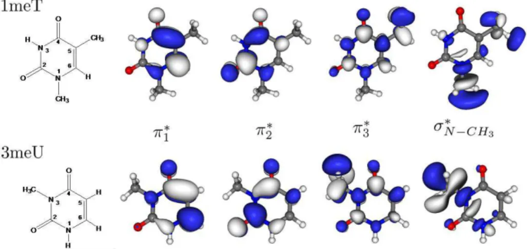
Einzel lens; d) XY Deflecting plates; e) Flight tube. Taken from[24]. ... 25 Figure 3. 5. Schematic of vacuum system. Taken from[24]... 27 Figure 4.1 Structure and virtual orbitals of π* character as well as of σ* character along the N-CH3 coordinate of the neutral 1meT and 3meU molecules obtained at the HF/6-31G* level of theory...34 Figure 4. 2. Plot of the 3meU SVOEs for the second and third π* orbitals (red) as well as of the σ*
orbital (blue), presented in Fig. 4.1, along the N-CH3 stretching coordinate relative to the neutral ground state energy (black). VOEs and ground state energy have been obtained at the HF/6-31G* level of theory; scaling of VOEs has been done according to description in text. ... 35 Figure 4. 3 Plot of the 1meT SVOEs for the second and third π* orbitals (red) as well as of the σ*
orbital (blue), presented in Fig. 4.1, along the N-CH3 stretching coordinate relative to the neutral ground state energy (black). VOEs and ground state energy have been obtained at the HF/6-31G* level of theory; scaling of VOEs has been done according to description in text. ... 35 Figure 4. 4 Negative ion TOF mass spectra for potassium collisions with 3meU at 14, 14.5, 15.5, 30
and 50 eV in the lab frame. Arbitrary units result from the anionic signal divided by the potassium beam current and accumulation time. ... 38 Figure 4. 5 Negative ion TOF mass spectra for potassium collisions with 1meT at 14, 19 and 100 eV
XII
Figure 4. 6 DEA resonance profiles for formation of a) (3meU-CH3)-, b) (3meU-CH4)-, c)(1meT-CH3)- and d) (1meT-CH4)-. Measurements shown in a), b) and d) were derived utilizing the Nier type ion source (energy resolution ~1 eV) while for c) the electron monochromator was used (resolution ~0.1 eV). ... 43 Figure 4. 7 Molecular structure of thymine, uracil, 1-methyl- thymine (1-meT), 3-methyl-uracil (3-meU), and partly deuterated thymine (thymine-d4). ... 51 Figure 4. 8. Time-of-flight mass spectrum showing the different anions formed in collisions of neutral
potassium atoms with 1-methyl-thymine (1-meT) at 66.1 eV collision energy. ... 53 Figure 4. 9. H–/D– ion yield as a function of the collision energy. The vertical dotted lines indicate the
mean values of the H–=D– position of the centre of the different resonances for N1, N3, C6 , and CH3 positions obtained in DEA studies (from Fig. 3 in Ref. [11]). (a) H– formation from thymine methylated at the N1 position (1-meT) at 7.6, 9.0 and 66.1 eV; (b) H– formation from uracil methylated at the N3 position (3-meU) at 5.3, 7.4, and 64.4 eV; (c) H– and D– formation from partly deuterated thymine (thymine-d4) at 7.4 and 64.9 eV. ... 54 Figure 5. 1 Mass spectra of cations produced in 1-4 keV H– collisions with gas-phase nitromethane. The data has normalized such that the intensities of the CH3NO2+ (parent cation) peaks are equal...63 Figure 5. 2 Mass spectra of cations produced in 4 keV H–, O–, and OH– collisions with gas-phase
nitromethane. The data has been normalized such that the intensities of the CH3NO2+ (parent cation) peaks are equal... 64 Figure 5. 3 Mass spectra of cations produced in 4 keV O– and OH– collisions with gas-phase methanol.
The data has been normalized such that the intensities of the CH3OH+ (parent cation) peaks are equal... 66 Figure 5. 4 Mass spectra of cations produced in 4 keV H–, O–, and OH– collisions with gas-phase
ethanol. The data has been normalized such that the intensities of the C2H5OH+ (parent cation) peaks are equal. ... 67 Figure 5. 5 Mass spectra of cations produced in 4 keV H–, O–, and OH– collisions with gas-phase water.
The data has been normalized such that the intensities of the H2O+ (parent cation) peaks are equal. ... 68 Figure 5. 6 Mass spectra of anions produced in 1, 2 and 4 keV H– collisions with gas-phase
nitromethane. The data has been normalized to the parent anion (61 Th). ... 70 Figure 5. 7 Mass spectra of anions produced in 4 keV H–, O– and OH– collisions with gas-phase
nitromethane. The data has been normalized to the parent anion (61 Th). ... 71 Figure 5. 8 Mass spectra of anions produced in 4 keV O– and OH– collisions with gas-phase methanol.
The data has been normalized to 1 Th. ... 72 Figure 5. 9 Mass spectra of anions produced in 4 keV O– and OH– collisions with gas-phase ethanol.
XIII
Figure 5. 10 Mass spectra of anions produced in 4 keV H–, O– and OH– collisions with gas-phase water.The data has been normalized to 17 Th. ... 74 Figure 6. 1 Negative TOF mass spectra in potassium-D-Ribose collisions at: a) 50 eV; b) 75 eV; and c) at 100 eV collision energy in the lab frame. An insert with the dominant conformer is added[29]...85 Figure 6. 2 Branching ratios for the anions as a function of the collision energy. Error bars are within
the data points and so not visible. ... 86 Figure 6. 3 Structure of gas-phase dominant Cs and C2 conformers of THF, according to proposed
geometries as in refs. [6,25]. ... 105 Figure 6. 4 TOF negative ions mass spectra from collisions of potassium atoms with THF at: a) 30 eV;
b) 70 eV and c) 100 eV. An inset of a spectra at 20 eV is presented in a) in order to show the absence of the fragmentation channels at the corresponding available energy. ... 106 Figure 6. 5 Branching ratios (fragment anion yield / total anion yield) as a function of the collision
XIV
Tables
Table 4. 1 Scaled virtual orbital energies (SVOEs) from HF/6-31G* calculations for the optimized neutral equilibrium molecules (see Fig. 1), in eV. ... 36 Table 4. 2. Available energies for the different collision energies in potassium molecule experiments. ... 36 Table 5. 1 Summary of anion-molecule collisions studied using product cation mass spectrometry...62 Table 5. 2. Cations produced in the present collisions (1-4 keV H– impact, 4 keV O– impact, and 4 keV OH– impact) compared with examples of previous electron impact and photoionisation data. It is noteworthy that for all impact energies and projectiles the same major peaks were observed. E Impact energy
unspecified (typically ~70 eV); m Local maximum; * Only resolved for 4 keV anion impact; # Only
resolved for O– and OH– impact; † Similar intensities ... 66 Table 5. 3Anionic fragmentation products for the several target molecules. The different projectiles do not yield
different products. m Local maximum; * Only resolved for 4 keV anion impact; # Only resolved for O- and
OH- impact; † Similar intensities; a Extrapolated ... 69
Table 6. 1 Assignment of TOF mass spectrum anions in collisions of potassium atoms with D-Ribose. Comparisons are made with the available DEA studies to deoxyribose and fructose molecules. M means parent...86
Table 6. 2 Assignment of the anions produced by potassium impact on THF (C4H8O, m/z = 72). Comparisons are
XV
Acronyms and symbols
1-MeT 1-methil-thymine 3-MeU 3-methil-uracil a.m.u Atomic mass units a.u. Arbitrary units α Experimental factor
b Impact parameter
CE Charge Exchange CM Centre of Mass
CTSR Charge transfer to shape resonance DBS Dipole-bound state
DD Direct detachment
DEA Dissociative electron attachment DFT Density functional theory DNA Deoxyribonucleic acid
DR D-Ribose
DSB Double strand Breaks
ECM Energy in the centre of mass framework
EA Molecular electron affinity
e- Single electron
ΔE Reaction energy Eeffec Effective energy
ELab Energy in the laboratory framework
E1;E2 Adiabatic potential curves
Ea Available energy EA Electron affinity
EAad Adiabatic electron affinity EAv Vertical electron affinity En,r electronic energy levels EThreshold Threshold energy
ETS Electron Transmission Spectroscopy FWHM Full-Width Half-Maximum
Hel Electronic Hamiltonian
H Hamiltonian
h Planck constant
XVI
H0 Hamiltonian for the internal motionH11;H22 Diabatic potential curves (diagonal coupling matrix elements)
H12;H21 Diabatic coupling terms (non-diagonal coupling matrix elements)
HF Hartree-Fock I Ionization potential IE Ionization energy
Khyper Hyperthermal potassium atom
K+
Hyper Hyperthermal potassium ion K+
Potassium cation
KTher Thermal potassium atom
k Electronic state
K Potassium
KE Kinetic energy
K0 Neutral potassium
LEE Low energy electrons
LUMO Lowest unoccupied molecular orbital
Mhyper Hyperthermal alkali atom
M+
Hyper Hyperthermal alkali ion
mp Projectile mass
mt Target mass
MTher Thermal alkali atom
M+
Thermal alkali ion m/z Mass to charge ratio MP Methylated pyrimidine base MO Molecular orbital
μ Dipolar moment
NB Nucleobase
p Landau-Zener probability PES Potential energy surface PID Proportional integral derivative Pt100 Platinum resistance thermometers Ψ(r,R) total wavefunction
QMS Quadrupole mass spectrometer π* πantibonding orbital
Rc1 First crossing radius
Rc2 Second crossing radius
XVII
R Nuclear coordinates
ΔR Characteristic length of the system R(t) nucleii classical trajectory
Rc Crossing radius
RET Rydberg Electron Transfer RNA Ribonucleic acid
Ro Fixed internuclear distance
r Vibrational coordinate of the diatomic molecule
SE Secondary electron SSB Single strand breaks
σ* σ antibonding orbital
Γ Resonance width
τ Autodetachment lifetime
Tn Kinetic energy operator of the nucleii
Te Kinetic energy operator of the electrons TEM Tissue equivalent material
T Thymine
tcol Collision time
Th Thomson (mass unit) THF Tetrahydrofuran
TNI Temporary negative ion TOF Time of Flight
tvib Vibration period
U0 Initial kinetic energy
U Uracil
V sum of all potentials between intervening particles in a system
VAE Vertical attachment energy
vbohr Bohr velocity
Vcov Covalent potential curve
V(q;R) Interaction potential
Vion Ionic potential curve
υ Radial velocity
v Velocity
Vcov(R,r) Covalent potential surface
Vion(R,r,a) Ionic potential surface
XVIII
Φk(r;R) adiabatic electronic wavefunctions1
1. Introduction
1.1. Radiation damage to biological tissue:
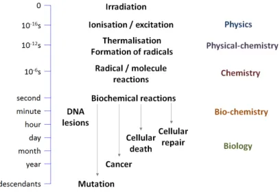
During recent decades, cancer research has gradually increasedits focus on the study of the fundamental molecular mechanisms that govern the appearance of mutagenic pathologies[1].In particular, a great interest lies in determining the role of ionizing radiation as a genotoxic agent, and in what way these mutagenic effects occur at the cellular and molecular level. Indeed, it soon became clear that ionizing radiation, comprised by X-, γ-, β- rays, as well as electrons and ions from other sources, are quite efficient agents in damaging the DNA structure, by inducing single and double strand breaks in the DNA helix[2]. These molecular mechanisms occur during the initial instants of the interaction of the ionizing radiation and are precursors to the myriad of (bio-)chemical mechanisms that eventually result in mutagenesis and formation of cancer tissues. In Fig. 1, a representation of the different stages of DNA damage, starting at the time of irradiation and evolving up to a time scale of decades, is presented. The initial instants of irradiation (in the order of 10-16 s) are dominated by the occurrence of physical processes, namely excitation and ionization by the incident radiation. The products of these processes eventually lead to other physico-chemical processes that in turn damage the DNA molecule. From here on, a chain reaction of natural mechanisms and processes will eventually result in mutagenic effects in the living tissue, with the final consequences being felt even as far as decades later in the form of cancer and other health problems.
Figure 1.1.Chronological schematic of DNA damage by incident radiation
As such, a better understanding of the nascent molecular mechanisms that trigger this chain reaction is critical in order to avoid these health problems.
2
interactions with DNA have shown that the previously mentioned mechanisms are not alone in their role as DNA damaging agents, and more importantly, that their relative importance pales in comparison to the damaging capability of LEEs[4,5]. Indeed, these pioneering studies show that LEE’s have a resonant-like behaviour in degrading these molecules[4] at energies lower than the ionisation threshold of such molecules (~10 eV), and even at sub-excitation energies(<3 eV)[6]. Furthermore, they demonstrate that processes of electron capture of the incident electrons to the molecular components of DNA are the main cause for the formation of single and double strand breaks in this energy range. This discovery presents itself as a major breakthrough in the way we view DNA damage by radiation damage since a whole new set of natural mechanisms that were largely ignored until now, have to be taken into account.1.2. Indirect DNA damage by electrons (dissociative electron attachment):
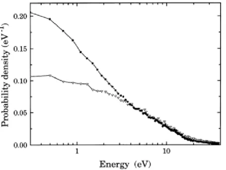
It is now well established within the scientific community that the main product of living tissue irradiation with ionizing radiation are LEEs. Indeed,it is known that for each MeV of deposited energy in living tissue, approximately 5x10⁴ secondary electrons are formed[7]. Furthermore, strong evidences show that these electrons possess an energy distribution ranging from virtually 0 eV up to 20 eV, as can be seen in Fig. 2. A glance at this distribution shows that the majority of the secondary electrons will be created with energies below the typical ionization energies of the relevant molecules (~10 eV), thereby further supporting the claim that the physical processes undergone (or triggered) by these LEEs at sub-ionization energies are dominant above ionization processes.
Figure 1.2. Secondary electron energy distribution for ejected electrons in 20 keV electron collisions with water. The triangles represent solely second generation electrons and dots represent all generations. Taken from ref.
3
These electrons are initially created by high-energy radiation interacting with the physiological medium and producing secondary electrons, among other species. These secondary electrons will subsequently gradually lose their kinetic energy by undergoing a series of different processes until reaching near-zero eV energies and becoming solvated in the physiological environment. Fig. 3 shows a schematic of this electron thermalization phenomenon.Figure 1.3. Thermalization of electrons after being created by interaction of ionizing radiation with the phyisiological environment [2].
Studies of electron interaction (near-0 to 20 eV) with Plasmid DNA show that electrons in these energy ranges are very efficient in inducing single and double strand breaks, as well as loss of helicity by the molecule. Given the resonant behaviour of the obtained profiles, DEA to the DNA constituents was proposed to be the main cause for damage of the DNA molecule. Following this seminal set of studies, several other DEA studies were performed, focusing mostly on biological relevant molecules, e.g., nucleobases, sugar units and amino-acids.
The mechanism of electron attachment consists of a resonant scattering of a low energy electron by a given molecule, resulting in a capture of the electron by said molecule and thus yielding a transient negative ion (TNI).In order for the capture to occur, the energy of the incident electron has to be the as that of the TNI. This mechanism can be seen as a resonant scattering, where the residence time of the electron in the vicinity of the molecule is much longer than its normal transit time. Furthermore, the scattering can be elastic, in which the energy of the incident electron is equal to the energy of the ejected electron, or inelastic, in which the energy of the incident electron is higher than the energy of the ejected electron. Regardless, this mechanism can be adequately understood in the context of Franck-Condon transitions, where the initial state is the neutral state and the final state is the anionic state that results from the capture of the incident electron. Indeed, in most cases the resulting molecular anion is unstable and can decay through one of the following pathways:
AB
AB h
AB
4
AB
*e
(1.2)
A B
(1.3)
Where e- represents the incident electron and ABrepresents a generic molecule. Equation 1.1 represents a radiative stabilization of the TNI into a stable version of the molecular anion. This decay occurs along time scales (10-12 s) significantly higher than the ones considered in the latter processes and thus will, in most cases, not be able to compete with the other channels, unless some external influence “forces” the electron to remain attached to the molecular framework for long enough for this mechanism to occur.
Equation 1.2 represents auto-detachment, i.e., the extra electron will be ejected from the TNI.As was approached above, the auto-detachment can be both elastic and inelastic which, in the case of the latter, will leave the resulting molecular anion in an electronic or vibrational excited state.The auto-detachment lifetime of the TNI will greatly depend on energy of the resonant processes, as well as on the geometry of the TNI. According to one of Heisenberg’s uncertainty principles, the relation between the auto-detachment lifetime and energy width of the final state is thus:
(1.4)
Where Γ is the energy width of the resonant anionic state, ћis the Plank’s constant andτ is the lifetime of the anionic state. One may, in effect,calculate the auto-detachment lifetime τ with this expression,only by knowing the width of the resonance Γ that allows the formation of that anionic state. This value can be obtained experimentally through some spectrometric and spectroscopic techniquesand theoretically by making use of electron scattering computations.
5
electron will also add energy to the molecule which also disrupts the cohesion of the molecule, resulting in bond breaking.In the context of DNA damage, dissociative electron attachment to DNA subunits has been demonstrated to be an extremely effective agent in damaging DNA[5]. However, it is not only the direct role of electrons that bears discussing, but also the indirect role. Indeed, low energy electrons present in the physiological medium may be also highly efficient in creating free radicals, which in turn have also been shown as being highly efficient in damaging DNA. In particular, radicals like OH˙ and O˙, produced from degradation of water have been shown to very damaging to DNA. As such, the sole study of free electron interaction processes in biologically relevant molecules does not entirely encompass the full extent of the damage capability of electrons to DNA. Indeed, it is now well known that presence of free electrons in the physiological medium is very limited and that these damaging agents mostly manifest in bound or quasi-bound states, namely solvated in this medium[11].
1.3. DNA damage by electron transfer:
As was already stated, the discovery that secondary LEEs are the main agents in DNA damage led to a significant interest in studying the interactions of these secondary species with biologically relevant molecules. In particular, extensive studies of dissociative electron attachment to gas-phase biological molecules like the DNA nucleobases, as well as the sugar units have provided a significant knowledge on how the fragmentation pathways of these molecules when electron capture occurs. However, these studies are still quite far in their ability to precisely mimic these processes in the physiological environment. One of the main reasons pertains to the fact that, since DNA is embedded in a physiological medium, the created LEEs will not act as free electrons, but rather have to be treated as being in a bound or quasi-bound state, namely solvated in water. As such, studies of electron transfer of “bound” electrons to biological relevant molecules can be a good stepping stone in providing additional information not obtainable throughgas-phase DEA studies. In particular, studies of atom-molecule collisions approach theseshortcomings in several ways: 1) in this type of collisions, the electron is not in a free state, but rather is bound to its donor, which can be seen as a gross approximation to what in effect happens to electrons in the physiological environment; 2) the presence of the donor projectile post-electron transfer can (and will be shown to) significantly change the fragmentation pathways of the concerned molecules.In these collisions, an electron donor (A) will, upon reaching a given distance from the molecule (AB), transfer its valence electron to the molecular target, resulting in a cationic donor and a molecular anion:
*
BC A BC
6
This molecular anion will, much like the case of (1.1), be a transient negative ion and the A+ BC system can proceed through various reaction pathways, in which the following are the most common:
BC A BC A BC
A *
(Non-dissociative Ionisation)
(1.6)
C B
A
(Dissociative Ionisation)
(1.7)
C
AB
(Associative Chemionisation)
(1.8)
A
++
B
++
C
-+
e
-(Dipolar Dissociation)
(1.9)
Pathway (1.6) represents the case where the electron acceptor is able to form a parent anion, which can have lifetimes in the order of microseconds. This pathway generally involves major structural differences between the neutral and anionic geometries, in order to minimize the repulsive character of the extra electron. A few cases of this process are known, where CH3NO2 and SF6 are two notable cases. It is interesting to note that, in the specific case of CH3NO2, the parent anion is obtained solely through electron transfer, which includes highly excited Rydberg atom donors [12]. Pathway (1.7) represents the fragmentation of the TNI, similarly to what happens in DEA. However, it is critical to note that, despite accessing the same initial resonances as in DEA, the TNI obtained in electron transfer may decay through different pathways, thereby potentially yielding different products. Such is the case of Nitromethane, where the formation of NO2‒ through DEA and electron transfer proceeds through accessing different initial anionic states. However, formation of the CH3NO2‒parent anion in electron transfer stems from initial access of the resonance through which NO2‒ is formed in DEA[13]. Apart from CH3NO2, no other electron transfer studies were performed where the parent anion was reported. Reaction (1.8) represents a capture of an element from the electron acceptor to the donor, it mostly comes in the form of proton transfer. This mechanism will occur mostly in conditions of reactive scattering, i.e., when the electron donor has near-zero eV kinetic energy and/or at below the threshold for ion-pair formation. Finally, (1.9) represents dipolar dissociation of the TNI, which has been observed in DEA (e.g. ref.[14]) but not discussed much in the context of electron transfer studies. It is worth noting that the electron transfer mechanism in atom-molecule collisions can be understood in two separate steps: initially, the valence electron of the donor projectile (A) is ionized, followed by a capture of the electron by the target molecule (BC). As such, from a thermodynamic point of view (at large atom-molecule distances),the endoergicity of the process is determined by the ionization energy of the donor projectile and the electron affinity of the molecular target, i.e.:
A EA
BC IEE
7
Where ΔE represents the reaction energy, IE(A) the ionization energy of the donor projectile A and EA(BC) the electron affinity of the molecular target BC. If the ionisation energy is higher than the electron affinity, then the process is endothermic. Another point to be made lies in the nature of the electron affinity of the molecular target. The electron affinity of a molecule can be described either as the adiabatic electron affinity, which is the transition energy between the ground state of the neutral and anionic state, or as the vertical electron affinity, which is the Franck-Condon transition from the neutral to the anionic state. Owing to convenience of using a donor projectile with a low ionization energy, the choice fell upon alkali atoms, in particular potassium, which is the element used throughout the studies presented in this thesis.Despite the possible analogies between electron transfer and DEA, the presence of an electron donor greatly affects the reaction pathways of the collision system. In particular, it has been shown that atom-molecule collisions can induce formation of a stable version of the parent anion[15]. Additionally, more recent measurements on electron transfer to DNA nucleobases highlight that ring breaking fragmentation pathways are much more prevalent than was initially assumed through DEA measurements[16]. Indeed, the presence of the cationic donor projectile post-collision is shown throughout the results of this thesis to greatly change the resulting fragmentation when comparing electron donation with free electron capture.
The main focus of this thesis consists of extending the already published studies on electron transfer to encompass the DNA sugar unit and possible substitutes, uridine, and also studying with greater care mechanisms of site and bond selectivity in pyrimidine nucleobases. Initially, through the use of a crossed molecular beam setup designed to study electron transfer in potassium-molecule collisions, sets of measurements of fragmentation patterns were performed for methylated versions of thymine and uracil with the main goal of studying site and bond selectivity from C-H against N-H sites[17]. Additionally, owing to the presence of the de-methylated parent anions in the mass spectra, further investigations on this fragment were performed, coupled with DEA measurements and supported byquantum chemical calculations[18]. Subsequently, in order to further expand these studies towards more complex molecules, deoxyriboseand its possible surrogate THF, were investigated in order to obtain additional information about the importance of the sugar unit as far as DNA damage is concerned.
9
2. Theory of electron transfer in Atom-molecule Collisions
In this chapter, an brief description of some generic concepts of electron transfer in atom-molecule collisions will be made, starting with the simplest case of atom-atom collisions. The concepts presented for this case will be further extended to atom-molecule collisions, where the most common example will be for diatomic molecules. It is critical to note that, given the complexity of the molecules studied throughout the work described in this thesis, the concepts discussed in this chapter will serve as an intuitive “guiding hand”, rather than models that can be used to precisely predict the behaviour of the studied collisions.
2.1. Atom-Atom collisions:
Collisions between two atoms can lead to two main processes. The first is elastic scattering, where the collision proceeds through a kinetic energy transfer from one atom to the other, yielding the same two atoms, albeit with different final kinetic energies. The second process is inelastic scattering, where the final electronic states of the colliding atoms can be different. The particular mechanism explored in this thesis is electron transfer, in which an electron is transferred from an atom to the other, with possible electronic excitation of the electron acceptor, i.e.:
*
B
A
B
A
(2.1)
A represents the electron-donating atom and B the electron-acceptor atom. The * means electronic excited. As is obvious from (2.1), the collision yields an ion-pair, in which it is possible to leave the target (or electron acceptor) electronically excited. Considering the interaction between two particles, the system will obey the time-dependent Schrödinger equation[20]:
dt
R
r
d
i
R
r
H
,
,
(2.2)
In which His the Hamiltonian operator and Ψ the total wavefunction of the atom-atom system.
R represents the nuclear coordinates and r represents the electronic coordinates. Furthermore, the
Hamiltonian H can be divided in three different components:
V
T
T
10
Where Tn is the kinetic energy operator of the nuclei, Te is the kinetic energy operator of the electrons and V is the potential sum between all of the intervening particles of the system. Given the sheer complexity inherent in considering all possible interactions encompassed in (2.3), the solution of (2.2) can only be achieved through approximations, the first being the Born-Oppenheimer approximation. This ubiquitously used approximation consists of separating the motion of the electrons from the motion of the nuclei. As such, it is possible to express the total wavefunction Ψ(r,R) as a complete orthogonal set of adiabatic electronic functions that depend parametrically on the nuclear coordinates R, in effect separating this wavefunction into a nuclear and electronic wavefunction[20–23], according to (2.4).
k
k k
r
R
R
R
r
,
;
.
(2.4)
Where Φk(r;R) is the adiabatic electronic wavefunctions and Ωk(R) is the nuclear wave function associated with each electronic state. As such, the usefulness of this approximation is twofold; a) it allows for considering the motion of the nuclei as a classical trajectory R(t), in which the nuclei will move as a function of the final state of the electrons; b) as a gross approximation, for all intents and purposes, it is plausible to consider the nuclei fixed when studying the behaviour of the electrons. This then leads to ignore the influence of Tn and defining a new Hamiltonian:
V
T
H
'
e
(2.5)
With which the following fixed-nuclei Schrödinger equation can be written[20,24]:
T
e
V
r
;
R
r
;
R
E
n,R
r
;
R
(2.6)
Where Φk(r;R) is the already mentioned adiabatic electronic wavefunction and En,R are the corresponding electronic energy levels. The next step lies in considering that the nuclei move slowly, insofar as the Born-Oppenheimer approximation is not violated. In this context, one can assume that the electronic state will adiabatically accompany the motion of the nuclei. In other words, as long as R(t) does not vary rapidly, we can adapt (2.6) from a fixed-nuclei solution to a time-dependent solution, in which the trajectory of the nuclei will still remain a parametric variable and the Born-Oppenheimer approximation still holds[24].
11
In other words, the main difference between (2.6) and (2.7) lies in, while the former describes a static situation (R is a fixed value), the latter describes a dynamic situation in which the nuclei coordinates change over time but always allow the electrons to reach their “equilibrium” positions.En(R) will represent continuous functions, rather than En,R which consists of sets of values.
Although Φk(r;R) is an eigenfunction of (2.3), it is not an eigenfunction of (2.5)[24]. However, through the use of perturbation theory[20], a set of eigenfunctions Θn(r;R) can be obtained through the use of Φk(r;R)[20]. The total wavefunction Ψ(r,R) can also be written as[20]:
t
n Rdt
E i n n n n n
n
r
R
a
r
R
e
a
R
r
,
,
,
.
0(2.8)
By forcing (2.8) to (2.2), the an coefficients can be calculated, thereby obtaining full knowledge of Ψ(r,R) in the adiabatic framework[20]. Ignoring Tn and taking into account (2.7), one can obtain the following system of coupled equations:
j dt E E i j n j n t n je
R
v
a
a
0(2.9)
where,
R
v
R
v
n j
(2.10)
v is the nuclear radial velocity, ΔR is a characteristic length of the system, which can be seen
as a measure of the non-adiabatic coupling between the adiabatic states[20,21,23,24]. By applying the Heisenberg uncertainty principle to this system, one can obtain:
1
.
v
R
E
(2.11)
if this condition is verified, it means that the adiabatic states are quasi-stationary and that
En(R) represents the adiabatic potential energy surfaces (PESs) that govern the motion of the
12
width between the energy levels is comparable to the energy uncertainty, thereby allowing for non-adiabatic transitions between non-adiabatic states. A class of these non-non-adiabatic transitions occur at a pseudo-crossing of adiabatic PES. In the context of atom-atom scattering, a transition from one adiabatic state to another will mean the transfer of an electron from one atom to the other, designated by ion-pair formation[20–24].The former demonstration is the treatment of electron transfer in atom-atom collisions from an adiabatic point of view. Below, an approach through a diabatic framework will be presented. This approach, albeit through a different pathway, will arrive at similar conclusions as the adiabatic one. The collision system A + B can be characterized by two stationary states |φi> and |φc>, the ionic and covalent states, respectively. These states arise from considering that the relative velocity of the two atoms is high enough to not allow enough time for any kind of interaction between them. As such, these states are eigenfunctions of the non-perturbed Hamiltonean H0, of which the eigenvalues H11 and H22 are eigenvalues[20,24]. The lifetime of |φi> and |φc>are supposed to be much larger than the collision time. This means that, in effect, when the system is in either of these states, it will remain so until acted upon. As such, only the following possibilities may hold:
B
A
B
A
(2.12)
B
A
B
A
(2.13)
Where (2.12) is the ionic curve, whose wavefunction is |φi> and (2.13) represents the covalent curve, described by |φc>. As such, the Schrödinger equation for these two wavefunctions will be[20]:
i i
H
H
0
11
(2.14)
c c
H
H
0
22
(2.15)
However, if one considers the atoms as moving slowly, the interaction between the nuclei can be seen as a perturbation W, which can induce transitions between the non-perturbed states, which in effect is a transfer of a valence electron from one particle to the other. The perturbed Hamiltonean Ha
= H0 + W, will therefore be defined as[20]:
E
H
a(2.16)
E
13
Where θ+ and θ- represent the possible eigenfunctions of the perturbed Hamiltonean Ha. Assuming that the W matrix only possesses non-diagonal terms, one can write H in the following way[20]:
22 21 12 11 0H
H
H
H
H
W
H
(2.18)
Where <θj|H|θk> = Hjk. In (2.18), H11 and H22 represent the diabatic energies and H12 and H21 the adiabatic coupling elements between the ionic and covalent diabatic states. However, [H] from (2.18) is diagonizable within the adiabatic bases of (2.16) and (2.17), which makes it possible to express these elements in terms of E+ and E-, in the following way[20]:
212 2
22 11
22
11
2
4
1
2
1
H
H
H
H
H
E
(2.19)
In summary, there are therefore two ways with which to describe the electron transfer process; from an diabatic point of view, the diabatic states presented in (2.14) and (2.15), correspond to the diabatic curves H11(r) and H22(r), in which adiabatic transitions (electron transfer) are induced by the H12 coupling term. On the other hand, from an adiabatic point of view, the PES correspond to the states described in (2.16) and (2.17), with E+(r) and E-(r) as their corresponding eigenstates, and non-adiabatic (i.e. diabatic) transitions between these states are induced by a so-called non-non-adiabatic coupling similar to the one described in (2.10). The coupling term H12, which can be seen as a “measure” of the electron transfer probability, can be estimated from semi-empirical or even completely empirical approximations. A possible formalism that can be used to calculate the non-adiabatic (electron transfer) transition probability was initially developed as a solution to a general quantum mechanical problem by Landau, Zener and Stuckelberg[20,22]:
R Rcc H H dR d v R H
e
p
22 11 2 12 2 (2.20)
14
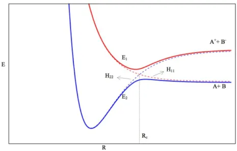
Figure 2.1Adiabatic and diabatic Potential Energy Surfaces for a general atom-atom collision. Dashed red linerepresents the diabatic ionic curve, dashed blue line is the diabatic covalent curve. The full lines represent the resulting adiabatic curves. Adapted from [24].
As can be seen, both the diabatic and adiabatic curves are similar for internuclear coordinates away from the crossing radius, where, for this particularcase an avoided crossing is observed. The asymptotic energy value between the covalent and ionic curves is given by:
EA
I
E
(2.21)
Where I is the ionization potential of the donor atom and EA is the electron affinity of the acceptor atom. If one makes the approximation of stating that the crossing radius Rc is large, then Van der Waals and induction forces can be disregarded, thereby making Vcov zero and Vion a purely coulombic interaction. This leads to Rc being approximated to:
EA
IE
R
c
14
.
41
(2.22)
with Rc in Å and ΔE in eV.
15
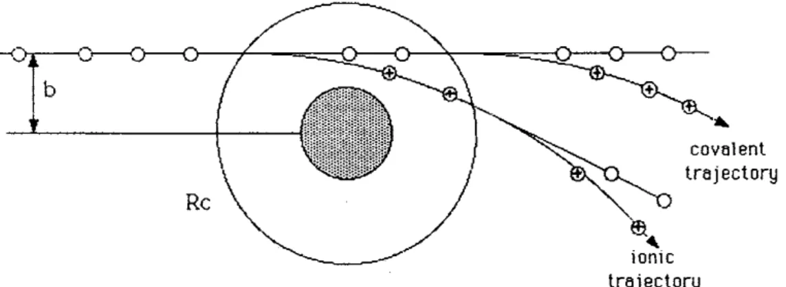
Figure 2.2. Schematic of atom-atom scattering with b representing the impact parameter, the shaded area representing the repulsive part of the nuclear potential. When the electron is not transferred at the first crossing,then the trajectory is known as covalent. When the electron is transferred at the first crossing, the trajectory is named ionic. Adapted from[23].
According to Fig. 2.2, if the impact parameter b is smaller than Rc, there will be then 4 possible processes. If the electron is transferred at the first crossing, then the coulombic interaction will create a significant deflection in the trajectory of the cation. Even if the electron is transferred back to the now-cationic electron donor (reneutralization) at the second crossing, the deflection will still be quite significant. In contrast, if there is no electron transfer at the first crossing, the trajectory of the electron donor will not be affected, regardless of a possible electron transfer at the second crossing. Both of thesepossibilities have been designated by covalent scattering, as can be seen in Fig. 2.1. At this point, it is worth noting that, for low radial velocities, the electron transfer probability is increased and as such, the ionic scattering, as described in Fig. 1, will tend to dominate relatively to covalent scattering. The probability of reneutralization will still be possible, however, as far as the work of this thesis is concerned, detection of neutral particles is not possible and as such, it is not possible to infer about this reaction pathway. A more thorough review on this subject can be obtained in ref.[23] and references therein. Furthermore, studies of potassium energy loss at different angles can provide information on whether the reaction channel is covalent or ionic (see e.g.[12,13]).
Until now, the presented formalism only holds for atom-atom collisions. However, some of the principles will still apply for atom-molecule collisions. Below a small discussion regarding this will be presented.
2.2. Atom-molecule collisions:
16
models considered above do not easily encompass these processes. Indeed, atom-molecule collisions can no longer be completely described by uni-dimensional potential energy curves.Another consideration is that, in order to develop a fully encompassing model of atom-molecule collisions, multi-dimensional potential hypersurfaces have to be considered, in which the various nuclear coordinates of the molecule will have to be taken into account. For this purpose, some models for simple diatomic molecules were developed (discussed in ref. [23] and references therein). However, these models cannot be used in polyatomic molecules, owing to the sheer number of possible processes that can occur. Indeed, the aforementioned models are not applicable to the molecules that are studied throughout this thesis and as such, it is neither possible, nor convenient, to make use of them. Moreover, it is not the main aim of this thesis to provide precise information on the effect of these processes.
The main intent in this chapter is therefore to provide a more qualitative perspective of the electron transfer process in these collisional systems. However, through this perspective, it is possible to obtain some information about the general behaviour of these collisional systems by carefully analysing the data and complementing it with electron scattering techniques and quantum chemical calculations, namely those related to DEA. However in the subsequent chapters we will make a discussion highlighting the major differences between these for each set of the relevant molecules.
As was already discussed in the previous chapter, dissociative electron attachment, as is detailed in (1.1) consists of the capture of a free electron by the molecular target, after which, the resulting transient negative ion can proceed through different reaction pathways. This process can be tentatively compared to what occurs in atom-molecule collisions insofar as the electron in these collisions has two main major differences: 1) the electron is initially in a ‘bound’ state; 2) the resulting capture of the electron will also result in the formation of a cationic species in the close vicinity of the now-anionic molecular target. As will be shown, these differences will greatly influence the reaction and fragmentation pathways of the TNI when comparing DEA to electron transfer by atom-molecule collisions.
17
E
a=
m
mm
m+
m
K.
a
.
E
lab-
IE
(2.23)
Where Ea is the available energy (in eV) for the process to occur, mKis the projectile’s mass, mMis the molecule’s mass, Elab is the kinetic energy of the projectile relative to the lab frame, α is an experimental correction factor and IE is the ionization energy of the donating projectile. It is critical to note two main points regarding this formula. The first point pertains to the implicit assumption (for the sake of simplicity) that the velocity of the acceptor molecule (which is at a thermal energy) is negligible in comparison to the velocity of the hyperthermal projectile beam.
The second point pertains to the experimental parameter that is taken into account in (2.23). This parameter stems from the non-linearity of the acceleration fields of the initially cationic projectile before it resonantly charge-exchanges. Studies on this effect have already been performed [25] and recent simulations also confirm this value. Furthermore, all studies assuming this formula appear to yield Ea values that are congruent with what is expected. This adimensional value (~0.90) does not appear to change appreciably with the applied accelerating potential.
Regarding (2.23), it implies that, as long as Ea is higher than the energy of the resonant anionic state that one wants to access, then it can be accessed. On the other hand, anionic resonant states with energies above Ea are not accessible. Through this rationale, measurements which demonstrated site and bond selectivity in pyrimidine bases yielding H- formation were performed [17]. Additionally, by tuning the potassium kinetic energy to values where Ea is below the accessible resonant states, it has been shown in several biologically relevant molecules that indeed, the accessed resonances appear to be the same as in DEA, despite the fragment yields being different. This was shown for other methylated derivatives of pyrimidine bases [18]. It is worth noting that one can take this rationale one step further, i.e., by gradually increasing Ea, it is possible to indirectly derive resonance profiles similar to the ones obtained through DEA. This is true as long as the lowest covalent and anionic states in the collision complex are involved.
19
3. Experimental setups
The main focus of this chapter pertains to the characterization of the two experimental setups used throughout the main work performed during this thesis. The main core of the work was performed in the Atomic and Molecular Collisions Laboratory, CEFITEC, FCT/UNL in an experimental setup designed for time-of-flight (TOF) mass spectrometric studies of negative ions that are a result of electron transfer in neutral atom-molecule collisions, with specific emphasis on biologically relevant molecules. It is worth noting that most of the system’s components were constructed and developed in our laboratory specifically devoted to this sort of collisions. In the next sub-chapters, each of these will be discussed.
Additionally, some research work was also performed in anion collisions with simple organic molecules. This work was performed in the Centre for Plasma Physics, Queen’s University Belfast, under the supervision of Prof. Robert W. McCullough. The system used for these measurements was fully usable during the time at Queen’s University Belfast so no experimental development was required.
We now discuss and present the main characteristics of both experimental setups.
3.1 Neutral atom collisions experimental setup:
20
The second region comprises the extraction system of the time-of-flight (TOF) mass spectrometer, the molecular target oven and a Langmuir-Taylor detector for beam monitoring. Depending on the sample, typical working pressures are of the order of 6×10⁻⁶ mbar (6×10⁻⁵ Pa). In the second chamber are installed a set of heating lamps to both avoid condensation of samples and to allow for baking of the chamber. Below, each of these components will be discussed in more detail.21
3.1.1. Projectile beam:
In Fig. 3.1, a schematic of the beam creation system is depicted, where all the major components can be visualized. Additionally, Fig. 3.2 shows a schematic explaining the formation of the neutral potassium beam. The hyperthermal potassium beam is obtained through a resonant charge exchange process between hyperthermal cationic potassium atoms and thermal neutral potassium atoms, briefly according to the following equation:
0 0
hyp ther
ther
hyp
K
K
K
K
(3.1)
Where K+
hyp represents the initial hyperthermal potassium cation beam and K0ther the neutral potassium vapour in the charge exchange chamber (b in Fig. 3.2), while the products of the reaction are the desired K0
hyp and the K+ther. Initially, a cationic potassium source creates a beam of K+ ions, which are then accelerated to the desired energy. This ion source is assembled as is shown in Fig. 3.2 in order to guarantee correct electrical and thermal isolation between the source and the rest of the metal body. More technical information can be found elsewhere[24].
Solid potassium is managed and inserted inside the charge exchange oven,. This oven will then be heated to approximately 160ºC (433 K) by two cartridge heaters and the temperature controlled by two PT100 resistors. The heating will create a potassium vapour, diffusing K0
ther into the
connected charge exchange chamber. The temperature of the charge exchange chamber is kept at a slightly higher temperature than the oven, in order to avoid condensation in the connection between the oven to the chamber.
The K+
hyp that is created by the ion source will then pass through the charge exchange
chamber (a) in Fig. 3.2), where it will resonantly charge exchange with the K0
ther atoms inside it, yielding K0
hyp. The K+hyp that do not charge exchange are deflected by the deflecting plates, thereby
producing a beam of K0
22
Figure 3. 2. Charge exchange beam formation. Taken from[24].The deflecting plates basically consist of two parallel plates, in which a positive voltage is applied to one of them, while the other is either connected to ground, or to an electrometer. The deflecting plates is; 1) provide deflection of all unwanted charged particles; 2) by connecting one of the plates to an electrometer, a relative current of the K+
hyp that did not charge exchange can be obtained. This value
can be used to monitor if the ion source is working properly and to have an indication of the charge-exchange efficiency, which is related to the fact that if there is still enough potassium vapour in the charge exchange chamber. In order to ensure an effective deflection of the undesired K+
hyp, the applied voltage to the deflecting plate is changed accordingly to the beam kinetic energy. Previous studies on optimal values have been already performed and are shown elsewhere[24].
3.1.2. Langmuir-Taylor surface detector:
The main purpose for this detector is to monitor the potassium neutral beam. It will allow for a relative measure of the beam in order to ascertain its stability for the same beam energy and relative intensity for different beam energies. The Langmuir-Taylor detector is essentially a high-purity iridium filament (>99%), surrounded by a stainless steel cylindrical collector. This collector has two wide holes along the beam axis and the filament is set slightly above the main section of the beam, in order to minimize tampering with it.
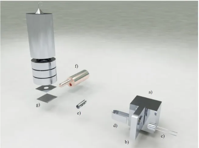
![Figure 3. 2. Charge exchange beam formation. Taken from[24].](https://thumb-eu.123doks.com/thumbv2/123dok_br/16538008.736615/40.892.183.713.109.424/figure-charge-exchange-beam-formation-taken-from.webp)
![Figure 3.3.Picture of the solid sample oven. a) Molecular target oven; b) heating lamp; c) Stainless steel support and alignment system; d) radiation deflector.Taken from[24].](https://thumb-eu.123doks.com/thumbv2/123dok_br/16538008.736615/41.892.114.780.627.1100/figure-picture-molecular-heating-stainless-alignment-radiation-deflector.webp)
![Figure 3. 4. Photograph of the extraction system. a) Extraction region; b) Acceleration region; c) Einzel lens; d) XY Deflecting plates; e) Flight tube.Taken from[24].](https://thumb-eu.123doks.com/thumbv2/123dok_br/16538008.736615/43.892.109.786.107.424/figure-photograph-extraction-extraction-acceleration-einzel-deflecting-flight.webp)
![Figure 3. 5. Schematic of vacuum system.Taken from[24].](https://thumb-eu.123doks.com/thumbv2/123dok_br/16538008.736615/45.892.170.716.108.519/figure-schematic-of-vacuum-system-taken-from.webp)