Ana Catarina Malheiro Tinoco
julho de 2015
Chemical Functionalization in Bionanoparticles
UMinho|20 15 Ana Cat arina Malheir o Tinoco Chemical F unctionalization in Bionanopar ticles
Universidade do Minho
Escola de Ciências
Ana Catarina Malheiro Tinoco
julho de 2015
Dissertação de Mestrado
Mestrado em Biofísica e Bionanossistemas
Chemical Functionalization in Bionanoparticles
Universidade do Minho
Escola de Ciências
Trabalho efetuado sob a orientação do
Professor Doutor Artur Cavaco-Paulo
e coorientação do
III
Acknowledges/Agradecimentos
Durante o decorrer deste ano, de uma maneira direta ou indireta, várias pessoas contribuíram para a realização desta tese de mestrado.
Agradeço ao meu orientador, Professor Doutor Artur Cavaco-Paulo, por me ter aceitado no seu grupo de investigação, pela oportunidade que me deu para a realização deste projeto de investigação e por todo o apoio e incentivo dado no decorrer deste ano.
Ao meu co-orientador, Doutor Artur Jorge Araújo Magalhães Ribeiro, um enorme obrigada pela orientação deste projeto, pela constante disponibilidade para me ajudar/esclarecer dúvidas e por todo o apoio incondicional prestado no decorrer deste ano. Obrigada pela paciência, pela preocupação, pelos conselhos e por tudo o que me ensinaste, de certeza que me vai ser útil no futuro.
Obrigada ao Professor Doutor Pier Parpot e ao Doutor César Oliveira por toda a ajuda e disponibilidade demonstrada para as análises por HPLC-MS e à Professora Doutora Andreia Gomes pela cedência do seu laboratório para a realização dos ensaios em linhas celulares.
Agradeço ainda a todas as pessoas que constituem o grupo de investigação em que me inseri, pela boa receção e pelo ambiente acolhedor que me proporcionaram. Obrigada por toda a ajuda, pelo apoio e por todos os momentos de alegria.
Por fim, mas o mais importante, agradeço imenso à minha família e aos meus amigos que me acompanharam sempre, sem nunca duvidar das minhas capacidades. Obrigada aos meus amigos pela amizade e por todos os momentos de descontração que me proporcionaram, pelo apoio, pelos conselhos e opiniões, por me conseguirem arrancar sorrisos nos dias mais difíceis. Agradeço de uma maneira que as palavras não conseguem descrever, aos meus maravilhosos pais e irmão que, acima de tudo, sempre me apoiaram e sempre estiverem presentes para me ajudarem quando necessário.
V
Abstract
Asparaginase is an enzyme that has been extensively used for the treatment of acute lymphoblastic leukemia (ALL) and other lymphoproliferative malignancies. This enzyme catalyzes the hydrolysis of asparagine into aspartic acid and ammonia, resulting in leukemic cancer cells death. One of the side-effects of asparaginase therapy is hyperammonemia that is caused by an abrupt elevation of ammonia levels in plasma after asparaginase therapy and can lead to cerebral edema, herniation, coma, and even death.
The main objective of the work was the development of asparaginase immobilized nanoparticles with the capacity to retain at surface the free ammonia, reducing the levels of circulating ammonia and avoiding hyperammonemia.
The BSA particles were produced by two high-energy emulsification methods, ultrasounds and high pressure homogenization. In terms of physical characteristics, the best nanoparticles were the ones prepared by homogenization, since the particles where in the nano-range (lower than 200 nm) and exhibit a lower PDI. Relatively to the activity, all the particles produced by ultrasounds almost lost the asparaginase activity after storage for two months at 4 ºC. For this particles, two surfactants were tested, Poloxamer 407 and zein, in order to stabilize the particles. Generally with the surfactants, it was observed an improvement on asparaginase activity during the first month, still the enzyme activity decreased almost to zero after two months. All the particles prepared by homogenization, except the BSA:Asparaginase:Pol407 D, also lost the activity after two months at 4 ºC. The BSA:Asparaginase:Pol407
D were the only particles that stabilize the enzyme and retain 87 % of the initial activity two months after immobilization.
The development of a new HPLC-MS method to quantify the asparagine and aspartic acid together with the ammonia quantification by Nesslerization, confirmed the ability of the particles with asparaginase to retain the ammonia resulting from the asparaginase activity. This capacity to retain ammonia could be used to avoid hyperammonemia during the treatment of ALL.
The cytotoxic effect of the particles prepared by ultrasounds and tested regarding their ability to retain ammonia, was evaluated using the mouse leukemic macrophage cell line RAW 264.7. The particles which displayed more cytotoxicity to the cells were the ones with zein on the formulation; while the BSA:Asparaginase B particles did not show any cytotoxic effect with a cellular viability around 93 % for all tested concentrations. Generally, the cells incubated with the particles had cellular viability always superior to 73 %.
VII
Resumo
A asparaginase é uma enzima que tem sido vastamente utilizada para o tratamento da Leucemia Linfoblástica Aguda (ALL) e outras doenças linfoproliferativas. Esta enzima catalisa a hidrólise da asparagina em ácido aspártico e amónia, resultando na morte das células leucémicas. Um dos efeitos colaterais desta terapia é a hiperamonemia, causada por uma elevação abrupta dos níveis de amónia no plasma sanguíneo, podendo resultar em edemas cerebrais, coma ou até morte.
O principal objetivo deste trabalho foi o desenvolvimento de nanopartículas onde a asparaginase pudesse ser imobilizada e com a capacidade de reter a amónia livre, reduzindo assim os níveis de amónia no plasma e evitando a hiperamonemia.
As partículas de BSA foram produzidas por dois métodos de alta-energia, os ultrassons e a homogeneização de alta-pressão. Em termos de características físicas, as melhores partículas foram as preparadas por homogeneização, uma vez que apresentavam um tamanho inferior a 200 nm e exibiam um baixo PDI. Relativamente à atividade, todas as partículas preparadas por ultrassons apresentavam uma atividade da asparaginase muito baixa ou nula após dois meses a 4 ºC. De modo a estabilizar estas partículas, foram testados dois surfactantes, Poloxamer 407 e zeína. Globalmente, a adição de surfactantes melhorou a atividade da asparaginase durante o primeiro mês, dimunuindo quase até zero após dois meses. Todas as nanopartículas preparadas por homogeneização, exceto as BSA:Asparaginase:Pol407 D, também perderam a atividade após dois meses a 4 ºC. As nanopartículas
BSA:Asparaginase:Pol407 D demostraram ser as únicas partículas capazes de estabilizar a enzima,
retendo 87 % da atividade inicial mesmo dois meses após imobilização.
O desenvolvimento do método de HPLC-MS para quantificar simultaneamente a asparagina e ácido aspártico, em conjunto com a quantificação de amónia pelo método de Nessler, confirmaram a capacidade das partículas com asparaginase em reter a amónia resultante da atividade desta enzima. Esta capacidade para reter a amónia livre pode ser utilizada para evitar a hiperamonemia durante o tratamento da ALL.
A citotoxicidade das partículas preparadas por ultrassons, as mesmas testadas relativamente à sua capacidade em reter amónia, foi avaliada com a linha celular de macrófagos de ratinho RAW 264.7. As partículas que demostraram uma maior citotoxicidade foram aquelas que incluíam zeina na sua formulação; as partículas BSA:Asapraginase B não demonstraram qualquer citotoxicidade, apresentando uma viabilidade celular superior a 93 % para todas as concentrações testadas. De um modo geral, as células incubadas com todas as partículas apresentaram uma viabilidade celular superior a 73 %.
IX
Table of Contents
List of Figures ...XIII List of Tabels ... XXI List of Abreviations ... XXIII
Chapter 1: Introduction ... 1
1.1. Enzymes Immobilization ... 3
1.1.1. Enzymes Immobilization Techniques ... 4
1.1.1.1. Covalent Enzyme Immobilization ... 4
1.1.1.2. Enzyme Immobilization based on adsorption ... 5
1.1.1.3. Immobilization by Enzyme Entrapment ... 6
1.1.1.4. Immobilization by Enzyme Encapsulation ... 7
1.1.1.5. Cross-linking Enzyme Immobilization ... 7
1.2. Protein PEGylation ... 9
1.2.1. PEG characteristics ... 9
1.3. Asparaginase ... 10
1.3.1. Preparations of Asparaginase available for therapy ... 11
1.3.2. Asparaginase Types and Structure ... 12
1.3.3. Acute Lymphoblastic Leukemia ... 13
1.3.4. Asparaginase Mechanism of Action ... 14
1.3.5. Toxicity of Asparaginase to Normal Cells ... 16
1.3.5.1. Hyperammonemia ... 18
1.4. Protein as a Surfactant Molecule ... 19
1.4.1. Zein as a surfactant ... 21
Chapter 2: Motivation and Objectives ... 25
2. Motivation and Objectives ... 27
Chapter 3: Materials and Methods ... 29
Materials ... 31
X
3.1. Determination of the purity degree of asparaginase ... 33
3.2. Enzymatic Assay of Asparaginase ... 33
3.2.1. Ammonia quantification by Nesslerization ... 34
3.2.2. Asparagine and Aspartic Acid quantification by HPLC-MS without derivatization ... 34
3.3. Bovine Serum Albumin based Nanoparticles ... 35
3.3.1. Nanoparticles prepared by sonication... 35
3.3.2. Nanoparticles prepared by homogenization ... 36
3.3.3. Nanoparticle characterization ... 36
3.3.4. Particle formation efficiency ... 37
3.4. SDS - Denaturing Electrophorese ... 37
3.4.1. Staining of the electrophoretic gel ... 38
3.5. Effect of BSA on asparaginase ... 38
3.6. Cellular viability assay ... 39
3.6.1. Cell culture maintenance ... 39
3.6.2. Cell viability assessed by MTT assay ... 39
Chapter 4: Results and Discussion ... 41
4.1. Kinetic study of free E. coli Asparaginase II ... 43
4.1.1. Study of Asparaginase II purity by SDS-PAGE and MALDI-TOFF ... 43
4.1.1.1. Study of purity by MALDI-TOF ... 43
4.1.1.2. Study of purity by SDS-PAGE ... 44
4.1.2. Determination of asparaginase kinetic parameters ... 45
4.1.3. Effect of temperature on the activity of asparaginase ... 52
4.1.4. Determination of asparaginase absolute activity over time ... 53
4.2. Effect of BSA on free Asparaginase ... 55
4.3. Effect of storage time, reaction buffer, protease inhibitor, antibiotic and immobilization on asparaginase activity... 59
4.4. Asparaginase Immobilization in Particles prepared by Ultrasounds and High Pressure Homogenization ... 64
4.4.1. Particles prepared by Ultrasounds... 64
XI
4.4.1.2. BSA:Asparaginase:Pol407 B and BSA:Asparaginase:Pol407 D Particles ... 69
4.4.1.3. BSA:Asparaginase:Zein B and BSA:Asparaginase:Zein D Particles ... 73
4.4.1.4. BSA:Asparaginase:Zein:Pol407 B and BSA:Asparaginase:Zein:Pol407 D Nanoparticles ………..76
4.4.2. Nanoparticles prepared by Homogenization ... 79
4.4.2.1. BSA:Asparaginase B and BSA:Asparaginase D Nanoparticles ... 79
4.4.2.2. BSA:Asparaginase:Pol407 B and BSA:Asparaginase:Pol407 D Nanoparticles ... 83
4.4.3. Comparison of Asparaginase activity on different nanoparticles... 86
4.4.3.1. Effect of the addition of Poloxamer 407 ... 86
4.4.3.2. Effect of the addition of zein ... 89
4.4.3.3. Effect of the Technique used for Particle Synthesis on Asparaginase Activity and Particle Properties ………..91
4.5. Determination of Asparaginase Activity by HPLC-MS ... 93
4.5.1. Protocol Optimization... 93
4.5.2. Calibration Curves ... 99
4.5.3. Determination of Asparaginase Activity by Aspartic Acid Quantification by HPLC-MS ... 100
4.6. Nanoparticles capacity to retain ammonia ... 102
4.7. Nanoparticles cytotoxicity by MTT assay ... 103
Chapter 5: Conclusion and Future Perspectives ... 107
5. Conclusion and Future Perspectives ... 109
Chapter 6: Bibliography ... 113
XIII
L
IST OFF
IGURESFigure 1 – Effect of multipoint immobilization on enzymes stability via short spacer arms
(adapted from Mateo et al., 2007). 5
Figure 2 – Enzyme immobilization by surface adsorption and electrostatic interaction (adapted
from Ronkainen, 2010). 6
Figure 3 – Enzyme entrapment on a gel solution (adapted from Cao, 2006). 6 Figure 4 – Enzyme encapsulation and free movement of substrates and products across the
membrane pores (adapted from Cao, 2006). 7
Figure 5 – Formation of a solid aggregate of enzymes by cross-linking immobilization (adapted
from Barbosa et al., 2014). 8
Figure 6 – Reaction of degradation of asparagine and glutamine, catalyzed by Asparaginase
(adapted from Fu and Sakamoto, 2007). 10
Figure 7 – Structural features of bacterial-type asparaginases. a) the quaternary structure of the periplasmatic E. coli asparaginase II. The green (subunit A) and red (subunit C) or gray (subunit B) and blue (subunit D) monomers form the intimate dimer. The arrows correspond to the two-fold axes defining the 222 symmetry of this homotetramer; b) the active site of E. coli asparaginase II with an asparagine substrate molecule modeled according to the complex enzyme-substrate. The dotted red lines represent hydrogen bonds while the green ones symbolize a potential line of attack of the active-site Thr residues on the substrate’s amide C atom (adapted from Michalska and Jaskolski, 2006).
13
Figure 8 – Biosynthesis of asparagine by asparagine synthetase (adapted from El-Naggar et al.,
2014). 14
Figure 9 – Schematic representation of asparagine mechanism of action (adapted from Narta
et al., 2007). 15
Figure 10 – a) A possible nine-helical zein protein structural model for α-zeins b) a possible model for arrangement of zein proteins within a plane as well for the stacking of molecular planes (adapted from Argos et al., 1982).
22 Figure 11 – a) A possible structural model for α-zeins (Z22), where the helical segments are
aligned to form a 13 nm long asymmetric cylindrical structure. The sides of the cylinder correspond to the surfaces of hydrophobic helices, while the upper and lower surfaces are connected by glutamine bridges, which are hydrophilic (adapted from (Corradini et al., 2014)).
23
Figure 12 – Representation of the enzymatic conversion of MTT to formazan. 40 Figure 13 – MALDI-TOF spectrum of lyophilized asparaginase stored at 4 ºC. 44 Figure 14 – Analysis of asparaginase purity. 15 % SDS-PAGE stained with Coomassie Blue. Lane
A: 10 μg of asparaginase; Lane B: 5 μg of asparaginase; Lane C: Protein molecular weight marker; Lane D: 15 μg of asparaginase.
XIV
Figure 15 – Calibration curve prepared with ammonium sulfate for the determination of asparaginase activity. The absorbance of the solutions after Nesslerization were measured at 436 nm.
45 Figure 16 – Michaelis-Menten plot for different concentrations of asparagine (0.001; 0.005;
0.010; 0.025 and 0.050 g/L). The reaction was performed for 30 min at 37 ºC with asparagine concentrations between 0 and 200 mM; in 50 mM Tris buffer, pH 8.6. The absorbance of the solutions after Nesslerization were measured at 436 nm.
47
Figure 17 – Michaelis-Menten plot for different concentrations of asparagine (0.001; 0.005; 0.010; 0.025 and 0.050 g/L). The reaction was performed for 30 min at 40 ºC with asparagine concentrations between 0 and 200 mM; in 50 mM Tris buffer, pH 8.6. The absorbance of the solutions after Nesslerization were measured at 436 nm.
47
Figure 18 – Michaelis-Menten and Lineweaver-Burk plot for an asparaginase concentration of 0.050 g/L. Reaction was performed for 30 min at 37 ºC in 50 mM Tris buffer, pH 8.6. The absorbance of the solutions after Nesslerization were measured at 436 nm.
49 Figure 19 – Effect of asparaginase concentration and asparagine concentration on the specific
activity of asparaginase. The reaction was performed for 30 min at 37 ºC with asparagine concentrations between 0 and 200 mM; in 50 mM Tris buffer, pH 8.6. The absorbance of the solutions after Nesslerization were measured at 436 nm.
51
Figure 20 – Effect of temperature on the velocity of the hydrolysis of asparagine, at 30 or 40 ºC. The reaction was performed for 30 min at 37 and 40 ºC with asparagine concentrations between 0 and 200 mM; in 50 mM Tris buffer, pH 8.6. The absorbance of the solutions after Nesslerization were measured at 436 nm.
52
Figure 21 – Effect of the enzyme concentration on asparaginase activity over time. Five concentrations of asparaginase were tested: 0.001, 0.005, 0.010, 0.025 and 0.050 mg/mL. The reaction was performed for 240 min at 37 ºC with 100 mM asparagine, in 50 Mm Tris buffer, pH 8.6. Samples were taken at 30 min intervals, for a total of 240 min. The absorbance of the solutions after Nesslerization were measured at 436 nm.
54
Figure 22 – Effect of the asparagine concentration on asparaginase activity over time. The reaction was performed for 240 min at 37 ºC with 0.010 g/L of asparaginase in 50 mM Tris buffer, pH 8.6. The concentrations of asparagine varied between 5-200 mM. Samples were taken at 30 min intervals, for a total of 240 min. The absorbance of the solutions after Nesslerization were measured at 436 nm.
55
Figure 23 – Asparaginase activity over time of several solutions (A - 0 % BSA/100 % asparaginase solution; B – 10 % BSA/90 % asparaginase solution; C – 20 % BSA/80 % asparaginase solution; D – 30 % BSA/70 % asparaginase solution; E – 40 % BSA/60 % asparaginase solution; F – 50 % BSA/50 % asparaginase solution; G – 60 % BSA/40 % asparaginase solution; H – 70 % BSA/30 % asparaginase solution; I – 80 % BSA/20 % asparaginase solution; J – 90 % BSA/10 % asparaginase solution; K – 98 % BSA/2 % asparaginase solution) with different proportions of BSA and asparaginase. The reaction was performed for 240 min, at 37 ºC, with 0.010 g/L of asparaginase in 50 mM Tris buffer, pH 8.6. The absorbance of the solutions after Nesslerization were measured at 436 nm.
XV
Figure 24 – Effect of storage time at 4 ºC in asparaginase stability measured for several BSA/Asparaginase ratio solutions. Samples correspond to the first day of storage at 4 ºC. 15 % SDS-PAGE stained with Coomassie Blue.
58 Figure 25 – Effect of storage time at 4 ºC in asparaginase stability measured for several
BSA/Asparaginase ratio solutions. Samples correspond to day 22 of storage at 4 ºC. 15 % SDS-PAGE stained with Coomassie Blue.
58 Figure 26 – Effect of the storage at 4 ºC on asparaginase activity over time. The reaction was
performed with 0.001 g/L of asparaginase, 100 mM of asparagine for 240 min, at 37 ºC, in 50 mM Tris buffer, pH of 8. 6. The absorbance of the solutions after Nesslerization were measured at 436 nm.
59
Figure 27 – Effect of PBS on the activity of asparaginase over time, after storage at 4 ºC. The reaction was performed with 0.001 g/L of asparaginase, 100 mM of asparagine for 240 min, at 37 ºC, in 50 mM Tris buffer, pH of 8.6. The absorbance of the solutions after Nesslerization were measured at 436 nm. Data were analyzed by one way-ANOVA: * p-value≤0.05, ● p-value≤0.01; ♦ p-value≤0.001; ■ p-value≤0.0001
60
Figure 28 – Effect of BSA in the absence of antibiotic and protease inhibitor on the activity of asparaginase over time, after storage at 4 ºC. The reaction was performed with 0.001 g/L of asparaginase, 100 mM of asparagine for 240 min, at 37 ºC, in 50 mM Tris buffer, pH 8.6. The absorbance of the solutions after Nesslerization were measured at 436 nm. Data were analyzed by one way-ANOVA: * p-value≤0.05, ● p-value≤0.01; ♦ p-value≤0.001; ■ p-value≤0.0001.
61
Figure 29 – Effect over asparaginase activity of BSA in the absence of antibiotic and protease inhibitor. 15 % SDS-PAGE stained with Coomassie Blue. Lane A) BSA Solution; Lane B) Molecular weight marker; Lane C) Aqueous solution of asparaginase – 0 days at 4 ºC; Lane D) Aqueous solution of asparaginase – 35 days at 4 ºC; Lane E) Solution of 0.2 mg/mL asparaginase and 9.8 mg/mL BSA – 0 days at 4ºC; F) Solution of 0.2 mg/mL asparaginase and 9.8 mg/mL BSA – 35 days at 4 ºC.
62
Figure 30 – MALDI-TOF spectrum of a solution of BSA and asparaginase, in PBS, after storage
during 35 days at 4 ºC. 62
Figure 31 – Effect of the addition of antibiotic on asparaginase activity over time, after storage at 4 ºC. The reaction was performed with 0.001 g/L of asparaginase, 100 mM of asparagine for 240 min at 37 ºC, in 50 mM Tris buffer, pH 8.6. The absorbance of the solutions after Nesslerization were measured at 436. Data were analyzed by one way-ANOVA: * p-value≤0.05, ● p-value≤0.01; ♦ p-value≤0.001; ■ p-value≤0.0001.
XVI
Figure 32 – Activity of asparaginase immobilized on BSA particles prepared by ultrasounds (BSA:Asparaginase B and BSA:Asparaginase D), and free asparaginase (10 mg/mL) on a PBS solution, after storage at 4 ºC. The reaction was performed with 0.001 g/L of asparaginase, 100 mM of asparagine for 240 min, at 37 ºC, in 50 mM Tris buffer, pH 8.6. The absorbance of the solutions after Nesslerization were measured at 436 nm. Data were analyzed by one way-ANOVA: * value≤0.05, ● value≤0.01; ♦ value≤0.001; ■ p-value≤0.0001.
65
Figure 33 – Effect on asparaginase activity of adding the enzyme at different stages during the process of particles synthesis by ultrasounds. BSA:Asparaginase B – enzyme added before the sonication cycles. BSA:Asparaginase D – enzyme added during the sonication cycles, after 69 s. Data were analyzed by one way-ANOVA: * value≤0.05, ● value≤0.01; ♦ p-value≤0.001; ■ p-value≤0.0001.
66
Figure 34 – Characterization of BSA:Asparaginase B particles, during storage at 4 ºC: A) Z-potential; B) Z-average and PDI. Data were analyzed by one way-ANOVA: * p-value≤0.05, ● p-value≤0.01; ♦ p-value≤0.001; ■ p-value≤0.0001, compared to the results obtained at day 1.
67
Figure 35 – Characterization of BSA:Asparaginase D particles, during storage at 4 ºC: A) Z-potential; B) Z-average and PDI. Data were analyzed by one way-ANOVA: * p-value≤0.05, ●p-value≤0.01; ♦ p-value≤0.001; ■ p-value≤0.0001, compared to the results obtained at day 1.
67
Figure 36 – Poloxamer 407 chemical structure (a=98, b=67). 69 Figure 37 – Activity of asparaginase immobilized on BSA:Pol407 particles prepared by
ultrasounds and free asparaginase (10 mg/mL) on a PBS solution, after storage at 4 ºC. The reaction was performed with 0.001 g/L of asparaginase, 100 mM of asparagine for 240 min, at 37 ºC, in 50 mM Tris buffer, pH 8.6. The absorbance of the solutions after Nesslerization were measured at 436 nm. Data were analyzed by one way-ANOVA: * p-value≤0.05, ● p-value≤0.01; ♦ p-value≤0.001; ■ p-value≤0.0001.
70
Figure 38 – Effect on asparaginase activity of adding the enzyme at different stages during the process of particles synthesis. BSA:Asparaginase:Pol407 B – enzyme added before the
sonication cycles. BSA:Asparaginase:Pol407 D – enzyme added during the sonication
cycles, after 69 s. Data were analyzed by one way-ANOVA: * p-value≤0.05, ● p-value≤0.01; ♦ p-value≤0.001; ■ p-value≤0.0001.
71
Figure 39 – Characterization of BSA:Asparaginase:Pol407 B particles, during storage at 4 ºC: A)
Z-potential; B) Z-average and PDI. Data were analyzed by one way-ANOVA: * p-value≤0.05, ● p-value≤0.01; ♦ p-value≤0.001; ■ p-value≤0.0001, compared to the results obtained at day 1.
XVII
Figure 40 – Characterization of BSA:Asparaginase:Pol407 D particles, during storage at 4 ºC: A)
Z-potential; B) Z-average and PDI. Data were analyzed by one way-ANOVA: * p-value≤0.05, ● p-value≤0.01; ♦ p-value≤0.001; ■ p-value≤0.0001, compared to the results obtained at day 1.
72
Figure 41 – Activity of asparaginase immobilized on BSA:Zein particles, prepared by ultrasounds, and free asparaginase (10 mg/mL) on a PBS solution, after storage at 4 ºC. The reaction was performed with 0.001 g/L of asparaginase, 100 mM of asparagine for 240 min, at 37 ºC, in 50 mM Tris buffer, pH 8.6. The absorbance of the solutions after Nesslerization were measured at 436 nm. Data were analyzed by one way-ANOVA: * p-value≤0.05, ●p-value≤0.01; ♦ p-value≤0.001; ■ p-value≤0.0001.
73
Figure 42 – Effect on asparaginase activity of adding the enzyme at different stages during the process of particles synthesis. BSA:Asparaginase:Zein B – enzyme added before the sonication cycles. BSA:Asparaginase:Zein D – enzyme added during the sonication cycles, after 69 s. Data were analyzed by one way-ANOVA: * value≤0.05, ● value≤0.01; ♦ p-value≤0.001; ■ p-value≤0.0001.
74
Figure 43 – Characterization of BSA:Asparaginase:Zein B particles, during storage at 4 ºC: A) Z-potential; B) Z-average and PDI. Data were analyzed by one way-ANOVA: * p-value≤0.05, ● p-value≤0.01; ♦ p-value≤0.001; ■ p-value≤0.0001, compared to the results obtained at day 1.
75
Figure 44 – Characterization of BSA:Asparaginase:Zein D particles, during storage at 4 ºC: A) Z-potential; B) Z-average and PDI Data were analyzed by one way-ANOVA: * p-value≤0.05, ● p-value≤0.01; ♦ p-value≤0.001; ■ p-value≤0.0001, compared to the results obtained at day 1.
75
Figure 45 – Activity of asparaginase immobilized on BSA:Zein:Pol407 particles, prepared by
ultrasounds, and free asparaginase (10 mg/mL) on a PBS solution, after storage at 4 ºC. The reaction was performed with 0.001 g/L of asparaginase, 100 mM of asparagine for 240 min, at 37 ºC, in 50 mM Tris buffer, pH 8.6. The absorbance of the solutions after Nesslerization were measured at 436 nm. Data were analyzed by one way-ANOVA: * p-value≤0.05, ● p-value≤0.01; ♦ p-value≤0.001; ■ p-value≤0.0001.
77
Figure 46 – Effect on asparaginase activity of adding the enzyme at different stages during the process of particles synthesis. BSA:Asparaginase:Zein:Pol407 B – enzyme added before the
sonication cycles. BSA:Asparaginase:Zein:Pol407 D – enzyme added during the sonication
cycles, after 69 s. Data were analyzed by one way-ANOVA: * p-value≤0.05, ● p-value≤0.01; ♦ p-value≤0.001; ■ p-value≤0.0001.
77
Figure 47 – Characterization of BSA:Asparaginase:Zein:Pol407 B particles, during storage at 4
ºC: A) Z-potential; B) Z-average and PDI. Data were analyzed by one way-ANOVA: * p-value≤0.05, ● p-value≤0.01; ♦ p-value≤0.001; ■ p-value≤0.0001, compared to the results obtained at day 1.
XVIII
Figure 48 – Characterization of BSA:Asparaginase:Zein:Pol407 D particles, during storage at 4
ºC: A) Z-potential; B) Z-average and PDI. Data were analyzed by one way-ANOVA: * p-value≤0.05, ● p-value≤0.01; ♦ p-value≤0.001; ■ p-value≤0.0001.
78 Figure 49 – Activity of asparaginase immobilized on BSA nanoparticles prepared by
homogenization (BSA:Asparaginase B and BSA:Asparaginase D), and free asparaginase (10 mg/mL) on a PBS solution, after storage at 4 ºC. The reaction was performed with 0.001 g/L of asparaginase, 100 mM of asparagine for 240 min, at 37 ºC, in 50 mM Tris buffer, pH 8.6. The absorbance of the solutions after Nesslerization were measured at 436 nm. Data were analyzed by one way-ANOVA: * value≤0.05, ● value≤0.01; ♦ p-value≤0.001; ■ p-value≤0.0001.
80
Figure 50 – Effect on asparaginase activity of adding the enzyme at different stages during the process of nanoparticles synthesis by homogenization. BSA:Asparaginase B – enzyme added before the homogenization cycles. BSA:Asparaginase D – enzyme added after 5 homogenization cycles. Data were analyzed by one way-ANOVA: * value≤0.05, ● p-value≤0.01; ♦ p-value≤0.001; ■ p-value≤0.0001.
81
Figure 51 – Characterization of BSA:Asparaginase B nanoparticles, during storage at 4 ºC: A) Z-potential; B) Z-average and PDI. Data were analyzed by one way-ANOVA: * p-value≤0.05, ● p-value≤0.01; ♦ p-value≤0.001; ■ p-value≤0.0001.
82 Figure 52 – Characterization of BSA:Asparaginase D nanoparticles, during storage at 4 ºC: A)
Zeta-potential; B) Z-average and PDI of nanoparticles. Data were analyzed by one way-ANOVA: * p-value≤0.05, ● p-value≤0.01; ♦ p-value≤0.001; ■ p-value≤0.0001.
82 Figure 53 – Activity of asparaginase immobilized on BSA:Pol407 nanoparticles prepared by
homogenization (BSA:Asparaginase:Pol407 B and BSA:Asparaginase:Pol407 D), and free
asparaginase (10 mg/mL) on a PBS solution, after storage at 4 ºC. The reaction was performed with 0.001 g/L of asparaginase, 100 mM of asparagine for 240 min, at 37 ºC, in 50 mM buffer, pH 8.6. The absorbance of the solutions after Nesslerization were measured at 436 nm. Data were analyzed by one way-ANOVA: * value≤0.05, ● p-value≤0.01; ♦ p-value≤0.001; ■ p-value≤0.0001.
83
Figure 54 – Effect on asparaginase activity of adding the enzyme different stages during the process of nanoparticles synthesis by homogenization. BSA:Asparaginase:Pol407 B – enzyme added before the homogenization cycles. BSA:Asparaginase:Pol407 D – enzyme added after 5 homogenization cycles. Data were analyzed by one way-ANOVA: * p-value≤0.05, ● p-value≤0.01; ♦ p-value≤0.001; ■ p-value≤0.0001.
84
Figure 55 – Characterization of BSA:Asparaginase:Pol407 B nanoparticles, during storage at 4
ºC: A) Z-potential; B) Z-average and PDI. Data were analyzed by one way-ANOVA: * p-value≤0.05, ● p-value≤0.01; ♦ p-value≤0.001; ■ p-value≤0.0001.
85 Figure 56 – Characterization of BSA:Asparaginase:Pol407 D nanoparticles, during storage at 4
ºC: A) Z-potential; B) Z-average and PDI. Data were analyzed by one way-ANOVA: * p-value≤0.05, ● p-value≤0.01; ♦ p-value≤0.001; ■ p-value≤0.0001.
XIX
Figure 57 – Effect of the addition of Poloxamer 407 to the different nanoparticles prepared by ultrasounds: A) BSA:Asparaginase B; B) BSA:Asparaginase D; C) BSA:Asparaginase:Zein B; D) BSA:Asparaginase:Zein D. The reaction was performed with 0.001 g/L of asparaginase, 100 mM of asparagine for 240 min, at 37 ºC, in 50 mM Tris buffer, pH 8.6. The absorbance of the solutions after Nesslerization were measured at 436 nm. Data were analyzed by one way-ANOVA: * value≤0.05, ● value≤0.01; ♦ value≤0.001; ■ p-value≤0.0001.
87
Figure 58 – Effect on asparaginase activity of the addition of Poloxamer 407 to the nanoparticles prepared by homogenization: A) BSA:Asparaginase B; B) BSA:Asparaginase D. The reaction was performed with 0.001 g/L of asparaginase, 100 mM of asparagine for 240 min, at 37 ºC, in 50 mM buffer, pH 8.6. The absorbance of the solutions after Nesslerization were measured at 436 nm. Data were analyzed by one way-ANOVA: *value≤0.05, ● p-value≤0.01; ♦ p-value≤0.001; ■ p-value≤0.0001.
88
Figure 59 – Effect of Poloxamer 407 addition to the physical characteristics of the BSA:Asparaginase D nanoparticles, prepared by homogenization, one day after synthesis. 89 Figure 60 – Effect on asparaginase activity of the addition of zein to the different nanoparticles
prepared by ultrasounds: A) BSA:Asparaginase B; B) BSA:Asparaginase D; C) BSA:Asparaginase:Pol407 B; D) BSA:Asparaginase:Pol407 D. The reaction was performed
with 0.001 g/L of asparaginase, 100 mM of asparagine for 240 min, at 37 ºC, in 50 mM Tris buffer, pH 8.6. The absorbance of the solutions after Nesslerization were measured at 436 nm. Data were analyzed by one way-ANOVA: * value≤0.05, ● value≤0.01; ♦ p-value≤0.001; ■ p-value≤0.0001.
90
Figure 61 – Effect on asparaginase activity of the technique used for particles synthesis (ultrasounds or homogenizer). The reaction was performed with 0.001 g/L of asparaginase, 100 mM of asparagine for 240 min at 37 ºC, in 50 mM Tris buffer, pH 8.6. The absorbance of the solutions after Nesslerization were measured at 436 nm.
91
Figure 62 – Effect on Z-average (A) and PDI (B) of the technique used for the particles synthesis
- ultrasounds or homogenizer. 92
Figure 63 - Mass spectrum on positive ionization and chemical structure of A) asparagine; B) aspartic acid. Both amino acids were dissolved in acetonitrile 20 %. 94 Figure 64 – Chromatograms of aqueous solutions of A) aspartic acid and B) asparagine,
analyzed with a reverse phase C18 column and a mobile phase of 50 % water with formic acid and 50 % of acetonitrile.
95 Figure 65 – A) Chromatogram of an aqueous mixture of asparagine and aspartic acid, analyzed
with a reverse phase C18 column and a mobile phase of 50 % water with formic acid and 50 % of acetonitrile; B) Mass spectrum of the compounds with a retention time of 3.89 min.
96
Figure 66 – Chromatogram of an aqueous solution of A) aspartic acid; B) asparagine; C) mixture of asparagine and aspartic acid; analyzed with a hydro-C18 column and a gradient of elution of: 0-5 min, 80 % A + 20 % B; 5-15 min, 80 % A + 20 % B; 15-20 min, 40 % A + 60 % B; 20 min, 80 % A + 20 % B.
XX
Figure 67 – Chromatogram of an aqueous mixture of asparagine and aspartic acid when analyzed with a Hydro-C18 column, with ammonium bicarbonate and acetonitrile as eluents and a flow rate of 0.2 mL/min.
98 Figure 68 – Chromatogram of an aqueous mixture of asparagine and aspartic acid when
analyzed with a Hydro-C18 column, with ammonium bicarbonate and acetonitrile as eluents and a flow rate of 0.3 mL/min.
99 Figure 69 – Calibration curves of A) asparagine and B) aspartic acid, obtained by HPLC-MS with
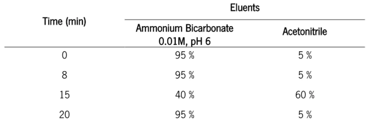
a Hydro-C18 column, with ammonium bicarbonate pH 6 (a) and acetonitrile (b) as eluents and a flow rate of 0.3 mL/min. The elution gradient was: 0-8 min: 95 % a + 5 % b; 8-15 min: 95 % a + 5 % b; 15-20 min: 40 % a + 60 % b; 20 min: 95 % a + 5 % b. 100 Figure 70 – Relative viability of RAW 264.7 macrophage cell line from leukemic mouse,
evaluated by MTT assay, after 24 h incubation in medium containing BSA, BSA:Asparaginase B, BSA:Asparaginase:Pol407 B, BSA:Asparaginase:Zein B, BSA:Asparaginase:Zein:Pol407 B nanoparticles and two concentrations of free asparaginase. The asparaginase concentrations correspond to the amount of enzyme present in the highest nanoparticles concentrations. Cells only incubated with medium were used as life control and cells incubated with 30 % DMSO as control of death. Data were determined in relation to the control cells. Results are the mean ± SD of triplicate of three independent experiments. Statistical significant differences from the control are indicated as: * p-value≤0.05, ● p-value≤0.01; ♦ p-value≤0.001; ■ p-value≤0.0001.
XXI
L
IST OFT
ABELSTable I - Chemical and pharmacological properties of different asparaginase preparations. 11
Table II – Reagents used in the work. 31
Table III – Equipment used in the work. 32
Table IV – Asparaginase and Asparagine concentrations for the enzymatic assay. 34 Table V - Mobile phase gradient for the HPLC-MS determination of asparagine and aspartic
acid. 35
Table VI – Steps for the execution of Lowry Method. 37 Table VII - Composition of resolving and stacking gel in a SDS-PAGE. 38 Table VIII – Percentage of asparaginase and BSA on PBS solutions. 39 Table IX - Kinetic parameters for the hydrolysis of asparagine by asparaginase. Reaction
performed at 37 ºC, in 50 mM Tris buffer, pH 8.6. 49
Table X - Kinetic parameters for the hydrolysis of asparagine by asparaginase. Reaction
performed at 37 and 40 ºC, in 50 mM Tris buffer, pH 8.6 53 Table XI - Half-life time of asparaginase, when prepared with different percentages of BSA on
PBS. 57
Table XII – pKa values of the ionizable groups of asparagine and aspartic acid. 98 Table XIII – Asparaginase activity determined by the quantification of aspartic acid by
HPLC-MS. 101
Table XIV - Study of the capacity of the nanoparticles to retain ammonia at surface by
XXIII
L
IST OFA
BREVIATIONS[S] Initial concentration of the substrate, ALL Acute Lymphoblastic Leukemia BSA Bovine Serum Albumin
CLEA Cross-Linked Enzyme Aggregates
DOC Deoxycholate
DMEM Dulbecco’s modified Eagle’s medium DMSO Dimethyl Sulphoxide
DTT Dithiothreitol FBS Fetal Bovine Serum
HEPES 4-(2-hydroxyethyl)-1-piperazineethanesulfonic acid
HPLC-MS High Performance Liquid Chromatography–Mass Spectrometry KM Michaelis-Menten constant
MALDI-TOF Matrix Assisted Laser Desorption Ionization Time-of-Flight mass spectroscopy. MTT 3-[4,5-dimethylthiazol-2-yl]-2,5 diphenyl tetrazolium bromide
PBS Phosphate-buffered saline PEG Polyethylene Glycol Pol407 Poloxamer 407
SDS Sodium Dodecyl Sulfate
SDS-PAGE Sodium Dodecyl Sulfate Polyacrylamide Gel Electrophoresis TCA Trichloroacetic Acid
TEMED Tetramethylethylenediamine TFA Trifloroacetic acid
v steady-state rate of the enzymatic reaction,
1
3
1.1. Enzymes Immobilization
By definition, enzymes are protein molecules that have the capacity to catalyze chemical and metabolic reactions important to sustain life, showing a high specificity for those processes. These biological catalysts have the potential to work in mild reactions conditions as neutral pH, atmospheric pressure and near room temperature. However, enzymes can be very expensive duo to the high cost of acquisition and purification, instability of their structures once they are isolated from their natural environment and their sensitivity both for process conditions and trace levels of some substances that can act as inhibitors. Enzymes present some main characteristics like its basic function is to increase the rate of a reaction, they act specifically with only one substrate converting it into products and some enzymes can be regulated from a low activity state to a high activity state and vice-versa (Mateo et al., 2007; Nisha et al. , 2012; Krajewska, 2004).
Recently, numerous efforts have been devoted to the development of insoluble immobilize enzymes that present many advantages for a variety of applications, comparing with their soluble counterparts. For example, as they can be easily reused multiple times for the same reaction with longer half-lives and less degradation, immobilized enzymes can reduce the productions costs. They can also be used as stable and reusable devices for analytical and medical applications, as selective adsorbents for purification of molecules, and as effective micro-devices for controlled release of proteins drugs. Thereby, the immobilization techniques provided a straightforward method of controlling reaction rate as well as reaction start and stop time (Spahn and Minteer, 2008; Cao, 2006; Ansari and Husain, 2012; Sheldon et al., 2005).
Enzyme immobilization is the process of confining the enzyme molecules to a phase different from the one that substrates and products are included, generally using inert polymers and inorganic materials as carrier matrices (Datta et al., 2012). To a successful approach, these matrices have to present some important features as inertness, physical strength, stability and ability to increase enzyme specificity/activity. They also have to reduce product’s inhibition, nonspecific adsorption and microbial contamination (Datta et al., 2012). If properly designed, immobilization can be a powerful tool to improve almost all enzyme properties like stability, activity, specificity, selectivity, reduction of reaction products’ inhibition and protection against autolysis (Mateo et al., 2007; Ansari and Husain, 2012).
In this way, immobilized enzymes may also exhibit much better functional properties than the corresponding soluble enzymes by very simple immobilization protocols (Mateo et al, 2007).
4
1.1.1. Enzymes Immobilization Techniques
Basically, three traditional methods of enzyme immobilization can be distinguished: cross-linking, binding to a support (carrier) and entrapment or encapsulation (Sheldon, 2007). Although the basic methods of enzyme immobilization can be categorized only into a few different methods, hundreds of variations have been developed that take in consideration combinations of these original methods, which can facilitate the design of robust immobilized enzymes for a variety of applications (Cao, 2006; Sheldon et al., 2005). A disadvantage of the immobilization methods that take in account the formation of an enzyme-carrier conjugate is a decrease in enzymes catalytic activity, resulting from the introduction of a large proportion of non-catalytic mass (Sheldon et al., 2005).
The stability of an immobilized enzyme can be influenced by many factors as the chemical and physical structure of the carrier, the conditions under which the enzyme is immobilized, the microenvironment in which the enzyme molecule is located, the properties of its interaction with the carrier, the binding position and the number of the bonds between the enzyme and the carrier (Spahn and Minteer, 2008). Moreover, the activity of the immobilized enzymes can be enhanced by the microenvironment effect, conformational change of the enzyme and flexibility of that conformational change, molecular orientation and binding mode (Cao, 2006; Krajewska, 2004).
1.1.1.1. Covalent Enzyme Immobilization
In general, covalent bonding of an enzyme to a carrier is based on a chemical reaction between the active amino groups located on the enzyme surface and a functional group that are attached to the carrier surface, or vice-versa. This type of immobilization, with a covalent bond between the enzyme and the carrier, provide the strongest linkages of all the immobilization methods, minimizing the leakage of enzyme from the matrix (Cao, 2006; Divya et al., 1998; Nisha et al., 2012).
The performance of the enzyme-carrier conjugate can be affected by the physical and chemical nature of the carrier, the conformation of the enzyme when immobilized, enzyme orientation and the number of bonds formed between the enzyme and the carrier. These properties can cause the freezing of the enzyme conformation due to the multipoint attachment and due to the fact that this kind of bond is irreversible (Cao, 2006).
Multipoint covalent attachment of enzymes on highly activated pre-existing supports via short spacer arms and involved many residues of the enzymes surface promotes a rigidification of the structure
5
of the immobilized enzyme (Figure 1). This rigidification can increase enzyme stability since it should reduce any conformational change involved in enzyme inactivation by heat, organic solvent and extreme pH (Mateo et al., 2002). A support suitable for protein multipoint immobilization should present a high superficial density of reactive groups that interact with groups frequently placed in the enzyme surface, so it is possible to achieve a strong multipoint covalent attachment. It also needs stable reactive groups that permit long enzyme-support reaction periods and a final inert surface after immobilization, by destroying or blocking the remaining reactive groups in the support without affecting the enzyme (Mateo et al., 2000; Mateo et al, 2007; Cowan and Wood, 1995).
1.1.1.2. Enzyme Immobilization based on adsorption
The adsorption method (Figure 2) involves the enzyme being physically adsorbed on the surface of a carrier matrix, often a polymer matrix (Spahn and Minteer, 2008). The enzyme immobilization via non-covalent bonding can be divided into hydrophobic adsorption between regions of the enzyme and the carrier; electrostatic adsorption that is based on the charge-charge interaction among the carrier and the enzymes; and non-specific physical adsorption which include Van der Waals forces, hydrogen bonds and hydrophilic interaction (Sheldon, 2007; Nisha et al., 2012).
Adsorption is one of the simplest methods of physical immobilization of enzymes with the added advantage of being inexpensive and mild to the enzyme. Others advantages of this technique is its Figure 1 – Effect of multipoint immobilization on enzymes stability via short spacer arms (adapted from Mateo et al., 2007).
6
reversibility, which enables not only the purification of proteins but also the reuse of the carriers, and the possible high retention of activity since there is no chemical modification in the enzyme (Diaz and Balkus, 1996). However, the immobilized enzymes prepared by adsorption tend to leak from the carriers, owing to the relatively weak interaction between the enzyme and the carrier, which can be destroyed by desorption forces such as high ionic strength and pH (Cao, 2006; Pierre et al., 2006).
1.1.1.3. Immobilization by Enzyme Entrapment
Enzyme entrapment refers to the confinement of the enzymes in a synthetic or natural polymeric networks formed by chemical or physical means, such as cross-linking or gelation, respectively (Figure 3). It can be achieved by gel or fiber entrapping and microencapsulation and these networks allow the retention of the enzyme inside the network but, simultaneously, the free movement of the substrates and the products, since it is a permeable membrane for those compounds (Cao, 2006; Nisha et al., 2012).
Figure 3 – Enzyme entrapment on a gel solution (adapted from Cao, 2006).
Figure 2 – Enzyme immobilization by surface adsorption and electrostatic interaction (adapted from Ronkainen, 2010).
7
1.1.1.4. Immobilization by Enzyme Encapsulation
Immobilization of enzymes by encapsulation is the method that incorporates the enzyme molecules within spherical semipermeable membranes, as liposomes, polymeric particles and microemulsion droplets. These membranes have a selective controlled permeability, since the enzyme molecules are physically confined to the interior of the membrane but the substrates and products are able to diffuse freely across the membrane if the their pore size is bigger than the size of both compounds (Figure 4). There are many factors that have the capacity to affect the activity of the encapsulate enzymes, for example the thickness of the membrane, the pore size, the processes used to form this conjugate and the properties of the enzyme (Cao, 2006; Nisha et al., 2012; Betancor and Luckarift, 2008).
1.1.1.5. Cross-linking Enzyme Immobilization
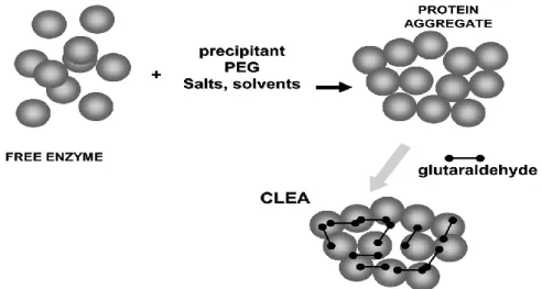
Cross-linking enzyme immobilization is a carrier-free method and results in the formation of aggregates connected by intermolecular linkages, being these aggregates composed by a covalent linkage between the enzymes molecules and bi-functional reagents with low molar mass. These bi-functional reagents has the ability to react rapidly with some groups located at the surface of the enzyme, as carboxyl and amino groups (Fernandes et al., 2010; Sheldon, 2007).
Generally, the precipitation of proteins as solid aggregates is possible by the simple addition of some compounds to the aqueous solutions of proteins, as salts, organic solvents or non-ionic polymers. These solid aggregates of proteins or enzymes are stabilized by non-covalent bonding, without perturbation of their tertiary structure that is, without denaturation. After the precipitation, it is possible to Figure 4 – Enzyme encapsulation and free movement of substrates and products across the membrane pores (adapted from Cao, 2006).
8
remove the aggregates from the solution where the enzymes precipitate and dissolve them in a new aqueous solution, purifying the enzymes. Considering this precipitation process, the pre-organized structure and catalytic activity of the aggregates can be maintained by cross-linking of these solid aggregates, making them permanently insoluble. These technique leads to the development of a new family of immobilized enzymes called CLEA: cross-linked enzyme aggregates (Figure 5) (Sheldon, 2007; Mateo et al., 2007; Cao et al., 2003).
Sometimes, cross-linking between the subunits of an enzyme also occurs, playing a critical role in multimeric proteins stabilization, as it prevents protein dissociation and helping on the reassociation of its subunits (Cao et al., 2000). There is another family of immobilized enzymes called CLECs (cross-linked enzyme crystals) which are significantly more stable to denaturation by heat, organic solvents and proteolysis than the corresponding soluble enzyme, allowing reactions at higher temperatures and in aqueous organic solvent mixtures. Comparing to immobilized or soluble enzymes, CLECs also have a higher activity per unit volume. Their operational stability, controllable particle size and ease of recycling, coupled with their high catalyst activities, made them ideally suited for industrial bio-transformations. Despite these attractive features, a disadvantage of CLECs is the need to crystallize the enzyme, an arduous procedure which require the enzyme in a high purity stage (Sheldon, 2007; Roy and Abraham, 2004; Govardhan, 1999).
Figure 5 – Formation of a solid aggregate of enzymes by cross-linking immobilization (adapted from Barbosa et al., 2014).
9
1.2. Protein PEGylation
PEGylation refers to the modification of a peptide, protein or non-peptide molecule by the covalent attachment of one or more polyethylene glycol (PEG) chains. The interest for this procedure has been rising since it allows the enhancement of the therapeutic and biotechnological potential of the compounds attached to PEG. When the PEG chain is properly linked to the protein, it can modify several of its characteristics while the enzymatic activity and the receptor recognition are maintained (Veronese, 2001; Veronese and Pasut, 2005).
1.2.1. PEG characteristics
PEG is synthesized by ring opening polymerization of ethylene oxide using methanol or water as initiator, to yield methoxy–PEG (mPEG–OH) or diol-PEG (HO–PEG–OH), respectively (Pasut and Veronese, 2007). Many studies report that the conjugated proteins have unchanged secondary and tertiary structures, although is normal to verify a reduction in enzyme activity after PEGylation due to the steric interference of polymer chains during the biological processes (Pasut and Veronese, 2012).
PEG has some key properties as great flexibility due to the absence of bulky substituents along the chain as well as the high hydration of the polymeric backbone. PEG also have high solubility in water and in many organic solvents, did not present toxicity to the cells and is approved by FDA for human use. When conjugated with proteins, PEG improves the conjugates bioavailability and their plasma half-life owing to the increased hydrodynamic volume that reduces the kidney clearance; it protects the proteins against degrading enzymes and prevents or reduce protein aggregation, immunogenicity and antigenicity (Harris and Chess, 2003; Pasut and Veronese, 2012). The reduction of immunogenicity is attributed to the shielding effect of polymeric chains around the protein since it prevents proteins interaction with degrading enzymes or antibodies (Pasut and Veronese, 2007; Pasut et al., 2004). Generally, the factors that affect the properties of the conjugate are structure, molecular weight, number of PEG chains attached to the polypeptide, the location of PEG linkages on the protein and the method used to make the PEG-polypeptide conjugation (Roberts et al., 2002).
10
1.3. Asparaginase
In 1953, Kidd noted the remarkable observation that guinea pig serum had antileukemia activity in mice. But, just in 1961, Broome attributed the cause of this antileukemia activity to depletion of asparagine by the enzyme asparaginase (Rytting, 2012). Since 1970, asparaginase has been used for the treatment of malignant hematopoietic diseases in children as a single agent or in combination with other chemotherapy agents. Those diseases include, principally, pediatric acute lymphoblastic leukemia (ALL), acute myelocitic leukemia, lymphosarcoma treatment and non-Hodgkin lymphomas (Muller and Boos, 1998; El-Naggar et al., 2014).
Although the successful treatment in the last years, a significant percentage of the ALL patients (10-25 %) relapse early, normally during the 3 years after the induction phase. In order to decrease this relapse, pharmacological changes have been made to try to improve the treatment of relapsed patients, however these changes have not been very successful basically due to acquired drug resistance (Avramis and Panosyan, 2005).
Asparaginase (EC 3.5.1.1) belongs to a group of homologous amidohydrolases family, characterized by catalyzing the hydrolysis reaction of the sidechain amide bond of asparagine, resulting in the formation of aspartate and ammonia (Figure 6) (Kumar and Sobha, 2012). The asparaginase reaction can be assayed by measuring the release of aspartate in a glutamate-oxaloacetate amidotransferase/malate dehydrogenase coupled enzymatic test and the release of ammonia in a simple Nessler test or by the disappearance of asparagine (Michalska and Jaskolski, 2006).
Heterologous enzymes are typically not suitable for human therapy, especially when a repeated or prolonged administration is required, once they are recognized as non-human by the human body and can result in adverse immunological reactions. Additionally, it is important that the therapeutic enzymes for cancer treatment present some pharmacological properties like: (i) must be highly specific to the Figure 6 – Reaction of degradation of asparagine and glutamine, catalyzed by asparaginase (adapted from Fu and Sakamoto, 2007).
11
substrate in order to avoid any undesired side effects, (ii) should exhibit a stability and high catalytic activity under physiological conditions to afford a reasonable dosing regimen; and (iii) must be amenable to formulation at the administration dose without adverse effects such as aggregate formation or inactivation (Cantor et al., 2012).
1.3.1. Preparations of Asparaginase available for therapy
The asparaginase is currently available in three different formulations: two are derived from Escherichia coli (native asparaginase and asparaginase conjugated with PEG) and one from Erwinia chrysanthemi. All three formulations share the same mechanism of action but have different pharmacokinetic properties (Table I), which do not make them easily switchable (Cantor et al., 2012; Rizzari et al., 2013).
Table I - Chemical and pharmacological properties of different asparaginase preparations Source Molecular
weight (kDa) KM (M) ASN KM (M) GLN
Isoeletric point (pH) Apparent 𝒕𝟏𝟐 (days) E. chrysanthemi 138 12 x 10-6 1.10x 10⁻³ 8.7 0.65±0.13 E. coli 141 10 x 10-6 6.25 x 10⁻³ 5.0 1.24±0.17 PEG-E. coli 145 10 x 10-6 -- 5.0 5.73±3.24
Asparagine (ASN); Glutamine (GLN)
Data according to Muller and Boos, 1998 and Asselin et al., 1993
Asselin et al (1993) found that the serum half-life of E. coli asparaginase activity was 1.24±0.17 days, approximately the double of the half-life of E. chrysanthemi activity (0.65±0.13 day). They also reported that the serum half-life of E. coli asparaginase activity increased to 5.73±3.24 days when the enzyme was conjugated with PEG. There are also differences on the time required to the serum asparaginase return to normal concentrations, being necessary 4 days for E. chrysanthemi asparaginase versus 11 days for E. coli asparaginase (Duval, 2002; Muller and Boos, 1998).
The antileukemic activity of asparaginase can be influenced by many factors. Some of them are biochemical factors like the rate of hydrolysis and the KM of the enzyme for asparagine and glutamine,
and others are pharmacological factors as serum clearance of the enzyme and tumor cell resistance to asparaginase. The antileukemic activity is also affected by the host immunological effects of
anti-12
asparaginase antibody formation, the contributions from the nutrient intake and the augmented asparagine ‘input’ from the de novo biosynthesis of asparagine by the liver (Avramis and Panosyan, 2005).
1.3.2. Asparaginase Types and Structure
Generally, bacteria produce two distinct asparaginases: type I and type II. Type I asparaginase is cytoplasmic, display high KM values relatively to asparagine and show also glutaminase activity. Type II
asparaginase is periplasmic, exhibits low KM values against asparagine owing to the high affinity to this
molecule and have low activity towards glutamine. Additionally, E. coli synthesizes a third type of asparaginase, asparaginase type III or EcAIII, that is classified as a plant-type asparaginase once this protein has a 70% amino acid sequence similarity to the asparaginase from plants, and belong to the Ntn-hydrolases family. Because type I has a much lower affinity for asparagine, only type II asparaginase has the anti-tumor properties and is used for the treatment of malignant hematopoietic diseases (Cappelletti et al., 2008; Kumar and Sobha, 2012; Michalska and Jaskolski, 2006). Although both E. coli and E. chrysanthemi asparaginase II have glutaminase activities, it only represent 3-9% of the total asparaginase activity once the KM value for glutamine is 100 times higher than that for asparagine (Aslanian and
Kilberg, 2001).
The functional form of E. coli asparaginase II exists as a homotretramer with a molecular mass between 133 and 141 kDa and each monomer composed by 300-350 amino acids (326 according to Protein Data Bank) (Cappelletti et al., 2008; Kozak et al., 2002). This enzyme has a nearly ideal 222 symmetry and is composed of four identical subunits bound principally by non-covalent forces (Figure 7), where the subunit A is connected to the subunit C (dimer A/C) and the subunit B is connected to the subunit D (dimer B/D). The asparaginase tetramer has four active sites and can be classified as a dimer of dimers because the dimer A/C is linked to the dimer B/D and each interface of the intimate dimer has two active sites (Kozak et al., 2002; Muller and Boos, 1998; Michalska and Jaskolski, 2006). Although the dimers contains all the functional groups and the structural elements to create a complete active site environment, the enzyme is only active and functional when the tetramer is complete by the approximation of the dimer A/C and the dimer B/D (Khushoo et al., 2004; Fu and Sakamoto, 2007).
13
1.3.3. Acute Lymphoblastic Leukemia
Leukemia is a malignant cancer of the blood and bone marrow, is a cancer of white blood cells, the cells that normally fight infections. This disease is characterized by an uncontrolled increase and excessive multiplications of malignant and immature lymphoblast in bone marrow, which alters the normal blood cells function and, in many instances, can lead to death (Kwan et al., 2009; Jain et al., 2012). Acute lymphoblastic leukemia (ALL) is seen in both children and adults, but is more common Figure 7 – Structural features of bacterial-type asparaginases. a) the quaternary structure of the periplasmatic E. coli asparaginase II. The green (subunit A) and red (subunit C) or gray (subunit B) and blue (subunit D) monomers form the intimate dimer. The arrows correspond to the two-fold axes defining the 222 symmetry of this homotetramer; b) the active site of E. coli asparaginase II with an asparagine substrate molecule modeled according to the complex enzyme-substrate. The dotted red lines represent hydrogen bonds while the green ones symbolize a potential line of attack of the active-site Thr residues on the substrate’s amide C atom (adapted from Michalska and Jaskolski, 2006).
14
between the ages of 2 and 5 years. The origin of ALL is considered to be multi-factorial, including endogenous or exogenous exposures, genetic predisposition, and fortuitous (Hiroto et al., 2013). There are different types of treatment to ALL and they include steroids, chemotherapy, radiation therapy and intensive combined treatments including steam cell or bone marrow transplants. Despite the variety of drugs available today, their efficacy in treatment of cancers is doubtful and the side effects caused by these chemotherapeutics agents are really aggressive, which can include infertility, secondary neoplasm, nausea and immunosuppression (Jain et al., 2012). Among the antitumor drugs described previously, there is another chemotherapeutic agent in pediatric oncotherapy especially for ALL that is the bacterial enzyme asparaginase. This enzyme has been employed as the most effective chemotherapeutic agent in pediatric ALL and improved the survival rate of pediatric ALL to approximately 90 % in recent trials (El-Naggar et al., 2014).
1.3.4. Asparaginase Mechanism of Action
Asparagine is an amino acid required for the synthesis of protein molecules and survival of the cells. This amino acid can be synthesized within the healthy cell by an enzyme called asparagine synthetase, so it is not considered an essential amino acid in normal cells. This enzyme catalysis the conversion of aspartic acid and glutamine into asparagine and glutamic acid (Figure 8), where glutamine is the donor of amino group and aspartate is transaminated to asparagine in an ATP-dependent reaction. On the other hand, asparagine can also be absorbed from the diet, as soon as it is retained into the body and made available to the cells (Cantor et al., 2012; El-Naggar et al., 2014; Fu and Sakamoto, 2007).
However, the availability of asparaginase is different on tumor cells. These cells cannot synthesize sufficient asparagine due to the absence or very low expression of asparagine synthetase and due to the Figure 8 – Biosynthesis of asparagine by asparagine synthetase (adapted from El-Naggar et al., 2014).
15
inability of tumor cells to increase asparagine synthetase activity after asparaginase administration. Thus, the malignant cells are dependent on serum levels of asparagine for their proliferation and survival. In order to keep their rapid malignant proliferation, tumor cells use both extracellular asparagine and intracellular synthesized asparagine, which is limited to satisfy their large asparagine demand (Savitri et al., 2003; Kumar and Sobha, 2012; Wang et al., 2003).
Therefore, asparagine is an essential amino acid for the growth of tumor cells, whereas the growth of normal cells is independent on its requirement since this amino acid can be synthesized by their own enzyme asparagine synthetase, in sufficient quantities for their metabolic needs (El-Naggar et al., 2014; Wetzler et al., 2007). In this way, administration of asparaginase deprives tumor cells of their extracellular source of asparagine which leads to tumor cells death. The death of tumor cells happens because they are dependent of blood serum asparaginase and without it, cells are unable to complete protein synthesis (Figure 9) (Cantor et al., 2012).
Some mechanisms of clinical relevance responsible for the tumor cells death are the arrestment of cell cycle in G1 phase, the induction of apoptosis and the cytotoxicity caused by the depletion of asparagine. There are a lot of studies on the mechanisms of action and the occurrence of resistance phenomena to the treatment with asparaginase. This studies demonstrate that a treatment response may only be expected if the malignant cells are unable to increase their expression of asparagine synthetase to an extent that can provide enough asparagine for the cell survival. If the tumor cell respond to the Figure 9 – Schematic representation of asparagine mechanism of action (adapted from Narta et al., 2007).
16
treatment with an increase on the synthesis of asparagine, it will be observed a decrease in sensitivity of this cells to asparaginase (Kumar and Sobha, 2012; Muller and Boos, 1998).
1.3.5. Toxicity of Asparaginase to Normal Cells
Despite the distinctive mechanism of action of asparaginase, which shows relative selectivity against the malignant cells and not against the normal cells, some patients experience some negative side effects during asparaginase therapy (El-Naggar et al., 2014).
The immunogenicity of exogenous proteins rises with increasing molecular weight and higher complexity of the structure. So proteins like asparaginase are highly immunogenic, since they are composed by a large number of subunits and they have complex quaternary structure. Clinical experience has shown that the application of the asparaginase leads to the formation of specific antibodies to this molecule in many patients (Cantor et al., 2012; Fu and Sakamoto, 2007; Muller and Boos, 1998). In this way, the efficiency of asparaginase has been compromised by a high rate of immunological reactions that can include enzyme inactivation without any clinical manifestation as well as anaphylactic shock; rapid clearance of the enzyme from the blood stream; shortening the enzyme half-life and development of anti-asparaginase antibodies, that neutralize anti-asparaginase activity (El-Naggar et al., 2014; Fu and Sakamoto, 2007). Patients with anti-asparaginase specific IgG antibodies do not have sufficient active enzyme for the depletion of asparagine until a therapeutic effect is verified and, in some cases, asparaginase activity may be undetectable. It is clear that there will be a loss of the therapeutic effect if the enzyme is inactivated and levels of asparagine are inadequate (Rizzari et al., 2013; Wang et al., 2003).
In case of toxicities arising from inhibited proteins, normal tissues with high rates of protein synthesis like pancreas, liver and coagulation systems are the most affected by this therapy, resulting those effects from an impairment of the protein synthesis (El-Naggar et al., 2014; Muller and Boos, 1998). The changes of the coagulation system related to asparaginase manifest themselves in a decrease of several coagulation factors and coagulation inhibiting proteins. The changes affecting the proteins of the coagulation system have a significant clinical impact since they can induce bleeding as well as enhance the probability of thromboembolic events. The occurrence of thrombotic complications in patients who were carry out the treatment with asparaginase varies from less than 10 % to greater than 30 %, with the incidence of thrombus formation increased with the increasing of the age population (Cantor et al., 2012). Other toxic effects related to protein inhibition include abnormalities on lipid metabolism and on
17
hemostasis, hypoalbuminemia, hyperglycemia due to a decrease in serum insulin and a decrease in lipoproteins, pancreatitis, and hepatoxicity (Fu and Sakamoto, 2007; Kumar and Sobha, 2012).
Additionally, the secondary effects may be associated with life-threatening complications when the central nervous system is involved (Muller and Boos, 1998). An impairment of central nervous system functions is demonstrated as convulsions, irritability, tension, depression, confusion, visions, disorientation, somnolence or even coma and this symptoms are principally due to hyperammonemia (Fu and Sakamoto, 2007; Muller and Boos, 1998).
Hypersensitivity reactions to asparaginase preparations are not rare and the risk to have a severe allergic reaction depend on prior exposures to the enzyme and concomitant medications, for example with corticosteroids. The signs and symptoms of clinical hypersensitivity include rash, urticaria, laryngeal edema, hypotension, bronchospasm and occasionally anaphylactic shock (Cantor et al., 2012; Rizzari et al., 2013). This allergic reactions become more prominent and occur in approximately 5–10 % of patients after repeat exposures to asparaginase and while patients are not receiving corticosteroids. Once patients demonstrate signs of clinical hypersensitivity to this enzyme, they could not continue the same treatment. Although, there are available asparaginases from different bacterial strains that present only limited cross reactivity, which make possible the treatment continuation by selecting an enzyme from a different biological source. The continuation of the treatment also can be possible by using the enzyme linked to PEG (Muller and Boos, 1998; Rizzari et al., 2013).
asparaginase show some advantages comparatively to asparaginase. Although PEG-asparaginase is less immunogenic than the enzyme alone, it still causes severe allergic reactions in patients who get repeated doses of PEG-asparaginase during treatment. However, patients who have allergic reactions to asparaginase or PEG-asparaginase may continue the treatment with the asparaginase from E. chrysanthemi, since antibodies to PEG-asparaginase and asparaginase from E.coli do not cross-react with E. chrysanthemi asparaginase (Cantor et al., 2012; El-Naggar et al., 2014).
Anti-asparaginase antibodies are the main cause of resistance to the treatment with asparaginase. Asparaginase resistance can be symptomatic when the patients exhibit signs of clinical hypersensitivity, or asymptomatic without any signs. Asymptomatic resistance, also called silent inactivation, is more dangerous than clinical hypersensitivity, as there are no clinical signs of allergy to alert against asparaginase resistance. In vitro studies have demonstrated an adaptive mechanism by human leukemia cells that are resistant to asparaginase treatment, where they increase their expression of asparagine synthetase in order to survive against the therapy (Aslanian and Kilberg, 2001; Rizzari et al., 2013).