Comparison of Two Self-Etching Primers and Effect of
Saliva Contamination on Shear Bond Strength of
Orthodontic Brackets
Joana Godinho*, Sofia S. A. Oliveira**, Luís Jardim***
Resumo:Introdução: A contaminação salivar é uma das principais causas de insucesso na cimentação de brackets. Os self-etching primers, recentemente introduzidos em ortodontia, reduzem o tempo de execução da técnica e a probabilidade de contaminação. Objectivos: Determinar o efeito da contaminação salivar na resistência adesiva a forças de corte de brackets ortodônticos, cimentados com dois self-etching primers. Materiais e Métodos: Foram cimentados 100 brackets em premolares humanos (10 amostras por grupo), utilizando 2 adesivos (Transbond Plus e First Step) e 5 condições de super-fície: 1) Condição ideal, esmalte seco; 2) Contaminação salivar antes do primer; 3) Contaminação salivar e secagem antes do primer; 4) Contaminação salivar depois do primer; 5) Contaminação salivar depois do primer, secagem e reaplicação do primer. As amostras foram armazenadas 7 dias em água a 37ºC e sujeitas a 500 ciclos de termociclagem. A área de adesão foi observada ao microscópio óptico para determinar o tipo de falha adesiva. Resultados: A análise de variância demonstrou diferenças significativas entre as forças de resistência adesiva, sendo o Transbond Plus superior ao First Step. A contaminação não influenciou significativamente a resistência adesiva. Conclusões: 1) A resistência adesiva foi mais elevada com o Transbond Plus em comparação com o First Step, independentemente da condição. 2) Para ambos os self-etching primers, a adesão não foi significativamente afectada pela presença de saliva ou pelos procedimentos de descon-taminação. 3) No grupo do First Step foi encontrado um maior número de falhas adesivas na interface dente / resina composta.
Palavras-Chave:Self-etching primers; Saliva; Contaminação; Força de cisalhamento; Índice de Adesivo Residual
Abstract: Introduction: Saliva contamination during the bonding procedure is a common cause of bracket bond failure. By combining acid and primer in one component, self-etching primers (SEP) reduce the working time and the risk of contam-ination. Objectives: The purpose of this study was to investigate the effect of saliva contamination of the enamel surface on the shear bond strength of orthodontic brackets cemented with two self-etching primers. Materials and Methods: One hundred orthodontic brackets were bonded to human premolars with Transbond Plus Self-etching Primer (TPSEP, 3M Unitek, Monrovia, CA) or First Step (FS, Reliance Orthodontic Products, Itasca, IL) under the following enamel surface conditions: 1) dry enamel; 2) saliva contamination/ primer; 3) saliva contamination/ air drying/ primer; 4) primer/ saliva contami-nation; 5) primer/ saliva contamination/ air drying/ reapplication of the primer. Samples were stored for 7 days in water at 37ºC and submitted to 500 cycles of thermal stress. Bond failure sites were classified by an Adhesive Remnant Index score system. Results: Mean shear bond strengths were significantly higher in the TPSEP groups compared to the FS groups (p < .001). For the same adhesive, no significant differences were found between the enamel surface conditions. More adhesive failures were observed in the FS groups. Conclusions: Brackets bonded with TPSEP had the highest shear bond strength values, under the different enamel surface conditions. For both SEPs, bond strengths were not significantly affect-ed by the enamel surface conditions. The FS groups failaffect-ed more frequently at the enamel / resin interface.
Key-words:Self-etching primers; Saliva; Contamination; Shear-peel bond strength; Adhesive Remnant Index
(Godinho J, Oliveira SSA, Jardim L. Comparison of Two Self-Etching Primers and Effect of Saliva Contamination on Shear Bond Strength of Orthodontic Brackets. Rev Port Estomatol Cir Maxilofac 2007;48:197-203)
*Assistente estagiária, Unidade de Ortodontia da Faculdade de Medicina Dentária da Universidade de Lisboa **Professora auxiliar, Departamento de Biomateriais da Faculdade de Medicina Dentária da Universidade de Lisboa
***Professor catedrático, Coordenador do Curso Pós-graduado de Especialização em Ortodontia da Faculdade de Medicina Dentária da Universidade de Lisboa. Presidente do Colégio de Ortodontia da Ordem dos Médicos Dentistas
Conventional direct bonding of orthodontic brackets to the enamel surface involves three different agents: an enamel condi-tioner, a primer, and an adhesive resin. Besides being time-consu-ming this procedure requires a dry environment, which some-times can be difficult to achieve (e.g. hard-to-reach areas, partial-ly erupted or surgicalpartial-ly exposed teeth).(1)Moisture
contamina-tion is the most common reason for bond failure with composi-te resins.(2-6)Debonded brackets are inconvenient, delay
ment, require extra-appointments and might compromise treat-ment outcomes.
The recently introduced self-etching primers combine etching and priming in one single component with the advantages of saving time, and reducing both the technique-sensitiveness and the chances for contamination.(7-11)Since these products are
effec-tive in bonding to enamel they have been used for direct adhe-sion of orthodontic brackets.(12-16)
Transbond Plus Self-etching Primer (TPSEP, 3M Unitek, Monrovia, CA, USA) was the first self-etching primer commer-cialized for orthodontic purposes, and the one that has been mainly reported in the literature. The molecule that etches and primes the tooth simultaneously is formed when phosphoric acid and a methacrylate group are combined to generate a metha-crylated phosphoric acid ester.(11,17)The single product is applied
to dried enamel and gently evaporated with a stream of air to facilitate solvent evaporation. Different studies have found that TPSEP can achieve adequate bond strength levels when applied to a dry enamel surface.(18-20)Saliva contamination, both before
and after the application of TPSEP has been reported in the lite-rature. Contamination after the self-etching primer resulted in a significantly lower bond strength.(1,21)However, when saliva
was applied before the primer, no significant differences were found.(1)A decontamination procedure was recently reported by
Zeppieri et al,(11)in which TPSEP was reapplied after saliva
conta-mination. No significant differences were found among the bond strengths obtained with dry, contaminated and decontaminated enamel surfaces.
To date, the influence of saliva contamination on the self-etching primer First Step (FS, Reliance Orthodontic Products, Itasca, IL, USA) and the effect of air drying as a decontamination proce-dure have not been reported in the literature. The purposes of this in vitro study were: 1- to compare the shear bond strength of brackets bonded with two self-etching primers, TPSEP and FS; 2 - to assess the effect of different contamination and deconta-mination procedures on the bond strength of the same self-etching primers as in 1.
One hundred human premolars with intact buccal enamel were collected and stored in a bacteriostatic solution (0.5% chlo-ramine-T) at 4ºC as recommended by the ISO standards.(22)Less
than 6 months had elapsed between extraction and the bonding experiment. The buccal surfaces were cleaned and polished with non-fluoridated pumice paste applied with a rubber prophyla-ctic cup on a slow-speed hand piece for 10 seconds, rinsed for 5 seconds and dried with an oil - and moisture-free air spray for 5 seconds.
Orthodontic stainless steel premolar brackets with a 0.018 inch slot (Mini Diamond Twin, Lot 03J418J; SDS Ormco, Orange, CA, USA) were used in this study. The average bracket base surface (mean value of ten brackets area) was determined to be 9.48 mm2.
The specimens were randomly divided into ten groups and bonded according to one of the protocols described below. When applicable, the enamel surface was contaminated with 1.5 µL of whole, unstimulated fresh human saliva, spread with a micro-brush with two strokes. Saliva was collected from one donor who was instructed to brush the teeth and not to eat for one hour before the saliva was collected.
Experimental groups (Figure 1) were divided as follows: Group 1, the enamel surface was simultaneously etched and
primed with Transbond Plus Self-etching Primer (TPSEP, Lot 130809-L6; 3M Unitek, Monrovia, CA, USA), rubbed with the applicator brush for 5 seconds and then evapo-rated with a gentle air burst for 2 seconds, as recom-mended by the manufacturer.
Group 2, contamination with saliva was performed as described above and TPSEP was applied as in group 1. Group 3, contamination with saliva was performed and the tooth
was dried for 5 seconds. TPSEP was applied as in group 1. Group 4, TPSEP was applied as in group 1, and then the surface
was contaminated with saliva.
Group 5, TPSEP was applied as in group 1, and then the surface was contaminated with saliva, and dried for 5 seconds. TPSEP was reapplied with the same steps.
Group 6, the enamel surface was simultaneously etched and primed with First Step (FS, Lot 211110; Reliance Orthodontic Products, Itasca, IL, USA), rubbed with the applicator brush for 5 seconds and then evaporated for 5 seconds, as recommended by the manufacturer. Group 7, contamination with saliva was performed as described
above and FS was applied as in group 6.
Group 8, contamination with saliva was performed and the tooth was dried for 5 seconds. FS was applied as in group 6.
Group 9, FS was applied as in group 6, and then the surface was con-taminated with saliva.
Group 10, FS was applied as in group 6. The tooth was contami-nated with saliva and dried for 5 seconds. First Step was reapplied with the same steps.
Orthodontic brackets were bonded by one investigator with Transbond XT composite resin (Lot 2FP; 3M Unitek, Monrovia, CA, USA) near the center of the buccal surface with sufficient pressure to express excess adhesive, which was then removed with a sharp scaler. The composite resin was light-cured for 10 seconds on the mesial side and 10 seconds on the distal side (total cure time 20 seconds) with an Ortholux LED Curing Light (3M ESPE Minneapolis, MN, USA). All samples were stored in distilled water at 37ºC for 7 days in an incubator (Memmert, GmbH+Co., 8540 Schwabach, Germany). During this period the specimens were subjected to 500 cycles of thermal stress between 5ºC and 55ºC (20 seconds each bath) in distilled water with 5 seconds dwell time.
Three retentive sulcus were created on the buccal and lingual aspects of each root. The roots were embedded in self-curing polymetilmethacrylate. Steel cylinders (12 mm height / 13 mm diameter) were used as casts for the acrylic. A stainless steel 0.018 x 0.025 wire was used to align the buccal surface of each tooth perpendicular to the bottom of the mold. The methacry-late was cured in a Polyclav vessel (Dentaurum, D-7530, Pforzheim, Germany) for 10 minutes, under a pressure of 1.5 bar and a temperature of 40ºC.
Shear bond strength tests were performed on a universal testing machine (model 4502, Instron Ltd, Bucks, HP12 3SY, UK). A wire loop was placed under the gingival wings of the ortho-dontic brackets so that the shear force was applied parallel to the long axis of the tooth. A 1 KiloNewton (KN) load cell set at a crosshead speed of 1 mm/min was used. The maximum load necessary to debond each tooth (N) was divided by the bracket surface area (mm2) to calculate the shear bond strengths (MPa).
After debonding, the teeth and brackets were examined at x20 magnification with a stereomicroscope (Nikon SMZ-2, Nikon Europe BV, P.O.B. 7609, The Netherlands). Any adhesive remai-ning after removal of the bracket was assessed according to the Adhesive Remnant Index of Årtun and Bergland.(23)This scale
ranges from 0 to 3; a score of 0 indicates that no adhesive remai-ned on the tooth in the bonding area, 1 indicates that less than half of the adhesive remained on the tooth, 2 indicates that that more than half of the adhesive remained on the tooth, and 3 indicates that all the adhesive remained on the tooth, with a distinct impression of the bracket mesh (Figure 2).
Statistical analysis was performed using Statview 4.5 (Abacus Concepts, Berkeley, CA, USA). Descriptive statistics including the mean, standard deviation, minimum and maximum values were calculated for each of the ten test groups. Data was evaluated by a two-way analysis of variance (ANOVA). Differences between experimental groups were performed with post hoc analysis using the Student-Newman-Keuls multiple range test at · = 0.05 level of significance.
The chi-square test was used to determine significant diffe-rences in the ARI scores among the different groups.
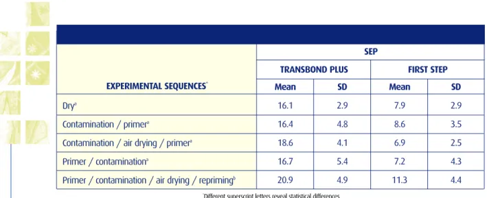
Figure 1 - Schematic representation of the different bonding sequences
EXPERIMENTAL SEQUENCES*
FIRST STEP TRANSBOND PLUS
SEP
*Different superscript letters reveal statistical differences
Table 1 - Descriptive Statistics for the shear bond strengths (MPa) of the experimental groups
Drya
Contamination / primera
Contamination / air drying / primera
Primer / contaminationa
Primer / contamination / air drying / reprimingb
Mean 16.1 16.4 18.6 16.7 20.9 SD 2.9 4.8 4.1 5.4 4.9 Mean 7.9 8.6 6.9 7.2 11.3 SD 2.9 3.5 2.5 4.3 4.4 EXPERIMENTAL GROUPS GROUPS ARI SCORES
Table 3 - Frequency distribution of the adhesive remnant index of the experimental groups
Transbond Plus, dry
Transbond Plus , contamination / primer
Transbond Plus, contamination / air drying / primer Transbond Plus, primer / contamination
Transbond Plus, primer / contamination / air drying / repriming First Step, dry
First Step, contamination / primer
First Step, contamination / air drying / primer First Step, primer / contamination
First Step, primer / contamination / air drying / repriming 1 2 3 4 5 6 7 8 9 10 0 -6 5 8 6 4
Table 2 - Statistical analysis: Two/way ANOVA
Sum of squares 2187.042 241.315 48.449 1498.042 Mean Square 2187.042 60.329 12.112 16.645 F-Value 131.394 3.624 .728 SEP
Enamel surface condition SEP * Enamel surface condition Residual DF 1 4 4 90 F-Value <.0001 .0087 2.911 1 7 6 5 3 2 4 5 2 4 5 2 3 4 5 7 7 -1 3 -1
-Mean shear bond strengths of the 10 groups tested are presented in Table I. Two-way ANOVA (Table II) showed signifi-cant differences between the two self-etching primers (p < .0001) and between the enamel surface conditions (p < .05). No signi-ficant interaction was found.
For all the enamel surface conditions tested, higher bond strength values were obtained with TPSEP. When the different enamel surface conditions were compared, significantly higher shear bond strengths were obtained in the groups where the self-etching primer was reapplied after saliva contamination (groups 5 and 10).
The frequency distributions of ARI scores for the 10 groups tested are listed in Table III. Chi-square test results indicated signi-ficant differences between the experimental groups (χ2= 74.82,
p < .0001). There was a greater frequency of ARI scores of 1 and 2 in the TPSEP groups (ie, more residual adhesive was left on the enamel surface after debonding). On the other hand, in the FS groups, there was a higher frequency of ARI scores of 0 and 1 (ie, less residual adhesive was left on the enamel surface after debonding).
New products combining etching and priming in one single component were recently introduced in orthodontics and have been subject of intensive research. Different self-etching primers have been evaluated, mostly under non-contaminated condi-tions.(24,25)
The present study compared two self-etching primers desig-ned for orthodontic purposes and evaluated the influence of sali-va contamination and different decontamination procedures on the bracket shear bond strengths. To the best of our knowled-ge, the effect of saliva contamination on the self-etching primer First Step has not been previously reported.
For each experimental group ten premolars were tested. The reduced sample size was taken into consideration when performing the statistical analysis, therefore the two self-etching primers and the five different enamel surface conditions were compared, with fifty and twenty teeth in each comparison respec-tively.
For all enamel surface conditions tested, the mean shear bond strengths in the FS groups were significantly lower than the ones in the TPSEP groups (p < .0001). Similar findings were reported by Trites et al(25)in a study where the FS and the TPSEP
were used in ideal conditions, after storage periods of 24 hours, 30 days and 3 months.
In this study the self-etching primers were not significantly affected by the presence of small amounts of saliva. These findings agreed with a previous report by Zeppieri et al(11)which
demons-trated that reapplying TPSEP when moisture contamination is detected provides an acceptable bond strength. The present data also demonstrated that significantly higher shear bond strength is obtained with the reapplication of the self-etching primers, even after saliva contamination. Since moisture contamination can occur without being noticed by the clinician, the application of two coats of self-etching primer seems to be useful, and could be performed even when there is no evidence of contamina-tion. More research is needed to determine the validity of this idea.
In orthodontics practice, less residual adhesive on the enamel surface after debonding is advantageous, since it reduces the time required to clean the teeth. Some studies have reported more failures at the enamel/resin interface with self-etching primers, which means less adhesive left on the tooth.(11,14,26 ) This
may be caused by the thinner and less uniform resin tags obtai-ned with these adhesive systems in comparison to conventio-nal etching and priming, decreasing the mechanical interlocking between resin and enamel.(26)
The present results demonstrated differences between the failure modes of the two self-etching primers. In FS groups, there was a higher frequency of failures at the enamel/resin interfa-ce, meaning less residual adhesive on the teeth. In brackets bonded with TPSEP the failures were mostly combined, leaving resin both on the enamel and the bracket surfaces. These distinct patterns might be explained by the differences in shear bond strengths between the two self-etching primers: TPSEP was supe-rior to FS independently of the enamel surface condition consi-dered. A favorable failure mode of brackets leaving less adhe-sive on the enamel is only an advantage if the bond strength achieved is clinically adequate. However, it must be remembe-red that to date, the minimum clinically effective bond strength value is not known.
Within the limitations of this in vitro study, the following conclusions were drawn:
1. Brackets bonded with Transbond Plus Self-etching Primer showed the highest shear bond strengths, under the various enamel surface conditions.
2. For both materials tested, higher shear bond strengths were
RESULTS
DISCUSSION
obtained when the self-etching primers were reapplied after saliva contamination.
3. The First Step groups failed more often at the enamel/resin interface, leaving less residual resin on the enamel surface.
1 - Cacciafesta V, Sfondrini MF, De Angelis M, Scribante A, Klersy C. Effect of water and saliva contamination on shear bond strength of brackets bonded with conventional, hydrophilic, and self-etching primers. Am J Orthod Dentofacial Orthop 2003;123:633-40. 2 - Zachrisson BJ. A posttreatment evaluation of direct bonding in orthodontics. Am J Orthod 1977; 71:173-89.
3 - Silverstone LM, Hicks MJ, Featherstone MJ. Oral fluid contamination of etched enamel surfaces: an SEM study. J Am Dent Assoc 1985;110:329-92.
4 - Xie J, Powers JM, McGuckin RS. In vitro bond strength of two adhesives to enamel and dentin under normal and contaminated conditions. Dent Mater 1993;9:295-9.
5 - Gwinnett AJ. Bonding of restorative resins to enamel. Int Dent J 1988;38:91-6.
6 - Powers JM, Finger WJ, Xie J. Bonding of composite resin to contaminated human enamel and dentin. J Prosthodont 1995;4:28-32. 7 - Chigira H, Koike T, Hasegawa T, Itoh K, Wakumoto S, Hayakawa T. Effect of the self etching dentin primers on the bonding
effi-cacy of a dentin adhesive. Dent Mater 1989;8:86-92.
8 - Barkmeier WW, Los SA, Triolo PT. Bond strengths and SEM evaluation of Clearfil Liner Bond 2. Am J Dent 1995;8:289-93. 9 - Nakabayashi N. Dentinal bonding mechanisms. Quintessence Int 1991; 22:73-4.
10 - Grubisa H, Heo Giseon, Raboud D, Glover KE, Major PW. An evaluation and comparison of orthodontic bracket bond strengths achieved with self-etching primer. Am J Orthod Dentofacial Orthop 2004;126:13-9.
11 - Zeppieri IL, Chung CH, Mante FK. Effect of saliva on shear bond strength of an orthodontic adhesive used with moisture-insen-sitive and self-etching primers. Am J Orthod Dentofacial Orthop 2003;124:414-9.
12 - Bishara SE, Gordan VV, VonWald L, Olson ME. Effect of an acidic primer on shear bond strength of orthodontic brackets. Am J Orthod Dentofacial Orthop 1998;114:243-7.
13 - Bishara SE, VonWald L, Laffoon JF, Warren JJ. Effect of a self-etch primer/adhesive on the shear bond strength of orthodontic brackets. Am J Orthod Dentofacial Orthop 2001;119:621-4.
14 - Bishara SE, Gordan VV, VonWald L, Jakobsen JR. Shear bond strength of composite, glass ionomer, and acidic primer adhesive systems. Am J Orthod Dentofacial Orthop 1999;115:24-8.
15 - Bishara SE, Oonsombat C, Ajlouni R, Denehy G. The effect of saliva contamination on shear bond strength of orthodontic bracke-ts when using a self-etch primer. Angle Orthod 2002; 72:554-7.
16 - Yamada R, Hayakawa T, Kasai K. Effect of using self-etching primer for bonding orthodontic brackets. Angle Ortho~d 2002;72:558-64. 17 - Aljubouri YD, Millett DT, Gilmour WH. Laboratory evaluation of a self-etching primer for orthodontic bonding. Eur J Orthod
2003;25:411-5.
18 - Arnold RW, Combe EC, Warford JH. Bonding of stainless steel brackets to enamel with a new self-etching primer. Am J Orthod Dentofacial Orthop 2002;122:274-6.
19 - Larmour CJ, Stirrups DR. An ex vivo assessment of a bonding technique using a self-etching primer. J Orthod 2003;30:225-8. 20 - Dorminey JC, Dunn WJ, Taloumis LJ. Shear bond strength of orthodontic brackets bonded with a modified 1-step
etchant-and-primer technique. Am J Orthod Dentofacial Orthop 2003;124:410-3.
21 - Rajagopal R, Padmanabhan S, Gnanamani J. A comparison of shear bond strength and debonding characteristics of conven-tional, moisture-insensitive, and self-etching primers in vitro. Angle Orthod 2004;74:264-8.
22 - Dental Materials. Testing of adhesion to tooth structure. ISO/TS 11405: 2003.
23 - Årtun J, Bergland S. Clinical trials with crystal growth conditioning as an alternative to acid-etch enamel pre-treatment. Am J Orthod 1984; 85:333-40.
24 - Bishara SE, Oonsombat C, Ajlouni R, Laffoon JF. Comparison of the shear bond strength of 2 self-etch primer/adhesive systems. Am J Orthod Dentofacial Orthop 2004;125:348-50.
25 - Trites B, Foley TF, Banting D. Bond strength comparison of 2 self-etching primers over a 3-month storage period. Am J Orthod Dentofacial Orthop 2004;126:709-16.
26 - Oliveira SSA, Pugach MK, Hilton JF, Watanabe LG, Marshall SJ, Marshall GW. Dentin Smear Layer Variations and Adhesion of a Self-Etching Primer. Dental Mater 2003; 19:758-67.