UNIVERSIDADE DA BEIRA INTERIOR
Ciências da Saúde
DHT and E2 as modulators of apoptotic signaling
in rat Sertoli cells
Vera Lúcia Silveira Simões
Dissertação para obtenção do Grau de Mestre em
Ciências Biomédicas
(2º ciclo de estudos)
Orientador: Prof. Doutor Pedro Fontes Oliveira
iii O conteúdo do presente trabalho é da exclusiva responsabilidade do autor:
________________________________________ Vera Lúcia Silveira Simões
v
Agradecimentos
Ao professor Doutor Pedro Fontes Oliveira, orientador deste trabalho, agradeço a oportunidade dada na participação deste estudo, o apoio, a partilha de conhecimentos e as valiosas contribuições para este trabalho.
Queria também agradecer ao Dr. Marco Alves pela sua disponibilidade, atenção e transmissão de conhecimentos, que serão essenciais para esta nova etapa que se avizinha.
Dirijo também uma palavra de apreço ao Luís Rato por toda a ajuda que me foi prestada e a todo o grupo de trabalho, especialmente à Ana e Tânia, que me acompanharam nesta longa caminhada.
Ao Filipe, Tiago, Liliana e todos os meus amigos que contribuíram para que esta fase se concluísse com êxito, obrigada pela paciência, carinho e apoio.
O meu maior agradecimento é dirigido aos meus pais, por terem sido o contínuo apoio em todos estes anos, ensinando-me, principalmente, a importância da construção e coerência dos meus próprios valores. Assim como aos meus irmãos.
vii
Resumo
A apoptose é um evento regulador importante na homeostase testicular e na otimização da produção de espermatozoides. As células de Sertoli formam a barreira hemato-testicular, criando um microambiente especial onde as células germinativas se desenvolvem e estão sob controle hormonal estrito. De facto, os estrogénios e os androgénios são conhecidos por desempenharem um papel importante no funcionamento das células de Sertoli, melhorando a sua sobrevivência in vitro e impedindo a progressão da apoptose.
Neste trabalho estudou-se a influência do 17β-estradiol (E2) e da 5α-dihidrotestosterona (DHT) nas vias de sinalização apoptótica em culturas de células de Sertoli de ratos imaturos. Para isso, foram selecionados pontos chave da via apoptótica que interagem com a mitocôndria e foram avaliados os níveis de expressão de mRNA e/ou dos níveis de proteína de vários desses marcadores apoptóticos, tais como o p53, o membro pró-apoptótico da família Bcl-2 designado por Bax, o factor de indução de apoptose (AIF) e as caspase-9 e caspase-3. A actividade da caspase-3 foi também avaliada como um marcador da fase final da apoptose.
Pudemos verificar que tanto o E2 como a DHT diminuíram os níveis de transcritos de mRNA do p53, Bax, caspase-9 e caspase-3. Os níveis proteicos de AIF foram também reduzidos após o tratamento com DHT, enquanto que nas células tratadas com E2, se verificou uma diminuição nos níveis proteicos da caspase-9 clivada. A actividade da caspase-3 apresentou uma acentuada diminuição após o tratamento hormonal quer com E2, quer com DHT.
Em conjunto, estes resultados demonstram que o E2 e a DHT actuam como moduladores in vitro da sinalização apoptótica em células de Sertoli de ratos imaturos, sugerindo que, nessas células, os androgénios e os estrogénios podem ser capazes de modular vias independentes da apoptose, uma vez que parecem regular diferencialmente a expressão de factores pró-apoptóticos distintos.
Palavras-chave
Resumo Alargado
O processo de apoptose é um tipo de morte celular que participa activamente nos processos de diferenciação, crescimento e desenvolvimento dos tecidos, pois permite manter o equilíbrio entre a taxa de proliferação e degeneração, ajudando na manutenção dos vários constituintes do corpo. A manutenção das células de Sertoli, que são um constituinte importante para o bom desenvolvimento da espermatogénese, é crucial para que se formem espermatozóides viáveis. Assim, a apoptose das células de Sertoli torna-se um processo essencial para que estas desempenhem o seu papel na espermatogénese.
O presente estudo tem como objectivo estudar o efeito de hormonas esteroides sexuais nas vias de sinalização apoptótica em culturas de células de Sertoli de ratos imaturos. Foram analisados os níveis de mRNA do p53, Bax, caspase 9 e caspase 3 por RT-PCR, e por Western Blot foram analisados os níveis proteicos de Bax, do factor indutor de apoptose (AIF) e da caspase 9 clivada. Para além disso, e com o objectivo de analisar um marcador final da apoptose, foi também analisado a actividade da caspase 3, sendo a partir deste ponto, o processo apoptótico irreversível. Foram utilizadas para este estudo culturas primárias de células de Sertoli de rato imaturos tratadas com 17β-estradiol (E2) ou 5α -dihidrotestosterona (DHT).
Neste estudo demonstramos que ambos E2 e DHT agem como moduladores da via de sinalização apoptótica em culturas de células de Sertoli de ratos imaturos. O E2 e DHT foram capazes de diminuir os níveis de mRNA do p53, Bax, caspase 9 e caspase 3. A DHT actuando sozinho foi capaz de diminuir os níveis proteicos do AIF, assim como o E2 foi capaz de diminuir os níveis de proteína da caspase 9 clivada, o que poderá sugerir que os androgénios e os estrogénios podem ser capazes de modular vias independentes no processo de apoptose. No entanto, será necessário realizar mais estudos para uma plena elucidação dos mecanismos por de trás desse fenómeno. Contudo tanto a DHT como o E2 foram capazes de reduzir a actividade da caspase 3, um marcador da fase final da apoptose, que confirma claramente a sua acção anti-apoptótica.
Em conclusão, este estudo permitiu um melhor conhecimento sobre a influência dos androgénios e dos estrogénios no processo de apoptose das células de Sertoli, processo este crucial para manter a homeostasia entre as células de Sertoli e as células germinativas e, por consequência, no desenvolvimento da espermatogénese. Assim, estes resultados apontam novos caminhos para uma melhor compreensão de algumas das causas de infertilidade masculina, nomeadamente as relacionadas com desregulação hormonal.
ix
Abstract
Apoptosis is an important regulatory event in testicular homeostasis and optimization of sperm production. Sertoli cells (SCs) form the blood-testis barrier creating a special microenvironment where germ cells develop and are under strict hormonal control. Estrogens and androgens are known to play critical roles in SCs functioning, improving their in vitro survival by preventing apoptotic progression.
Herein, the influence of 17β-estradiol (E2) and 5α-dihydrotestosterone (DHT) on the
apoptotic signaling pathways of immature rat cultured SCs was studied. For that purpose key points of the apoptotic pathway that interact with the mitochondria were chosen and the mRNA expression and/or protein levels of several apoptotic markers such as p53, the pro-apoptotic Bcl2 family member Bax, the apoptosis-inducing factor (AIF) and caspase-3 and 9 were evaluated. Caspase-3 activity was also evaluated as an endpoint marker of apoptosis.
E2 and DHT down-regulated the mRNA transcript levels of p53, Bax, caspase-9 and caspase-3. The protein levels of AIF were reduced after DHT treatment while E2-treated cells decreased the cleaved caspase-9 protein levels. The apoptotic endpoint Caspase-3 activity presented highly decreased levels after hormonal treatment.
Taken together, these results showed that E2 and DHT act as apoptotic signaling modulators in in vitro immature rat SCs suggesting that androgens and estrogens may be capable of modulating independent pathways of the apoptotic event by regulating different pro-apoptotic factors.
Keywords
Publications
Simões VL, Alves MG, Martins AD, Dias TD, Rato L, Socorro S, Oliveira PF (2012) Regulation of Apoptotic Signalling Pathways by 5α-dihydrotestosterone and 17β-estradiol in Immature Rat Sertoli Cells. (Submitted)
Martins AD, Alves MG, Simões VL, Dias TD, Rato L, Moreira, PI, Socorro S, Cavaco JE, Oliveira PF (2012) 17β-estradiol and 5α-dihydrotestosterone modulate transporters and enzymes of glucose metabolism in cultured immature rat Sertoli cells. (Submitted)
xi
Table of Contents
I. INTRODUCTION ... 1
1.THE TESTIS:FORM AND FUNCTION ... 2
1.1 Spermatogenesis ... 3
1.2 Hormonal Regulation of Spermatogenesis ... 4
1.3 Sertoli Cells ... 6
2.APOPTOSIS ... 9
2.1 Apoptotic Activation Pathways ... 9
2.2 Regulatory Mechanisms of Apoptosis ... 12
3.APOPTOSIS IN MALE GONADS ... 12
3.1. Apoptosis in Sertoli Cells ... 13
II. AIM OF THE PROJECT ... 15
II. MATERIALS AND METHODS ... 17
1-CHEMICALS... 18
2-ANIMALS AND TISSUES ... 18
3-SERTOLI CELL CULTURE ... 18
4-EXPERIMENTAL GROUPS ... 19
5-RT-PCR ... 19
6-WESTERN BLOT ... 20
7-CASPASE-3 ACTIVITY ASSAY ... 21
8-STATISTICAL ANALYSIS ... 21
IV. RESULTS ... 22
1.E2 DOWN-REGULATES MRNA TRANSCRIPT LEVELS OF APOPTOTIC SIGNALING MARKERS ... 23
2. MRNA TRANSCRIPT LEVELS OF APOPTOTIC MARKERS ARE DOWNREGULATED AFTER DHT TREATMENT ... 24
3.E2 DECREASE CLEAVED CASPASE-9 PROTEIN LEVELS ... 25
4.AIF PROTEIN LEVELS ARE HIGHLY DECREASED AFTER DHT-TREATMENT ... 26
5.CASPASE-3 ACTIVITY IS HIGHLY DECREASE AFTER HORMONAL TREATMENT ... 26
V. DISCUSSION ... 28
xiii
List of Figures
Figure 1 - Structure of the testis ... 2 Figure 2 – Cellular changes occurring during spermatogenesis. ... 3 Figure 3 - Feedback regulation of the hypothalamic-pituitary-testicular axis in males. ... 5 Figure 4 - Mitochondrial control over apoptosis. ... 10 Figure 5 - Effect of E2 on p53, Bax, caspase 9 and caspase 3 mRNA levels in rat Sertoli cells. ... 23 Figure 6 - Effect of DHT on p53, Bax, caspase 9 and caspase 3 mRNA levels in rat Sertoli cells. ... 24 Figure 7 - Effect of E2 on Bax, AIF and cleaved caspase 9 protein levels in rat Sertoli cells. ... 25 Figure 8 - Effect of DHT on Bax, AIF, and cleaved caspase 9 protein levels in rat Sertoli cells. ... 26 Figure 9 - Effects of E2 and DHT on caspase-3 activity. ... 27
xv
List of Tables
Table 1 - Oligonucleotides and Cycling Conditions for PCR Amplification of p53, Bax, Caspase 9, Caspase 3 and 18S. ... 20
xvii
Abbreviations and Symbols
ABP – Androgen binding protein AIF - Apoptosis inducing factor
Apaf-1 - Apoptotic protease activating factor 1 AR – Androgen receptor
BSA - Bovine Serum Albumin BTB - Blood-testis-barrier Ca2+ - Calcium
CREB – Cyclic AMP–Responsive Element Binding Protein DAB - 3,3’ Diaminobenzidine Hydrochloride
DDT - 2,2-bis(4-Chlorophenyl)-1,1,1-trichloroethane DHT – 5α-Dihydrotestosterone
DMEM: Ham’s F12 - Dulbecco’s Modified Eagle Medium /Ham’s Nutrient Mixture F12 DNA - Deoxyribonucleic acid
dNTPs - deoxynucleotide triphosphates E2 - 17β-estradiol
EDTA - Ethylene Diamine Tetra Acetic Acid ERα - Estrogen receptor alfa
Erβ - Estrogen receptor beta FBS - Fetal Bovine Serum
FSH – Follicle-Stimulating Hormone GnRH - Gonadotropin-Releasing Hormone GPER - G protein-coupled estrogen receptor 1
HBSS - Hank’s Balanced Salts Solution ITS - Insulin-Transferrin-Sodium Selenite LH - Luteinizing Hormone
LH-R - Luteinizing Hormone receptor MAPK - Mitogen-Activated Protein Kinase
MIS - Mullerian Inhibiting Substance
M-MLV RT - moloney murine leukemia virus reverse transcriptase mRNA – Messenger ribonucleic acid
p53 – protein 53
PKA – cAMP-dependent protein kinase
p,p’-DDE - 1,1-dichloro-2,2-bis(p-chlorophenyl)-ethylene PTP - Permeability Transition Pore
RT-PCR – reverse transcriptase polymerase chain reaction SCs - Sertoli cells
2
1. The Testis: Form and Function
The testes play two important functions, spermatogenesis and steroidogenesis (Holdcraft and Braun 2004). Anatomically, the testis consists in two divided compartments, the avascular seminiferous tubules and the vascularized interstitial tissue (Figure 1), that are all enclosed by a capsule called tunica albuginea (Sharpe 1983; O'Donnell, Robertson et al. 2001). The seminiferous tubules account for 90 % of the volume of the testis and are lined by a tubular wall layered by germ cells in the different stages of development (spermatogonia, spermatocytes, spermatids and spermatozoa) (Sharpe 1983; Akingbemi 2005). These seminiferous tubules are the functional unit of mammalian testis and the place where the spermatogenesis occurs (Arthur J. Vander 2001). Seminiferous tubules from the different areas of testis unite to form a network of interconnected tubes, the rete testis (Figure 1), a channel located at the vascular pole of the testis (Hermo, Pelletier et al. 2010). Small ducts called efferent ductules then leave the rete testis, piercing the fibrous cover of the testis, and leading into a single duct within epididymis (O'Donnell, Robertson et al. 2001). In turn, the epididymis drains into the vas deferens (Figure 1), a large thick-walled tube lined with smooth muscle (Arthur J. Vander 2001).
Figure 1 - Structure of the testis
Internal structure of the testis and relation of the testis to the epididymis. Adapted from Guyton (2006)
The seminiferous tubules, surrounded by a basement membrane (or basal lamina), are composed by a single type of somatic cell, the Sertoli cells (SCs) (Dym and Fawcett 1970). Myoid-like cells, called peritubular cells, surround the seminiferous tubules and secrete the basal lamina components (Meng, Holdcraft et al. 2005). The interstitial tissue consists of
Leydig cells, blood, lymph vessels and a few other cell types (in particular, macrophages), with variable amount of connective tissue (Akingbemi 2005). Leydig cells, also named interstitial cells, lie in a small connective tissue space between the tubules, and are responsible for the secretion of testosterone, as well as other steroids including estrogens (Arthur J. Vander 2001; O'Donnell, Robertson et al. 2001).
1.1 Spermatogenesis
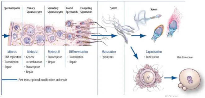
The process of spermatogenesis involves an array of complex biochemical, molecular, and cellular events, which can be arranged in distinct stages (Figure 2) (Mruk and Cheng 2004). These spermatogenesis stages are defined by the number of morphologically recognizable germ cell associations within the testis and vary according to species; in the rat there are 14 stages, in the mouse 12, and in the human there are 6 stages (O'Donnell, Robertson et al. 2001).
Figure 2 – Cellular changes occurring during spermatogenesis.
The stem cell population of the germinal cells lies on the basal lamina of the seminiferous tubules. The stages of spermatogenesis are represented by the three cell types, spermatogonia, spermatocytes and spermatids. A diploid spermatogonia which resides in the basal compartment of seminiferous tubules, divides mitotically to produce two diploid intermediate cells called primary spermatocytes. Each primary spermatocyte then moves into the adluminal compartment of the seminiferous tubules and duplicates its DNA and subsequently undergoes meiosis I to produce two haploid secondary spermatocytes, which will later divide once more into haploid spermatids. In the final stage of spermiogenesis occurs the maturation of spermatids into mature, motile spermatozoa. Adapted from du Plessis et al. (2011).
4
own numbers, and to cyclically produce primary spermatocytes, that undergo meiosis to produce haploid spermatids, which differentiate without further division into spermatozoa (Sharpe 1983; Johnson, Varner et al. 2000). The process of spermatogenesis can be divided in three major stages (Figure 2): mitotic proliferation of spermatogonia, meiosis of spermatocytes and spermiogenesis, the restructuring of spermatids into flagellated spermatozoa (Rudiger W. Schulz 2000; Walker 2010). In the first stage, the spermatogonia initiate to undergo mitotic division (a process that has its beginning at puberty) to produce a large number of germ cells, which will be available for entry into meiosis. (Arthur J. Vander 2001; O'Donnell, Robertson et al. 2001). These germinal stem cells include type A spermatogonia, intermediate spermatogonia (found only in rodents), and type B spermatogonia (O'Donnell, Robertson et al. 2001). Type A spermatogonia remains outside the blood-testis-barrier (BTB) and continue to multiply from puberty until death in order to maintain germ cell line. Type B spermatogonia migrate closer to the tubule lumen and differentiate into slightly larger cells called primary spermatocytes (Saladin 2001). In a second stage, primary spermatocytes replicate their DNA and hence initiate the first meiotic division of spermatogenesis form two secondary spermatocytes, from each primary spermatocyte (Arthur J. Vander 2001; O'Donnell, Robertson et al. 2001; Saladin 2001). Each secondary spermatocyte, in turn, undergoes a series of several mitotic divisions before it enters a meiotic prophase (Cooke and Saunders 2002). The last stage of spermatogenesis involves no further cell division but maturation of the sperm cells and is known as spermiogenesis. In this stage, occurs the formation and development of acrosome and flagellum, condensation of the chromatin, reshaping and elongation of the nucleus and removal of the cytoplasm before release of spermatid into the lumen of the tubule (Arthur J. Vander 2001; O'Donnell, Robertson et al. 2001; Saladin 2001). After the maturation of the spermatids, they are released from Scs into seminiferous tubule lumen prior to their passage to the epididymis via efferent ducts. This process is known as spermiation (O'Donnell, Robertson et al. 2001; O'Donnell, Nicholls et al. 2011).
1.2 Hormonal Regulation of Spermatogenesis
The hormonal control of the sexual functions in the male begins with secretion of gonadotropin-releasing hormone (GnRH) by the hypothalamus (Cheng, Wong et al. 2010). This hormone, in turn, stimulates the anterior pituitary gland to secrete two other hormones, called gonadotropic hormones: luteinizing hormone (LH) and follicle-stimulating hormone (FSH) (Figure 3). These gonadotropic hormones are the major endocrine regulators of spermatogenesis (Arthur J. Vander 2001; O'Donnell, Robertson et al. 2001; Arthur C. Guyton 2006; Cheng, Wong et al. 2010; Araujo and Wittert 2011)
The initiation and maintenance of quantitatively normal spermatogenesis and thus full fertility, by action of FSH and LH, needs a proper balance of the
hypothalamus-pituitary-testis axis (Figure 3) (Rudiger W. Schulz 2000; O'Donnell, Robertson et al. 2001). The target of LH is the Leydig cell via activation of the LH receptor (LH-R) and is the primary stimulus for the secretion of androgens that is testosterone. Leydig cell are located between the seminiferous tubules in the interstitial area of the testis, from where androgens diffuse into the seminiferous tubules, acting via androgen receptors to control spermatogenesis (Sharpe 1983; Rudiger W. Schulz 2000; O'Donnell, Robertson et al. 2001; Araujo and Wittert 2011).
Figure 3 - Feedback regulation of the hypothalamic-pituitary-testicular axis in males.
Note that GnRH stimulates the release of FSH and LH by the anterior pituitary gland. Feedback inhibition of LH secretion is a sex steroid-mediated event involving the testosterone, whereas FSH secretion has feedback regulation involving the Sertoli cell product inhibin. Stimulatory effects are shown by (+) and negative feedbacks inhibitory effects are shown by (-). GnRH, gonadotropin-releasing hormone; LH, luteinizing hormone; FSH,
follicle-6
FSH activates the proliferation of the SCs in the foetal and neonatal developmental stages and subsequently start the pubertal phase, that induces the mitotic activity of the spermatogonia and supports cellular differentiation, by meiotic divisions, until the round spermatid stage (van Alphen, van de Kant et al. 1988). FSH targets receptors within the SC to regulate spermatogenesis, by stimulating the production of numerous SC factors and synthesis of mRNA and protein in immature SCs, increases the secretion of specific proteins such as androgen binding protein, transferrin and inhibin (Gonzales, Risbridger et al. 1988).
This hypothalamus-pituitary-testis regulation of spermatogenesis is supported via negative feedbacks exerted by the components of this axis (Arthur C. Guyton 2006). It is important to point out that even though FSH and LH are produced by a single cell type, called gonadotropes, their secretion rates can be altered to different degrees by negative-feedback inputs (Figure 3) (Arthur J. Vander 2001). For instance, testosterone inhibits LH secretion in two ways: (1) by acting on the hypothalamus to decreased GnRH release and the decreased the amount of GnRH reaching the pituitary via hypophyseal portal circulation, resulting in less secretion of the gonadotropins; (2) by direct action on the anterior pituitary to inhibit LH release in response to any given level of GnRH. The decrease of FSH secretion is done directly on anterior pituitary and exerted by the protein hormone inhibin secreted by the SCs, without reducing LH and testosterone secretion. This process is the major source of feedback inhibition of FSH secretion and these cells are in all way the link between FSH and spermatogenesis (Arthur J. Vander 2001; Arthur C. Guyton 2006; Araujo and Wittert 2011).
1.3 Sertoli Cells
The SCs are the somatic constituents of the seminiferous tubules that extend from the base to the apex of the seminiferous epithelium and interact directly with developing germ cells (Mruk and Cheng 2004). They are in direct contact with the basement membrane, a modified form of the extracellular matrix present in the testis (Dym 1994). These cells are irregularly shaped, columnar cells that occupy a volume of approximately 17–19% in the seminiferous epithelium of adult rats (Russell, Ren et al. 1990). These sustentacular cells have a huge surface area, which permits them to sustain a vast number of developing germ cells at a Sertoli: germ cells ratio of approximately 1:50 in adult rat testis (Mruk and Cheng 2004). Each SC supports a maximum of germ cells, a number that varies between species and it is constant within a species
1.3.1 Sertoli Cells Functions
Since 1865, when Enrico Sertoli pursued the question of how the functions of the branched cells, named Sertoli cells, are linked to the formation of spermatozoa, scientists have called the SCs as the “nurse cells” because they provide nutrients and regulatory factors for the sustenance of germ cells (Sertoli 1865).
In the initial formation of the embryonic testis, SCs sequester the germ cells (gonocytes) inside of newly formed seminiferous tubules and inhibit the proclivity of these cells to enter into meiosis. Then SCs and germ cells undergo rapid proliferation (Griswold 1998; McLaren 2003; Phillips, Gassei et al. 2010). In puberty the cessation of mitosis of SCs occurs. The formation of tight junctions between adjacent SCs and the development of germ cells through meiosis and differentiation into spermatozoa generally occur as well during puberty (Griswold 1995; Griswold 1998; Alves, Oliveira et al. 2011).
As the germ cells move into the adluminal compartment, tight junctions between adjacent SCs form behind the germ cells creating a protective BTB (Meng, Holdcraft et al. 2005; Johnson, Thompson et al. 2008). This structure divides the seminiferous epithelium into basal and adluminal compartments and gives the required structural support for developing germ cells (Mruk and Cheng 2004; Johnson, Thompson et al. 2008; Cheng, Wong et al. 2010; Phillips, Gassei et al. 2010). The BTB creates a specialized environment, regulates the passage of molecules and serves as an immunological barrier (Mruk and Cheng 2004; Wong and Cheng 2005; Phillips, Gassei et al. 2010).
Hence, SCs are fundamental for the development of spermatogenesis because they produce specific products that are necessary for germ cell survival, and those products combine to form a unique and essential environment in the adluminal compartment providing structural support, nutrition and immunological protection to its progression, via direct contact with the germ cell and by controlling the environment milieu within the seminiferous tubules (Griswold 1995; Johnson, Thompson et al. 2008; Alves, Oliveira et al. 2011).
The production and secretion of a number of factors and proteins by SCs can be placed in categories according to their known biochemical properties and form the molecular basis for germ cell (Griswold 1998). The first category of secreted proteins includes the transport or bioprotective proteins that are secreted in relative high abundance and include metal ion transport proteins like transferrin and ceruloplasmin (Michael K. Skinner 1980; Michael K. Skinner 1983; Sylvester and Griswold 1994). The second category includes proteases and their inhibitors, which apparently are important in the process of remodeling tissue that occur during spermiation and movement of preleptotene spermatocytes into the adluminal compartment (Le Magueresse-Battistoni 2007). The last category of SC secretions includes the glycoproteins that form the basement membrane between the SCs and the peritubular cells (Davis, Papadopoulos et al. 1990; Raychoudhury, Irving et al. 1992; Richardson, Kleinman et al. 1995). A class of glycoproteins, also secreted by SCs, function as paracrine factors or growth factors that can be produced in very low abundance and still carry out their biochemical roles as such as Mullerian inhibiting substance (MIS), c kit ligand and inhibin (Griswold 1995). The production of a set of proteins that regulate and/or respond to pituitary hormone release is another important function of these cells influencing the mitotic activity of spermatogonia (Johnson, Thompson et al. 2008).
8 1.3.2 Hormonal Regulation of Sertoli Cells
SCs are an integral component of the mammalian seminiferous epithelium and their strategic position and structural relationships within the epithelium enable them to play an essential role in the maturation of the germ cells (Griswold 1998). Being a so important component for the spermatogenesis, their regulation is essential for a proper development of this process.
The somatic cells are the primary target for FSH and testosterone action in the mammalian testis (Tindall, Tash et al. 1981; Griswold 1998). These cells contain the majority of testicular plasma membrane receptors for these hormones, and thus are the major targets of the ultimate hormonal signals that regulate spermatogenesis (Tindall, Tash et al. 1981; Walker and Cheng 2005). The interaction of FSH with its specific cell surface receptors leads to stimulation of a number of intracellular events, including the activation of guanine nucleotide binding protein (G protein), adenylate cyclase and the cAMP-dependent protein kinase (PKA) pathway, that are involved in the control of metabolism, stress resistance and proliferation, in particular in connection with the available nutrient conditions (Laurent-Cadoret and Guillou 1995; Thevelein and de Winde 1999).
Testosterone and its derivates, such as dihydrotestosterone (DHT), are also crucial for the development of male reproductive tract. The genomic effects of androgens are mediated by the androgen receptor (AR) that belongs to the type I receptors of the nuclear receptor superfamily (Coleman and Smith 2001). In SCs, these hormones are responsible for a variety of actions, via the increase of the intracellular Ca2+ levels and activation of MAP kinase that
leads both for the phosphorylation of a transcription factor, the cyclic AMP–responsive element binding protein (CREB), which is an important transducer of FSH (Walker, Fucci et al. 1995) and testosterone (Fix, Jordan et al. 2004) signals to induce gene expression (Walker and Cheng 2005). They also bind to the androgen binding protein (ABP), a protein that is produced by SCs, following stimulation by FSH (Hansson, Weddington et al. 1975; Fritz, Rommerts et al. 1976). ABP serves as an excellent marker for SC function and binds specifically to testosterone, DHT, and 17β–estradiol (Tindall, Tash et al. 1981). ABP may serve as a carrier of androgen from testis to epididymis (French and Ritzen 1973).
SCs are also under the influence of estrogens as they express the estrogen receptor isoforms alfa (ERα) and beta (ERβ), nuclear hormone receptors and transcription factors that bonds to and are activated by estrogens (Saunders, Maguire et al. 1997; Atanassova, McKinnell et al. 1999; Cavaco, Laurentino et al. 2009). The presence of ERs in these cells seem to indicate that estrogens could play multiple roles in the development and/or function of the male reproductive and particularly in spermatogenesis. Although the role of estrogens in the development and physiology of male reproductive tract of and particularly in SCs physiology is still a matter of debate, endogenous estrogens appear to play a physiological role in the maturational development of SCs and potentially in the establishing of Sertoli cell - germ cells adhesion (MacCalman, Getsios et al. 1997).
Recent findings showed that sex steroid hormones (namely E2 and DHT) stimulation alters SCs metabolism and metabolism-related genes transcription. It has been described that DHT and E2 regulate glucose uptake and lactate production in human (Alves, Oliveira et al. 2011) and rat SCs (Rato, Alves et al. 2012). Moreover, DHT increased glucose uptake despite decreasing lactate synthesis, suggesting that this androgen may act on lactate synthesis (via LDH), and/or on its transport to extracellular medium (via MCTs) (Oliveira, Alves et al. 2011; Rato, Alves et al. 2012).
2. Apoptosis
Apoptosis is a fundamental biochemical cell death pathway characterized by several morphologic nuclear changes and plays a fundamental role in the maintenance of tissue homeostasis in the adult organism (Pothana Saikumar 1999; Fadeel and Orrenius 2005; Jourdain and Martinou 2009). This process occurs in a well-choreographed sequence of morphological events and when cells are exposed to various stimuli, they can interpret either as “good” or “harmful” and their path towards life and death (Jourdain and Martinou 2009).
After the activation of this cell death pathway, the dying cell undergoes nuclear and cytoplasmic condensation with blebbing of the plasma membrane, and eventually breaks up into membrane-enclosed particles called apoptotic bodies containing intact organelles, as well as portions of the nucleus (Gerschenson and Rotello 1992; Kiess and Gallaher 1998; Luo, Budihardjo et al. 1998; Pothana Saikumar 1999; Elmore 2007). These apoptotic bodies are then rapidly recognized, ingested and degraded by phagocytes or engulfed by macrophages or other adjacent healthy cells (Gerschenson and Rotello 1992; Elmore 2007).
Apoptosis occurs normally during development and aging and has a homeostatic mechanism to maintain cell populations in tissues (Monti, Troiano et al. 1992; Pothana Saikumar 1999). It also occurs as a defense mechanism such as in immune reactions or when cells are damaged by disease or noxious agents (Elmore 2007).
2.1 Apoptotic Activation Pathways
The mechanisms of apoptosis are highly complex and sophisticated, involving an energy dependent cascade of molecular events (Figure 4) (Elmore 2007). To date, research indicates that there are two main pathways involved in the apoptotic process: the extrinsic or death receptor pathway, occurring in response to activated death receptors present in the cell surface, and the intrinsic or mitochondrial pathway that occurs in response to signals originated from inside the cell (Fadeel and Orrenius 2005; Jourdain and Martinou 2009). Both involve caspase activation that leads to the cleavage of multiple intracellular substrates
10
(McConkey 1998). Thus, there are evidences that the two pathways are linked and that molecules in one pathway can influence the other (Igney and Krammer 2002).
The extrinsic signaling pathway plays a fundamental role in the maintenance of tissue homeostasis, especially in the immune system (Fadeel and Orrenius 2005) and involves transmembrane receptor-mediated interactions with death receptors that are members of the tumor necrosis factor (TNF) receptor gene superfamily (McConkey 1998; Locksley, Killeen et al. 2001). These death receptors play a critical role in transmitting the death signal from the cell surface to the intracellular signaling pathways (Ashkenazi and Dixit 1998).
Figure 4 - Mitochondrial control over apoptosis.
Both intrinsic and extrinsic pathways are shown. The signals originated from inside and outside the cell and the interactions between caspases, Bax, Bak, ROS and other proteins are represented. Adapted from Alves et al. (2009).
Stimulatory effect;
Translocation events;⊥
Inhibitory effect.Death receptors depend on signaling proteins that have a distinct set of modular protein motifs capable of homotypic interaction, including death domains and death effector domains that transmit signals from receptors to downstream effectors, such as the caspases (Aravind, Dixit et al. 1999). Ligation of death receptors, such as Fas (also known as APO-1 or CD95), is followed by the formation of the death-inducing signaling complex, resulting in the auto-catalytic activation of procaspase-8 (Kischkel, Hellbardt et al. 1995; Medema, Scaffidi et al. 1997; Salvesen and Dixit 1999). Once caspase-8 is activated, the cleavage of pro-apoptotic Bid, mediated by caspase-8, results in its translocation to mitochondria and cytochrome c
release in order to generate sufficient caspase activity to kill the cell (Figure 4) (Li, Zhu et al. 1998; Luo, Budihardjo et al. 1998).
The intrinsic signaling pathways that initiate apoptosis involves mitochondria and is triggered by many stimuli, such as cytotoxic stress, DNA damage and growth factor deprivation (Figure 4) (Jourdain and Martinou 2009). A set of cytotoxic stimuli and pro-apoptotic signal transducing molecules converge on mitochondria to induce outer mitochondrial membrane permeabilization (Decaudin, Marzo et al. 1998; Green and Kroemer 2004). This permeabilization is regulated by proteins from the Bcl-2 family (e.g. the pro-apoptotic protein Bax) and leads to the release of these pro-pro-apoptotic factors through the opening of a permeability transition pore (PTP), located in the outer mitochondria membrane (Schmitz, Kirchhoff et al. 2000). This pro-apoptotic molecule Bax, when activated, translocates to the outer membrane of mitochondria where it becomes an integral membrane protein (Gross, McDonnell et al. 1999; Pothana Saikumar 1999; Harris and Thompson 2000). The upregulation of Bax can be mediated by p53, a protein that act as a transcription factor involved in DNA repair (Chipuk, Kuwana et al. 2004). When activated, the p53 protein induces cell cycle arrest and is a key mediator of the apoptotic process. During stress signaling, p53 accumulates in the cells, and, when activated, initiates a cascade of events that results in apoptosis (Schwartz and Rotter 1998). The translocation of Bax to the outer membrane of mitochondria is followed by the release of many apoptosis promoting proteins that reside in the mitochondrial intermembrane space, including cytochrome c and apoptosis inducing factor (AIF). Both can activate caspases and endonucleases upon release into the cytosol (Figure 4) (Decaudin, Marzo et al. 1998; Harris and Thompson 2000). The release of cytochrome c from the intermembrane space of mitochondria leads to the formation of a complex formed by adapter protein Apaf-1 and the pro-caspase-9, called apoptosome (Schmitz, Kirchhoff et al. 2000; Elmore 2007; Ruwanpura, McLachlan et al. 2010). This ternary complex results in the activation of caspase-9 followed by sequential activation of effector caspase-3 and others (Pothana Saikumar 1999). In this point of the process, when the caspase cascade is initiated, the process of cell death cannot be reversed (Pothana Saikumar 1999). Mitochondria permeabilization can also lead to the release of the flavoprotein AIF from the mitochondria, that following its translocation to the nucleus, contributes to chromatin condensation and chromatinolysis, and lead to apoptosis (Cande, Vahsen et al. 2004; Vahsen, Cande et al. 2004).
Also belonging to the 2 family, the anti-apoptotic proteins, such as 2 and Bcl-xL, antagonize anti-apoptotic activity (Figure 4), therefore, interfering with the release of cytochrome c and AIF (Gross, Jockel et al. 1998). Anti-apoptotic members are initially integral membrane proteins found in the mitochondria, endoplasmic reticulum or nuclear membrane (Hockenbery, Nunez et al. 1990; Krajewski, Tanaka et al. 1993; Zhu, Cowie et al. 1996), in contrast with the pro-apoptotic members localize to cytosol prior to death signal
12
modification has been demonstrated to affect its anti-apoptotic activity (Haldar, Jena et al. 1995; Poommipanit, Chen et al. 1999).
2.2 Regulatory Mechanisms of Apoptosis
The process of apoptosis has an important role in the development and homeostasis of cell populations, and in the pathogenesis and expression of disease processes (Gerschenson and Rotello 1992). For this reason, an excessive or insufficient apoptosis may contribute for a pathogenesis of a wide variety of diseases related to ischemia, neurodegeneration, autoimmunity and viral infections, being also involved in the growth and regression of tumors (Pothana Saikumar 1999). Many factors and many levels of cell metabolism are involved in this death pathway. Viral infection, metabolic derangements, such as sudden changes in glucose concentrations, heat, irradiation, toxins and drugs, represent many of these factors that are capable of influencing the transition of a given cell to apoptosis (Kiess and Gallaher 1998).
Steroid hormones have a major role in the regulation of growth, development, homeostasis, and programmed cell death and, also, can induce or inhibit the process of cell death (Gould, Woolley et al. 1991; Henriksen, Hakovirta et al. 1995; Amsterdam, Dantes et al. 1997). Particularly, glucocorticoid hormones are responsible for the induction of apoptotic cell death in immature thymocytes and mature T cells (Wyllie 1980; Fletcher-Chiappini, Compton et al. 1993). Studies involving cells from ovary also indicate that sex steroids have an important role in the regulation of apoptotic cell death in these cells: whereas estrogens inhibit ovarian granulosa cell apoptosis, androgens seem to induce ovarian DNA fragmentation (Chun, Eisenhauer et al. 1996).
3. Apoptosis in Male Gonads
A stable germ cell population is determined by the balance of cell death (apoptosis) and cell multiplication, which is influenced by many biochemical factors as internal cues that control proper homeostasis of the testicular tissue, or external agents including testicular toxins, heat stress and chemotherapeutic agents (Shaha 2007; Stiblar-Martincic 2009; Ruwanpura, McLachlan et al. 2010). In addition, an imbalance of hormones can lead also to the apoptosis of germ cells (Dominique Royere and Reviers 2004; Shaha 2007). As known, the proliferation and differentiation of germ cells depends on the release of two hormones from the anterior pituitary gland, FSH and LH (Pareek, Joshi et al. 2007). The removal of these hormones induces germ cell apoptosis through indirect effects, since hormone receptors are present on the somatic cells in the testicular seminiferous tubule but not in the germ cells (Print and Loveland 2000).
The process of apoptosis occurs throughout spermatogenesis to maintain constant the number of cells that have undergone the process of meiosis (spermatocytes) and also to keep the homeostasis between SCs and germ cells (Levy and Seifer-Aknin 2001). Before the spermatogonia reaching maturity, up to 75% die in the process of programmed cell death (Stiblar-Martincic 2009). Male germ cell apoptosis occurs through two major pathways, involving either cell surface death receptors (extrinsic) or mitochondria (intrinsic) and depends on the type of stimuli they receive (Shaha 2007).
As said, SCs play an important role in the process of spermatogenesis by providing structural and nutritional support to germ cells. One of the ways that SCs control the germ cells population is through the Fas/FasL paracrine signal transduction system (extrinsic pathway) (Lee 1997; Dominique Royere and Reviers 2004). FasL is expressed by SCs and induces apoptosis when it binds with its receptor Fas, which is present in the surface of germ cells (Levy and Seifer-Aknin 2001; Stiblar-Martincic 2009). The binding of Fas/FasL activates the action of various caspases and causes the formation of apoptotic bodies resulting in the elimination of spermatocytes and spermatogonia cells (Levy and Seifer-Aknin 2001).
The intrinsic pathway of apoptosis involves the Bcl-2 group of proteins and different members of this group are involved in diverse situations (Shaha 2007). The pro-apoptotic protein Bax plays a fundamental role in the apoptosis of germ cell apparently during the first wave of spermatogenesis (Rodriguez, Ody et al. 1997). The anti-apoptotic members of the Bcl-2 family are also important in determining germ call fate (Print and Loveland 2000). For example, the anti-apoptotic protein Bcl-xL may regulate germ cell survival during the first wave of spermatogenesis, since it is expressed at high levels in testis during this period (Rodriguez, Ody et al. 1997).
3.1. Apoptosis in Sertoli Cells
The importance of apoptotic pathway in spermatogenesis has been well established. Since the SCs are important in the development of a successful spermatogenesis and the number of SCs can only determine a finite number of spermatozoa in the seminiferous tubules (Russell and Peterson 1984), any agent that impairs the viability of this cells may cause effect on spermatogenesis.
Some studies reported the apoptotic process in SCs. Shi and collaborators (2009) studied the influence of a xenobiotic agent, namely 1,1-dichloro-2,2-bis(p-chlorophenyl)-ethylene (p,p’-DDE). This metabolite is a derived of the organochlorine pesticide 2,2-bis(4-Chlorophenyl)-1,1,1-trichloroethane (DDT),it persists in the blood lipid and adipose tissue for several decades (Dyer 1995; Albanis, Hela et al. 1996) and may impair the normal reproductive functions in adulthood (Dyer 1995; Daxenberger 2002). Those authors suggested that exposure to p,p’-DDE might induce apoptosis of rat SCs through a FasL-dependent
14
cultured rat SCs through reactive oxygen species generation by the mediation of the release of cytochrome c into the cytosol and further the activation of caspase cascade (Song, Liang et al. 2008).
Le Goff and colleagues (2006) have also study the apoptosis of SCs and the influence of oxysterols, a derived from cholesterol, on the process. Oxysterol is an active molecule formed by the cholesterol oxidation (Schroepfer 2000) and may be derived from endogenous oxidation, mainly during cellular biosynthesis of 25-hydroxycholesterol (Russell 2000). This cholesterol derivative can cause cell death of several cell lines by apoptosis (Kolsch, Ludwig et al. 2001; Miyashita, Ozaki et al. 2002; Lim, Kang et al. 2003). In their work they showed that the apoptosis induced by 25-hydroxycholesterol decreases the Bcl-2 and increases the Bax expression in immature rat SCS and that 17β-estradiol (E2) induced a decrease of apoptosis caused by 25-hydroxycholesterol (Le Goff, Viville et al. 2006).
Dirami and colleagues (1995) have shown that a combination of peptide and steroid hormones and growth factors was able to significantly reduce apoptosis and increase cell survival of SCs in culture. However, in the absence of basement membrane, FSH and other regulators of SC function could not prevent SC apoptosis.
The role of E2 in the testicular development and his influence in the SCs apoptosis was also studied by Lucas and colleagues (2010; 2012). The influence of this hormone in a such important process, was investigated through the study of intracellular signaling events mediated by estrogen receptors (ERs) and G protein-coupled estrogen receptor 1 (GPER) involved in regulation of proliferation and apoptosis of rat SCs, via the activation of different signal transduction pathways, such as phosphorylation of the mitogen-activated protein kinase (MAPK). These reports demonstrated that the activation of this pathway, induced by E2, regulates proteins involved in proliferation and apoptosis of SCs, namely Bax and the anti-apoptotic proteins Bcl-2 and Bcl2l2. These authors demonstrated that E2 is involved in the upregulation of anti-apoptotic proteins Bcl-2 and Bcl2l2 and on the decrease of Bax expression. The CREB pathway was also investigated by these authors, since CREB phosphorylation has been linked to the activation of SC genes that potentially contribute to germ-cell survival (Scobey, Bertera et al. 2001). Their results showed a differential effect of E2 on the CREB-mediated transcription of pro- and anti-apoptotic genes of the same Bcl2 gene family. These results show that E2 modulate nuclear transcriptional events important for SC function and maintenance of normal testis development and homeostasis.
16
The aim of this work is to characterize the influence of 17β-estradiol (E2) and
5α-dihydrotestosterone (DHT) on the apoptotic signaling pathways in immature rat Sertoli cells cultures. We hypothesized that sex steroid hormones could have a role on the regulation of mitochondria related pro-apoptotic factors. So, we chose key points of the apoptotic pathway that interact with the mitochondria and evaluated mRNA expression of the p53, the pro-apoptotic Bcl2 family member Bax and the caspase-9 and caspase-3. The protein expression of caspase-9, Bax and the apoptosis-inducing factor (AIF) was also determined. Finally, Caspase-3 activity was evaluated as an endpoint marker of apoptosis.
18
1 - Chemicals
Hank’s Balanced Salts Solution (HBSS), Dulbecco’s Modified Eagle Medium Ham’s Nutrient Mixture F12 (DMEM: Ham’s F12), Ethylene Diamine Tetra Acetic acid (EDTA), Soybean Trypsin Inhibitor, DNAse, Collagenase type I, 17β-estradiol (E2), 5α-dihydrotestosterone (DHT), Bovine Serum Albumin (BSA), ExtrAvidin-Peroxidase Staining Kit, 3,3’-Diaminobenzidine Hydrochloride (DAB), trypsin-EDTA, Insulin-Transferrin-Sodium Selenite supplement (ITS supplement), TRI reagent and other drugs were obtained from Sigma Aldrich. (St. Louis, MO, USA). Fetal Bovine Serum (FBS) was obtained from Biochrom AG (Germany). Moloney Murine Leukemia Virus Reverse Transcriptase (M-MLV RT) and random hexamer primers were obtained from Invitrogen (CA, USA). dNTPs were obtained from GE Healthcare (Buckinghamshire, UK). 1x Buffer and Taq DNA Polymerase were obtained from Fermentas Life Sciences (Ontario, Canada). Polyclonal antibodies were obtained from Santa Cruz Biotechnology (Heidelberg, Germany) and Cell Signaling (Massachusetts, USA).
2 - Animals
Wistar male rats (Rattus norvegicus) were obtained from Charles River (Barcelona, Spain) and housed under a 12 h light-12 h darkness cycle, with food and water available ad libitum. Housing, maintenance and handling of animals comply with the “Guide for the Care and Use of Laboratory Animals”; published by the US National Institutes of Health (NIH Publication No. 85-23, revised 1996) and the rules for the care and handling of laboratory animals (Directive 86/609/EEC).
3 - Sertoli cell culture
Ten male Wistar rats (20-day-old) were sacrificed by cervical dislocation, the testis were immediately excised in aseptic conditions and washed two times in a 50 mL conical tube in 30 mL of ice cold HBSS containing 10000 U/mL of penicillin, 10mg/mL streptomycin and 25 μg/ ml amphotericin B (pH 7,4). SCs were isolated by slight modifications of the method previously described by Oliveira and collaborators (Oliveira, Alves et al. 2012). SC culture purity was assessed by the immunoperoxidase detection of specific markers, Anti-Mullerian hormone and Vimentin as described elsewhere (Steger, Rey et al. 1996). Shortly, cells were grown on 6 well culture plates, incubated overnight at 4ºC with primary polyclonal antibody and labeled streptavidin-biotin method using an ExtrAvidin-Peroxidase Staining Kit, giving a brown coloration to the SCs after reaction with diaminobenzidine. The cell nucleus was then stained with haematoxylin. Negative-control incubations were executed using PBS instead of
primary antibody. Cultures were examined by phase contrast microscopy and selected if cells contaminants were below 5% after 96h.
4 - Experimental groups
SCs were allowed to grow until reach 90-95% of confluence, and then washed thoroughly and the medium replaced by serum-free medium (DMEM:F12 1:1 with ITS supplement, pH 7.4). To evaluate the effects of sex hormones on mRNA and protein expression, SCs were treated during 50 hours with 100 nM of E2 or 100 nM of DHT. DHT was chosen as androgen family representative because is not conversable to E2 by the cells (Mahesh, Muldoon et al. 1975). The concentrations of the sex steroid hormones were chosen based on available data which reported that intratesticular interstitial fluid concentrations of those hormones are particularly higher than those of circulating plasma, reaching values up to 200 nanomolar (Turner, Jones et al. 1984; Setchell 2004). Control groups were treated with same amount of solvent (EtOH) used in DHT and E2 groups (<0,025% v/v). At the end of the treatment, the total number of cells per flask was determined with a neubauer chamber and cells were collected for RNA or protein extraction.
5 - RT-PCR
Total ribonucleic acid (RNAt) was extracted from isolated SCs using TRI reagent according to the manufacturer’s instructions. RNA concentration and absorbance ratios (A260/A280) were spectrophotometricaly determined (NanophotometerTM, Implen, Germany). RNAt (1μg)
was reverse transcribed in a final volume of 20 μL with 200 U of M-MLV RT according to the manufacturer’s protocol, using 250 ng of random hexamer primers and 0.5 mM of each dNTP. The resulting cDNA was used to amplify Bax, caspase-3, caspase-9 and p53 cDNA fragments using exon-exon spanning primer sets (Table 1). cDNA (1 μL) was amplified in a final volume of 25 μL, containing Reaction Buffer (2 mM MgCl2, 0.2 mM dNTP), 0.2μM of each primer and 0.625 U Taq DNA Polymerase. Optimal annealing temperature and the number of cycles needed for amplification phase of fragments are shown in Table 1. Expression of Bax, caspase-3, caspase-9 and p53 mRNA was normalized with 18S gene expression. Densities from each band were obtained with BIO-PROFIL Bio-1D Software from Quantity One (Vilber Lourmat, Marne-la-Vallée, France) according to standard methods. The band density acquired for each studied gene was then divided by the respective 18S band density and results are expressed as fold variation (induction/reduction) versus the control group.
20
Table 1 - Oligonucleotides and Cycling Conditions for PCR Amplification of p53, Bax, Caspase 9, Caspase 3 and 18S.
Gene Sequence (5'- 3') AT (ºC) Amplificon Size (bp) C
P53 Sense: CTG CCC ACC ACA GCG ACA GG 59 471 35
Antisense: AGG AGC CAG GCC GTC ACC AT
Bax Sense: CGC GTG GTT GCC CTC TTC TAC TTT 59 124 35 Antisense: CAA GCA GCC GCT CAC GGA GGA
Casp 9 Sense: TGC AGG GTA CGC CTT GTG CG 61 130 35
Antisense: CCT GAT CCC GCC GAG ACC CA
Casp 3 Sense: AGG CCT GCC GAG GTA CAG AGC 60 255 35 Antisense: CCG TGG CCA CCT TCC GCT TA
18 S Sense: AAG ACG AAC CAG AGC GAA AG 56 149 25
Antisense: GGC GGG TCA TGG GAA TAA
Abbreviations: AT - annealing temperature; C - Number of cycles during exponential phase of amplification; Casp - caspase.
6 - Western Blot
Western Blot procedure was performed as previously described (Alves, Machado et al. 2011). Briefly, total proteins were isolated from rat SCs using RIPAS buffer supplemented with protease and phosphatase inhibitors (1x PBS, 1%NP-40, 0,5% sodium deoxycholate, 0,1% SDS, 1mM PMSF, supplemented with 1% protease inhibitor cocktail and 100mM sodium orthovanadate). Protein concentration was determined by the Bradford micro assay. Proteins samples (50 µg) were fractionated in poliacrylamide gels and transferred to polyvinylidene difluoride membranes. The membranes were then incubated overnight a 4ºC with goat anti-Bax (1:5000, no. 2772, Cell Signaling Technology), rabbit anti-cleaved caspase-9 (Asp353) (1:1000, no. 9507, Cell Signaling Technology), or mouse anti-AIF (1:1000, no. 13116, Santa Cruz Biotechnology) primary antibodies. Mouse anti-actin primary antibody (1:1000, Sigma, Roedermark, Germany, A 5441) was used as protein loading control in different experimental conditions. The immune-reactive proteins were detected with goat anti-rabbit IgG-AP (1:5000, Santa Cruz Biotechnology Heidelberg, Germany, Sc 2007) or goat anti-mouse IgG-AP (1:5000, Santa Cruz Biotechnology Heidelberg, Germany, Sc 2008). Membranes were reacted with ECF detection system (GE, Healthcare, Weßling, Germany) and read with the BioRad FX-Pro-plus (Bio-Rad, Hemel Hempstead, UK). Using the Quantity One Software (Bio-Rad, Hemel Hempstead, UK) the densities from each band were obtained, according to standard methods. The obtained band density was divided by the respective actin band density and then normalized as percentage of the respective control.
7 - Caspase-3 activity assay
Caspase-3 activity was spectrophotometrically assessed by determining the cleavage of the respective colorimetric substrate as previously described (Alves, Machado et al. 2011). Briefly, 25 µg of proteins were incubated in assay buffer (25 mM HEPES, pH 7.5, 0.1% CHAPS, 10% sucrose and 10 mM DTT) with 100 µM of caspase-3 substrate (Ac-DEVD-pNA) for 2 h at 37º C. The caspase-3 activity was determined by detection of the chromophore p-nitroanilide, measured at 405 nm in a spectrophotometer. The method was calibrated with known concentrations of p-nitroanilide. The attained activities were expressed in percentage versus the control group.
8 - Statistical analysis
The statistical significance of mRNA and protein expression, and caspase-3 activity among the experimental groups was assessed by two-way ANOVA, followed by Bonferroni post-test. All experimental data are shown as mean ± SEM (n=5 for each condition). Statistical analysis was performed using GraphPad Prism 5 (GraphPad Software, San Diego, CA). P<0.05 was considered significant.
22
1.
E2 down-regulates mRNA transcript levels of apoptotic signaling markers
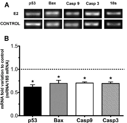
The treatment with E2 was followed by the analysis of gene expression of apoptotic signaling markers. p53 is a key regulator of apoptosis and can be translocated to the mitochondria, inducing apoptosis, or can localize directly in the sites of DNA damage and promote its proper repair (Speidel 2010). For this reason we evaluated the effect of E2 on the transcription of p53 in rat cultured SCs. The mRNA expression levels of p53 in E2-treated cells were significantly reduced. We observed a 0.62±0.05 fold reduction of p53 mRNA levels in E2-treated cells when compared with the control group (Figure 5).
A crucial event for initiating the intrinsic apoptotic pathway is the opening of a permeability transition pore that requires the activation of pro-apoptotic members, such as Bax, in a process mediated by p53 (Majors, Betenbaugh et al. 2007). Thus, we evaluated the mRNA levels of this pro-apoptotic protein and we also detected a significant decrease in E2-treated cells (0.70±0.07 fold reduction to control) (Figure 5).
After Bax activation and its translocation to the outer membrane of mitochondria, the release of pro-apoptotic factors occurs, leading to the cleavage of caspase-9 followed by sequential activation of the effector caspase-3. Thereby, we assessed the mRNA levels of caspase-9 and caspase-3 that were also significantly reduced (0.70±0.02 and 0.69±0.03 fold, respectively) in E2-treated cells (Figure 5).
Figure 5 - Effect of E2 on p53, Bax, caspase 9 and caspase 3 mRNA levels in rat Sertoli cells.
Panel A shows a representative agarose gel electrophoresis. Panel B shows pooled data of independent experiments, indicating the fold variation of mRNA levels found in cultures with 100 nM E2 when compared with cultures on control condition (dashed line). Results are
24
2. mRNA transcript levels of apoptotic markers are downregulated after
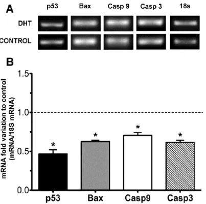
DHT treatment
Following the treatment of SCs with DHT, we analyzed the effect of this androgen in the transcript levels of signaling apoptotic markers. Starting with p53, we observed a pronounced decrease for the mRNA levels (0.47±0.05 fold reduction) (Figure 6). Bax activation is a key point of apoptosis as it leads to the release of pro-apoptotic factors. So we analyzed the effect of DHT in the gene transcript levels of this pro-apoptotic protein. The results showed a significant 0.63±0.02 fold decrease in the mRNA expression levels of Bax in DHT-treated cells (Figure 6).
The next apoptotic markers, caspase-9 and caspase-3, represent an endpoint in this process. We observed a significant decreased in mRNA levels of both caspase-9 and caspase-3 (0.70±0.04 and 0.62±0.03 fold reduction, respectively) (Figure 6) in DHT-treated cells.
Figure 6 - Effect of DHT on p53, Bax, caspase 9 and caspase 3 mRNA levels in rat Sertoli cells.
Panel A shows a representative agarose gel electrophoresis. Panel B shows pooled data of independent experiments, indicating the fold variation of mRNA found in cultures with 100 nM DHT when compared with cultures on control condition (dashed line). Results are expressed as means ± SEM (n=5 for each condition). Significantly differently results (p< 0,05) are indicated: * relatively to control.
3. E2 decrease cleaved
caspase-9 protein levels
After assessing the variation of mRNA levels in E2-treated SCs, we also evaluated the effect on the protein levels of some of the most relevant proteins involved in apoptotic signalling. Although we observed a significant decrease in the mRNA expression levels of Bax, these results were not followed by a significant decrease in the protein levels, as Bax protein levels in E2-treated cells did not present significant differences relatively to the control group (Figure 7).
The intrinsic apoptotic pathway involves also the release of caspase-independent death effectors such as apoptosis-inducing factor (AIF) (Vahsen, Cande et al. 2004). Its release represents an important stage in apoptosis but we found no differences regarding the protein expression of AIF in E2-treated when compared to the control SCs (Figure 7).
Caspase-9 triggers a caspase-signalling cascade to induce apoptosis (Zou, Yang et al. 2003). The binding of pro-caspase-9 to Apaf-1 leads to the autolytic cleavage of pro-caspase-9 (Zou, Yang et al. 2003), which allows the initiation of the caspases cascade (Twiddy and Cain 2007). Following the noted decrease on caspase-9 mRNA levels, in E2-treated cells, we also evaluated the protein levels that were consistent with those obtained for the mRNA levels, as we observed a significant 0.81±0.06 fold reduction to the control SCs (Figure 7).
Figure 7 - Effect of E2 on Bax, AIF and cleaved caspase 9 protein levels in rat Sertoli cells.
Panel A shows a representative western blot experiment. Panel B shows pooled data of independent experiments, indicating the fold variation of protein levels found in cultures with 100 nM E2 when compared with cultures on control condition (dashed line). Results are expressed as means ± SEM (n=5 for each condition). Significantly differently results (p< 0,05)
26
4. AIF protein levels are highly decreased after DHT-treatment
As said, during the apoptotic process, there is a release of potentially lethal proteins from the mitochondrial intermembrane space including caspase-independent apoptosis effectors such as AIF (Vahsen, Cande et al. 2004). This apoptotic marker has the nucleus of the cells as a target, where it participates in the degradation of DNA (Cande, Vahsen et al. 2004). The protein levels of AIF remained unaltered when treated with E2 (Figure 8), but DHT-treated cells presented a significant decrease (0.82±0.06 fold reduction) in AIF protein expression levels (Figure 8).
On the other hand, DHT treatment did not cause a significant alteration in Bax protein levels (Figure 8). Finally, we analysed the protein expression of cleaved caspase-9 in DHT-treated cells, concluding that the protein levels of cleaved caspase-9 were not significantly different from the control group (Figure 8).
Figure 8 - Effect of DHT on Bax, AIF, and cleaved caspase 9 protein levels in rat Sertoli cells.
Panel A shows a representative western blot experiment. Panel B shows pooled data of independent experiments, indicating the fold variation of protein levels found in cultures with 100 nM DHT when compared with cultures on control condition (dashed line). Results are expressed as means ± SEM (n=5 for each condition). Significantly differently results (p< 0,05) are indicated: * relatively to control.
5. Caspase-3 activity is highly decrease after hormonal treatment
Caspases are critical mediators of the apoptotic process. Activation of caspase-3 requires the proteolytic cleavage of its inactive precursor enzyme, pro-caspase-3, and this is
out by activated caspase-9 (Zou, Yang et al. 2003). The activation of caspase-3 turns the apoptotic pathway irreversible thus, following the alteration on the mRNA or protein expression of caspase-3 and 9, we measured caspase-3 activity in SCs subject to hormonal treatment (DHT and E2-treated groups) by measuring the cleavage of a specific substrate and the release of a chromophore (p-nitroanilide). We observed that caspase-3 activity was decreased in both E2 and DHT-treated cells (Figure 9). The results obtained for E2-treated cells showed a more striking effect, as we observed an intense decrease of caspase-3 activity of 47±9% relatively to that of the control group (Figure 9). Nevertheless, DHT-treated cells also showed a significant decrease in the activity of caspase-3 to 59±12% (Figure 9). These results are in agreement with and emphasize the alterations observed in caspase-3 mRNA levels.
Figure 9 - Effects of E2 and DHT on caspase-3 activity.
Results are presented as pooled data of independent experiments, indicating the percentage variation of activity levels found in cultures with 100 nM E2 (E2) or 100 nM DHT (DHT) when compared with cultures on control condition (dashed line). Results are expressed as mean ± SEM (n=5 for each condition). Significantly differently results (p< 0,05) are indicated: * relatively to control.
28
The role of SCs in the establishment of a successful spermatogenic event has been widely investigated but many questions are still unanswered. During their functioning, these cells suffer the influence of various endocrine and exocrine regulators (Griswold 1998; Sofikitis, Giotitsas et al. 2008). Sex steroid hormones are known as the key regulators of spermatogenesis and SCs are the mediators of their action (Carreau, Silandre et al. 2007). In fact, within the seminiferous tubules, SCs are known to express receptors for androgens and estrogens (Verhoeven and Cailleau 1988; Cavaco, Laurentino et al. 2009), rendering them as the primal targets for the hormonal signaling that regulates spermatogenesis.
Estrogens and androgens are known to play a critical role in preventing apoptosis in wide range of mammalian cells, namely in cells from the male reproductive tract (Erkkilä, Henriksén et al. 1997; Pelzer, Schumann et al. 2000; Pentikäinen, Erkkilä et al. 2000; Bialek, Zaremba et al. 2004; Laurentino, Gonçalves et al. 2011). In fact, apoptosis is a cellular event essential for the occurrence of a normal spermatogenesis as it permits the control of germ cells number (Tesarik, Guido et al. 1998). Nevertheless, as SCs can only accommodate the differentiation of a finite number of germ cells (Orth, Gunsalus et al. 1988), any agent that impairs the viability of SCs can cause deleterious effects on spermatogenesis, resulting in a lower fertility or even in infertility. So the enlightenment of the factors that can influence and modulate SCs apoptosis is of extreme relevance.
Recently, it has been described that E2 and DHT function as modulators of cell metabolism both in rat and in human SCs (Alves, Oliveira et al. 2011; Rato, Alves et al. 2012) and it has been also described that alterations of cell metabolism, particularly in glucose metabolism, are closely related with apoptosis (Majors, Betenbaugh et al. 2007). Decreases in intracellular ATP or increases in oxidative stress due to reduced glycolysis can lead to increased apoptosis (Moley and Mueckler 2000) and mitochondria seem to be the link between metabolic and apoptotic signaling and the source of many of the apoptotic proteins. Hence, following previously reported results on the effect of E2 and DHT in SCs metabolism, we hypothesized that sex steroid hormones could have an active participation on the regulation of these mitochondria related pro-apoptotic proteins.
The apoptotic pathway can be triggered due to several signals, and when a death signal occurs and the p53 protein is activated, a cascade of events is initiated resulting in apoptosis (Schwartz and Rotter 1998). p53 protein is known to induce the expression of pro-apoptotic members of the of Bcl-2 protein family, such as Bax (Terrones, Antonsson et al. 2004). When we assessed the effect of E2 or DHT on the expression of these apoptotic markers in rat SCs we could observe a significant down-regulation. Indeed, the results obtained showed that both sex steroid hormones were able to significantly decrease the mRNA levels of p53 and Bax, which is in agreement with previous results that describe that both estrogens and androgens are capable of preventing apoptosis.